Hyaluronic Acid in Musculoskeletal Rehabilitation: Comparison
Please note this is a comparison between Version 2 by Mona Zou and Version 3 by Mona Zou.

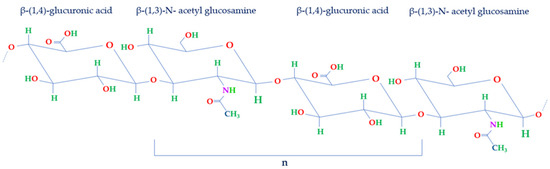
Hyaluronic acid (HA) naturally occurs as a biopolymer in the human body, primarily in connective tissues like joints and skin. Functioning as a vital element of synovial fluid, it lubricates joints, facilitating fluid movement and diminishing bone friction to protect articular well-being. Its distinctive attributes encompass notable viscosity and water retention capacities, ensuring flexibility and absorbing shock during motion. Furthermore, HA has gained significant attention for its potential benefits in various medical applications, including rehabilitation. Ongoing research explores its properties and functions, especially its biomedical applications in several clinical trials, with a focus on its role in improving rehabilitation outcomes. But the clinical and biochemical implications of HA in musculoskeletal rehabilitation have yet to be fully explored.

  • hyaluronic acid
  • active biomolecule
  • biomaterial
  • rehabilitation
  • HA injection
  • physical therapy

1. Hyaluronic Acid

Comprising a glycosaminoglycan (GAG), HA consists of a repetitive arrangement of disaccharide units (n). These units are structured with β-1,4-glucuronic acid (GlcA) and β-1,3-N-acetyl glucosamine (NAG), interconnected by means of β-1,3 and β-1,4 glycosidic linkages (Figure 1) [1][2].
Figure 1.
Structure of disaccharide repeating unit of HA. HA structure is made of repeated units of NAG and GlcA held together by β-1,3 and β-1,4 glycosidic bonds (n).

2. HA in the Rehabilitation of Musculoskeletal Diseases

The versatility of HA, its capacity to retain water, its compatibility with the human body, and its natural biodegradability all contribute to its widespread appeal in numerous therapeutic applications. These qualities continue to position HA as a highly effective therapeutic compound for addressing various medical conditions, including musculoskeletal diseases, and applications [3] (Figure 2). Consequently, a range of HA-based products have been developed and are readily available in the market. Furthermore, HA is not only employed on its own but also in conjunction with other bioproducts and therapies, further expanding its utility.
Figure 2. The efficacy of HA in the rehabilitation of musculoskeletal diseases.

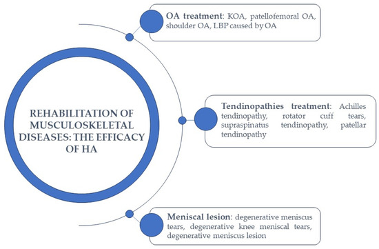
2.1. HA-Based Therapy in Osteoarthritis Treatment

As described above, HA is found in substantial amounts, particularly within cartilage and synovial fluid. It holds a vital function in the natural biomechanics of synovial fluid, governing tissue lubrication and elasticity [4][5].
The degradation of HA is a gradual process that can take place through enzymatic or nonenzymatic reactions. When HA undergoes degradation or its synthesis slows down, it leads to a decrease in its MW, which in turn affects various physical and chemical properties such as tissue volume, viscosity, and elasticity. HA is a molecule that is abundant in the knee joint, particularly in synovial fluid, but it is also present in articular cartilage [6].
It is crucial to emphasize that the decline in HA levels is not only linked to aging but also correlates with the progression of specific conditions, such as OA [4][5]. In this case, the viscoelastic properties of synovial fluid undergo substantial changes due to a reduction in both HA concentration and MW within the synovial fluid [7].
The pathogenic processes of OA induce modifications in the metabolism of type B synoviocytes, leading to the production of pathogenic HA [7][8]. This alteration in the quality of HA contributes to heightened mechanical strains within the joint, resulting in reduced lubrication capacity and, consequently, the stimulation of nociceptors that cause pain [9][10][11][12].
Notably, there exists a significant connection between joint movements and the active secretion of HA. The quantity of HA released appears to be influenced by both the frequency and duration of joint movements. This discovery suggests a biochemical mechanism that provides backing for the potential utilization of physical therapy in the management of OA [13][14][15].
Specifically, KOA emerges as a prevalent global joint condition, and its occurrence increases with age [16][17][18].
In particular, the knee joint presents a challenging biomechanical environment due to its avascular, aneural, and alymphatic nature. In this context, synovial fluid plays a crucial role as a lubricant with unique rheological properties [6]. Additionally, synovial fluid possesses the capability to scavenge free radicals and regulate intracellular activity and protein binding. The progression of KOA is intimately linked to the deterioration of synovial fluid’s lubricating function [6]. This deterioration results from the depolymerization of endogenous HA with high MW (ranging from 6500 to 10,900 kDa), transforming it into low MW HA (ranging from 2700 to 4500 kDa). HA with low MW leads to synovial fluid that exhibits significantly reduced mechanical and viscoelastic properties [6].
Moreover, treatment strategies for OA encompass not only surgical interventions but also efforts to reduce risk factors [19][20][21]. Additionally, they may involve participation in physical therapy and rehabilitation programs [22][23][24], as well as the use of oral corticosteroids or nonsteroidal anti-inflammatory drugs (NSAIDs), the use of oral dietary supplements [25][26], intra-articular injections of corticosteroids, HA, or platelet-rich concentrates [27][28][29][30][31][32][33][34][35][36][37].
Generally, the injection of high MW exogenous HA can be employed to mitigate the loss of synovial fluid properties caused by the endogenous depolymerization of natural HA. It is important to note that exogenous HA does not replace or restore the body’s own HA, but its presence in the joint induces symptom improvement in KOA over several months [38]. This improvement occurs because exogenous HA stimulates the synthesis of glycosaminoglycan and/or proteoglycan, allowing the synovial fluid to maintain its viscoelastic properties. Additionally, exogenous HA exhibits a significant anti-inflammatory effect, which, through secondary mechanisms, helps reduce pain.
FDA-approved HA injection products come with various physicochemical characteristics that can make one product more efficient or competitive than another. Among these characteristics, MW is a key consideration for clinicians, with options ranging from 500 up to 6000 kDa. Generally, the higher the MW, the longer the therapeutic efficacy [38].
Based on recent literature, exercise rehabilitation therapy continues to be a primary option for OA treatment [32][39]. Its crucial role lies in restoring muscle balance and proper load distribution, ultimately alleviating pain and enhancing function.
In particular, to assess pain in KOA, the most commonly used scales are the Visual Analog Scale (VAS) and the Western Ontario and McMaster Universities Osteoarthritis Index (WOMAC), in which the severity of pain is correlated with intra-articular changes. The radiological score Kellgren-Lawrence (KL) is associated with the presence of osteophytes and cartilage lesions and serves as an independent predictor for the VAS scale [40].
A recent meta-analysis of randomized controlled trials conducted by Liao et al. (2023) widely investigated the impact of combining intra-articular injections with rehabilitation therapy in patients suffering from KOA. Their specific emphasis was on assessing the combined therapy’s influence on pain levels, overall functional improvement, and walking capacity. Through this approach, they aimed to establish the most effective treatment option by ranking the effectiveness of each combined therapy regimen and exploring any factors that might influence treatment outcomes [41].
In the specific case of the combination of intra-articular injections of HA and physical therapy, it was observed that this treatment appeared to be more effective in reducing pain compared with physical therapy alone. Additionally, it allowed for better walking capacity in the short term, both compared with physical therapy alone and in comparison with other intra-articular injections [41].
In a blinded and randomized controlled study, Saccomanno et al. (2016) investigated the effectiveness of combining HA injections with personalized rehabilitation programs based on exercise-based rehabilitation (EBR) in patients with KOA [16]. A total of 165 participants with KOA were randomly assigned to three treatment groups: the first group received three intra-articular injections of high MW HA (Orthovisc 2 mL; 15 mg/mL; Anika Therapeutics Inc., Bedford, MA, USA); the second group underwent only rehabilitative exercises (detailed program of knee exercises); and the third group received both treatments, combining HA injections with an EBR program [16].
The analysis of the data revealed that all three treatment groups experienced decreased pain, improved flexibility, and enhanced functionality.
In particular, patients in group 1 showed the least pronounced treatment effect, which remained consistent over time. In contrast, patients in group 2 experienced notable declines in pain, stiffness, and functional results from the initial to the final follow-up. Finally, analysis of the WOMAC pain subscale revealed that patients in group 3 exhibited the most significant reduction in pain at the 1-month follow-up [16].
In a recent parallel randomized trial, Onu et al. (2022) conducted a study to evaluate the effectiveness of a HA-based product when used in combination with physical therapy for KOA treatment [42]. The research focused on a group of 52 patients who had been diagnosed with stage 2 KOA according to the KL scale, as determined through radiological examination of anterior-posterior knee X-rays [42]. In KL stage 2, patients typically exhibit osteophytes and narrowing of the joint space.
The patients were divided into two groups. The pilot group, consisting of 37 patients, received intra-articular HA injections (3 mL of Kombihylan, Ropharm, Romania) in combination with 10 consecutive sessions of physical therapy [42]. On the other hand, the control group was treated only with HA injections. In this case, a high-molecular-weight HA, 3 MDa, in the form of a viscoelastic solution obtained through bacterial fermentation of a Streptococcus strain, was used [42].
In particular, the physiotherapy treatment, in this case, involved several components: electrotherapy, specifically conventional transcutaneous electrical nerve stimulation (TENS) electroanalgesia; low-level laser therapy (LLLT); ultrasound (US) therapy; and physical therapeutic exercises (PTEs), which consisted of a 40 min session with moderate-intensity exercises, including a 5 min warm-up on a stationary bike, static quads with a 7 s hold, knee extensions over a roll with a 7 s hold, single-leg raises for 50 repetitions, step-ups for 50 repetitions, calf raises for three sets of 10–15 repetitions, and wall squats with a 5–10 s hold [42]. Also, neuroproprioceptive facilitation (PNF) techniques were incorporated into four movement patterns, and cryotherapy in the form of ice packs was applied at the conclusion of the physiotherapy session to cool down the affected knee [42].
The study observed that the WOMAC score, which assesses osteoarthritis severity, decreased for both groups of patients, but notably, the treated group showed significant improvements at the 3-month mark. This led to the conclusion that physical therapy consistently enhanced the quality of life for these patients. Additionally, it was demonstrated that VAS pain scores decreased from 5.7 to 2 in the treated group. Furthermore, the treated group experienced an increase in muscle strength [42].
Another multicenter study was conducted to assess the effectiveness of a comprehensive approach for treating and rehabilitating KOA. This approach combined physical therapy with HA injections, aiming to provide both clinical benefits and cost-effectiveness [43]. The rehabilitation program featured injectable HA and therapeutic physical exercises, including muscle strengthening, flexibility routines, and proprioception training [43].
Specifically, the study involved 553 patients with symptomatic KOA who had previously undergone unsuccessful pharmacological treatments. These patients were monitored over an 8-week period at 27 specialized centers across the USA. Guided by experienced physiotherapists, they participated in physical therapy sessions two to three times per week.
The results were promising, showing a significant reduction in knee pain, with a 59% decrease in all patients. The WOMAC score improved by 44% to 51%. Even patients with advanced stages of KOA (KL3–KL4) experienced a notable reduction in specific symptoms [43].
Moreover, the combined therapy program proved to be cost-effective over a 2-year monitoring period. It postponed the need for surgical knee treatments, underscoring the potential of rehabilitation through physical therapy and HA injections to slow down KOA progression while maintaining a favorable cost-effectiveness ratio [43].
Long-term outcomes measured included knee pain severity, medication use, knee operations, and health utility scores. The results were promising, with a reduction in knee pain severity during the 8-week program. At 1 and 2 years post-treatment, medication use remained common, and the utilization rates for total knee arthroplasty were 10.4% and 18.0%, respectively. Additionally, health utility scores improved significantly. This study demonstrated that the 8-week multimodal KOA treatment program provided meaningful improvements in KOA symptoms, even in advanced cases, and was cost-effective over a 2-year follow-up period [43].
A recent postmarket, single-blind, multicenter randomized controlled clinical trial was conducted to evaluate the treatment outcomes in a relatively young and active population of individuals with patellofemoral OA and/or tibiofemoral OA (NCT03281837). The trial aimed to compare the responses to two weekly intra-articular HA injections, administered with a 1-week interval between injections, of HYMOVIS 24 mg/3 mL (Fidia Pharma USA Inc., Florham Park, NJ, USA) in combination with a physical exercise program versus a physical exercise program alone (NCT03281837). In this study, 148 patients were divided into three groups: the first group received one injection of Hymovis per week for two consecutive weeks, along with at least 8 weeks of a physical exercise program; the second group underwent treatment with only the physical therapy program, without any additional intervention, while the last group included patients who were randomized to receive only the physical exercise program initially. If they did not respond to this intervention after 3 months, they had the option to cross over and receive two intra-articular weekly injections of Hymovis, with each injection given 1 week apart (NCT03281837). As of now, the results of this study have not been published, but it is hypothesized that the combined therapy approach, involving HA injections and physical exercise, will demonstrate its effectiveness in managing patellofemoral and tibiofemoral osteoarthritis (NCT03281837).

HA-Based Therapy in Low Back Pain

It is important to note that OA can manifest various symptoms, including lower back pain (LBP) [22], which is a highly prevalent issue. It is acknowledged that several underlying factors can contribute to LBP, encompassing conditions such as tumors, infections, fractures, and inflammatory disorders. Nonetheless, OA stands out as the most common cause. Additionally, mechanical and structural issues, such as lumbar spinal stenosis, spondylolysis, spondylolisthesis, and congenital deformities like scoliosis or hyperkyphosis, can contribute to LBP [22].
Current guidelines advocate exercise as the primary approach for addressing LBP [44][45]. Remarkably, rehabilitation utilizing the McKenzie method appears to yield superior short-term pain reduction and disability management in both acute and chronic LBP when compared with other physiotherapeutic approaches. A recent study demonstrated the effectiveness of oral viscosupplementation, involving a blend based on Fortigel (Gelita, Eberbach, Germany), including collagen peptides, Vitamin C, copper, manganese, and sodium hyaluronate, in conjunction with the McKenzie method kinesitherapy, compared with kinesitherapy alone in the treatment of chronic LBP due to osteoarthritis [22].
Specifically, in this randomized clinical trial, 60 patients were randomly allocated to two groups. Both groups underwent physiatric evaluations, encompassing medical history, physical exams, and imaging. Group A received a 3-week McKenzie rehabilitation program consisting of nine sessions, while group B received the same program in addition to a daily dietary supplement of Fortigel, which contains vitamin C (80 mg), sodium hyaluronate (50 mg, with 46 mg as HA), manganese (1 mg), and copper (0.5 mg), throughout the treatment period [22].
The findings indicated that both group A and group B experienced improvements in pain and disability scores (VAS and Oswestry Disability Index, ODI). Particularly, group B demonstrated a more significant reduction in VAS and ODI scores, and these scores remained relatively stable. It was also observed that both groups experienced an increase in the Short Form-12 (SF-12) physical dimension (PCS-12) score. However, even in this case, group B’s score remained stable [22].
This suggests that a combined treatment approach involving McKenzie back rehabilitation and oral supplementation with collagen peptides, HA, vitamin C, manganese, and copper can effectively reduce pain and motor disability and enhance the quality of life of patients suffering from chronic LBP due to OA [22].
In conclusion, these studies collectively demonstrate the effectiveness of HA-based products (injectables or oral-based formulations) when combined with physical and rehabilitative interventions in the treatment of OA and symptoms associated with it.
In this way, HA can alleviate pain and improve joint function, while physical therapy and rehabilitative exercises can further augment their effectiveness by strengthening the muscles around the affected joint, enhancing ROM, and supporting overall joint health.

2.2. HA-Based Therapy in Tendinopathies Treatment

Tendinopathies are a group of conditions that can affect tendons, and they can involve various processes, including inflammation, degeneration, or lesions of the tendon [46].
Tendon disorders are very common, and they often have a negative impact on patients’ quality of life [46][47]. The etiology of these conditions involves a multitude of factors, including mechanical overload, reduced blood flow, age, gender, and genetic, hormonal, and metabolic components [48][49][50][51]. Tendinopathic tendons display widespread structural alterations, such as increased tenocyte cell death, disruption of collagen fibers resulting in reduced production of collagen type I, an abnormal surge in type III collagen production, and ineffective formation of new blood vessels [48][52].
Patients commonly report pain localized at the affected tendon site, which intensifies during physical activity and daily life [53]. Treatment approaches for tendinopathy are a subject of ongoing debate; however, HA has been explored as a potential treatment option for some tendinopathies, particularly those involving inflammation and pain, since it is useful in tendon regeneration [54][55][56].
Specifically, tendon lesions are prevalent in sportsmen and physical workers [57], and they can manifest as either complete or partial lesions. Notably, there has been emerging research suggesting the potential of HA for treating such conditions attributed to its anti-inflammatory and lubricating properties.
In a prospective, open-label, multicenter clinical study led by Frizziero et al. (2019), 35 patients with symptomatic Achilles or patellar midportion tendinopathy were enrolled to assess the effectiveness of medium MW sodium hyaluronate (500–730 KDa) at a concentration of 20 mg/mL (Hyalotend, Fidia Farmaceutici, Abano Terme, Italy) [58].
The treatment involved administering HA via peritendinous ultrasound-guided injections placed between the paratenon and the tendon. Over the course of three consecutive weeks, each patient received a 2 mL injection weekly. The evaluation of outcomes occurred at the 90-day follow-up [58].
To assess functional improvement, Italian versions of the Victorian Institute of Sports Assessment-Achilles’ questionnaire (VISA-A) and the Victorian Institute of Sports Assessment-Patellar (VISA-P) were employed. The mean change from baseline in the VISA-A and VISA-P total scores was calculated, utilizing either the prevalence or the last observation carried forward (LOCF) approach [58].
In addition to these assessments, the study examined changes in pain using the Numeric Pain Rating Scale-11 (NRS-11), clinical parameters such as redness, warmth, swelling, tenderness on palpation, crepitus on motion, and accumulation of tissue fluid, as well as improvements in ultrasound parameters like the axial and sagittal thickness of the target tendon and neovascularization assessed via power Doppler using a 4-point scale. Other evaluations included the Patient Global Assessment (PGA), Clinical Observer Global Assessment (COGA), consumption of rescue medication (paracetamol), and assessment of Health-related Quality of Life using the EuroQoL EQ-5D-5L questionnaire [58].
The study concluded that the treatment was generally well-tolerated, with only one adverse event reported in the Achilles tendinopathy group, which was likely related to the injection procedure [58].
Total tendon lesions, where the tendon is completely severed, often require more invasive treatment compared with partial injuries, which is why HA is less commonly used in these cases.
Indeed, HA has shown its effectiveness in addressing even the most severe tendon disorders. A recent clinical trial, which has not yet published its results, aimed to compare the effectiveness of steroid, HA, platelet-rich plasma PRP, and a placebo (normal saline) in treating partial rotator cuff tears. This study also incorporated the same physical therapy protocol after injection (NCT04681937).
The trial likely followed a randomized controlled design, where 80 participants were divided into four groups: the first group received a subacromial injection of sodium hyaluronate (4 mL), followed by the same physical therapy regimen applied to all groups after injection; the second group received a subacromial injection of platelet-rich plasma (PRP) (4 mL) and underwent the same physical therapy; the third group received subacromial injections of methylprednisolone acetate (1 mL) along with the same physical therapy; and the last group received a placebo injection and followed the same physical therapy protocol (NCT04681937).
The objective of this study was to assess and compare several outcome measures, including the American Shoulder and Elbow Surgeons Shoulder Score (ASES), the Constant–Murley Shoulder Outcome Score (CMS), VAS for pain assessment, Subjective Shoulder Value (SSV), and ROM (NCT04681937).
Another recent prospective nonrandomized comparative study conducted by Huang et al. (2022) investigated the effectiveness of PRP and HA injections for the treatment of partial-thickness rotator cuff tears. Specifically, the study aimed to compare the outcomes of ultrasound-guided single PRP injection with three doses of HA injection (Hyruan Plus, LG Pharm Co., Ltd., Seoul, Korea), combined with postinjection rehabilitation, for managing partial-thickness rotator cuff tears. The study enrolled 48 patients who were divided into two groups: 24 patients received ultrasound-guided PRP intralesional and peritendinous injections along with rehabilitation exercises, while the remaining 24 patients received three doses of HA subacromial injections in addition to rehabilitation exercises [59].
In this case, therapeutic exercises included shoulder ROM exercises, flexibility exercises, scapular stabilization exercises, and shoulder girdle strengthening exercises [59].
The study assessed outcomes using the Shoulder Pain and Disability Index (SPADI), ROM measurements, VAS scores, and the Constant–Murley Shoulder Score (CMSS). These measurements were taken before the injection and at 1 and 3 months after the injection [59].
The results indicated that in the PRP group, SPADI scores, VAS scores, and CMSS showed significant improvements at both the 1-month and 3-month follow-ups. Furthermore, flexion and abduction ROM significantly increased at the 3-month follow-up. In the HA group, SPADI scores, VAS scores during overhead activities, VAS night pain, and CMSS also showed significant improvements at both the 1-month and 3-month follow-ups, with flexion and active abduction ROM significantly increasing in the third month [59].
These findings suggest that both PRP and HA injections, when combined with rehabilitation exercises, can effectively improve outcomes for patients with partial-thickness rotator cuff tears. However, the study emphasizes the need for further research to confirm these results and explore the long-term effects of these treatments [59].
In a study conducted by Flores et al. (2017), the efficacy and safety of peritendinous HA injections were investigated in patients with persistent supraspinatus tendinopathy. This study employed a parallel-group randomized controlled trial design [60]. Specifically, the researchers aimed to compare the therapeutic outcomes of treatment with HA as an adjuvant to physical therapy with those of physical therapy as the sole therapeutic intervention [60].
A total of 84 patients were randomly assigned to two study groups: the HA group received treatment with physical therapy in conjunction with a subacromial injection of HA (40 mg sodium hyaluronate/2 mL, MW 1.6 MDa) using OSTENIL TENDON (TRB CHEMEDICA AG, Feldkirchen/Munich, Germany), while the control group underwent physical therapy alone [60].
To assess treatment efficacy, the researchers used a VAS for pain and an Activities of Daily Living (ADL) scale. Other measures included the number of rehabilitation sessions required and the days needed for recovery, the Tampa Scale for Kinesiophobia (TSK), and the perception of efficacy and tolerability by both the physician and the patients. The patients were followed up for 90 days [60].
Overall, both the VAS and ADL scores exhibited a progressive decrease during the follow-up period, with no significant differences between the two groups. However, the TSK score showed a significant decrease in the HA group compared with the control group. Additionally, patients in the control group required more rehabilitation sessions and more days to return to their preinjury activity levels. Both patients and investigators perceived higher efficacy in the HA group than in the control group. Importantly, both treatments were found to be safe and well-tolerated [60].
This study demonstrated that subacromial HA injections, when combined with physical therapy, had a high efficacy in the treatment of supraspinatus tendinopathy. This approach led to an earlier return to preinjury activity levels and reduced the need for extensive rehabilitation sessions, which could be beneficial both for patients and the healthcare system [60].
In another preliminary open-label study, a total of 61 patients with varying tendon-related conditions were enrolled, including 14 with patellar tendinopathy [61]. The aim of the study was to demonstrate the effectiveness of high-molecular-weight HA (Suvenyl, Chugai Pharmaceutical Co., Ltd., Tokyo, Japan) [61].
During the study, patients received a single injection of HA, with a maximum volume of 2.5 mL, targeted at the attachment site of the affected tendon or ligament. In the case of patellar tendinopathy, HA was precisely injected into the proximal interface between the posterior surface of the patellar tendon and the infrapatellar fat pad [61]. This procedure was performed with the patient’s knee extended, and the needle was carefully inserted to reach the interface between the patellar tendon and the infrapatellar fat pad [61].
The outcomes of this trial revealed that a single HA injection had a significant positive impact on patellar tendinopathy. Notably, 50% of the patients with patellar tendinopathy experienced a substantial improvement of at least 50% in their VAS pain scores after receiving the injection compared with their baseline scores. This promising result suggests that HA could be a viable treatment option for individuals with patellar tendinopathy [61].
These recent trials have investigated the potential of HA as a viable treatment option for tendinopathies, especially those connected with inflammation and pain. The combined findings from these studies indicate that HA injections, whether administered on their own or in conjunction with suitable rehabilitation approaches, hold promise as effective treatment choices for a range of tendinopathies. In summary, HA appears to be a valuable addition to existing treatments for tendinopathies, potentially leading to better outcomes and an improved quality of life for individuals affected by these conditions.

2.3. HA-Based Therapy in Meniscal Lesions

The meniscus is a fibrocartilaginous disk-like structure located within the knee joint. An undamaged meniscus serves several crucial functions within the joint, including stabilizing the joint, distributing the load across the joint surface, absorbing shocks, providing lubrication, and facilitating nutrient supply [62][63].
Meniscal injuries, which are the second most common type of knee injury, carry a significant risk due to their potential to cause joint instability and reduced impact resistance, increasing the likelihood of developing degenerative osteoarthritis [64].
In particular, meniscal tears can be classified into two main types: traumatic and degenerative. Traumatic tears typically result from acute injuries or trauma and are more commonly observed in younger patients. In contrast, degenerative tears are more prevalent among older patients and typically occur due to gradual intrasubstance degeneration within the menisci over time [65]. Traditional treatments include physical therapy, pharmacologic approaches (including paracetamol, nonsteroidal anti-inflammatory drugs, and intra-articular corticosteroid injections) [66][67][68][69][70], and surgical interventions. However, there is ongoing research exploring the potential therapeutic role of HA in managing meniscal injuries [71].
A recent randomized clinical trial conducted by Başar et al. (2021) compared the effectiveness of two treatment approaches for patients with symptomatic degenerative meniscus tears: arthroscopic partial meniscectomy (APM) and physical therapy. The study also investigated the impact of HA injections on the outcomes of these treatments [72].
The trial included a total of 192 patients, who were randomly assigned to one of four groups. The first group received APM alone, the second group underwent APM followed by HA injections, the third group received only physical therapy, and the fourth group underwent physical therapy combined with HA injections. The physical therapy regimen in this study involved the use of TENS and low-intensity pulsed ultrasound [72].
During the exercise therapy, a program consisting of 104 progressive neuromuscular and strength exercises was administered. These exercises were conducted three times a week for 4 weeks initially and then continued for an additional 8 weeks, also with a frequency of three sessions per week. The exercise routine included single-leg strength training for both the injured and uninjured sides, encompassing both concentric and eccentric movements in positions with and without weight bearing. The program began with two sets of 15 repetitions and gradually progressed to three sets of 12 repetitions, followed by three sets of 8 repetitions, and concluded with four sets of 6 repetitions [72].

References

  1. Gallo, N.; Nasser, H.; Salvatore, L.; Natali, M.L.; Campa, L.; Mahmoud, M.; Capobianco, L.; Madaghiele, M. Hyaluronic acid for advanced therapies: Promises and challenges. Eur. Polym. J. 2019, 117, 134–147.
  2. Iaconisi, G.N.; Lunetti, P.; Gallo, N.; Cappello, A.R.; Fiermonte, G.; Dolce, V.; Capobianco, L. Hyaluronic Acid: A Powerful Biomolecule with Wide-Ranging Applications—A Comprehensive Review. Int. J. Mol. Sci. 2023, 24, 10296.
  3. Farì, G.; de Sire, A.; Fallea, C.; Albano, M.; Grossi, G.; Bettoni, E.; Di Paolo, S.; Agostini, F.; Bernetti, A.; Puntillo, F.; et al. Efficacy of Radiofrequency as Therapy and Diagnostic Support in the Management of Musculoskeletal Pain: A Systematic Review and Meta-Analysis. Diagnostics 2022, 12, 600.
  4. Gupta, R.C.; Lall, R.; Srivastava, A.; Sinha, A. Hyaluronic Acid: Molecular Mechanisms and Therapeutic Trajectory. Front. Vet. Sci. 2019, 6, 192.
  5. Kosiński, J.; Jarecki, J.; Przepiorka-Kosińska, J.; Ratajczak, M. Hyaluronic acid in orthopedics. Wiad. Lek. 2020, 73, 1878–1881.
  6. Onu, I.; Matei, D.; Galaction, A. Efficacy of intra-articular hyaluronic acid injections in the rehabilitation programme of Knee Osteoarthritis. BALNEO 2019, 225–230.
  7. Monticone, M.; Frizziero, A.; Rovere, G.; Vittadini, F.; Uliano, D.; LA Bruna, S.; Gatto, R.; Nava, C.; Leggero, V.; Masiero, S. Hyaluronic acid intra-articular injection and exercise therapy: Effects on pain and disability in subjects affected by lower limb joints osteoarthritis. A systematic review by the Italian Society of Physical and Rehabilitation Medicine (SIMFER). Eur. J. Phys. Rehabil. Med. 2016, 52, 389–399.
  8. Kobayashi, T.; Chanmee, T.; Itano, N. Hyaluronan: Metabolism and Function. Biomolecules 2020, 10, 1525.
  9. Li, C.; Cao, Z.; Li, W.; Liu, R.; Chen, Y.; Song, Y.; Liu, G.; Song, Z.; Liu, Z.; Lu, C.; et al. A review on the wide range applications of hyaluronic acid as a promising rejuvenating biomacromolecule in the treatments of bone related diseases. Int. J. Biol. Macromol. 2020, 165, 1264–1275.
  10. Gomis, A.; Pawlak, M.; Balazs, E.A.; Schmidt, R.F.; Belmonte, C. Effects of different molecular weight elastoviscous hyaluronan solutions on articular nociceptive afferents. Arthritis Rheum. 2004, 50, 314–326.
  11. Gomis, A.; Miralles, A.; Schmidt, R.F.; Belmonte, C. Nociceptive nerve activity in an experimental model of knee joint osteoarthritis of the guinea pig: Effect of intra-articular hyaluronan application. Pain 2007, 130, 126–136.
  12. Yang, X.; Liang, W.; Li, J.; Liu, P. A meta-analysis and systematic review of the therapeutic effects of arthroscopy combined with intra-articular injection of sodium hyaluronate in the treatment of knee osteoarthritis. Ann. Palliat. Med. 2021, 10, 9859–9869.
  13. Ingram, K.R.; Wann, A.K.; Angel, C.K.; Coleman, P.J.; Levick, J.R. Cyclic movement stimulates hyaluronan secretion into the synovial cavity of rabbit joints. J. Physiol. 2008, 586, 1715–1729.
  14. Castrogiovanni, P.; Di Rosa, M.; Ravalli, S.; Castorina, A.; Guglielmino, C.; Imbesi, R.; Vecchio, M.; Drago, F.; Szychlinska, M.A.; Musumeci, G. Moderate Physical Activity as a Prevention Method for Knee Osteoarthritis and the Role of Synoviocytes as Biological Key. Int. J. Mol. Sci. 2019, 20, 511.
  15. Clark, R.B.; Schmidt, T.A.; Sachse, F.B.; Boyle, D.; Firestein, G.S.; Giles, W.R. Cellular electrophysiological principles that modulate secretion from synovial fibroblasts. J. Physiol. 2017, 595, 635–645.
  16. Saccomanno, M.F.; Donati, F.; Careri, S.; Bartoli, M.; Severini, G.; Milano, G. Efficacy of intra-articular hyaluronic acid injections and exercise-based rehabilitation programme, administered as isolated or integrated therapeutic regimens for the treatment of knee osteoarthritis. Knee Surg. Sports Traumatol. Arthrosc. 2016, 24, 1686–1694.
  17. Arden, N.; Nevitt, M.C. Osteoarthritis: Epidemiology. Best. Pract. Res. Clin. Rheumatol. 2006, 20, 3–25.
  18. Murphy, L.; Schwartz, T.A.; Helmick, C.G.; Renner, J.B.; Tudor, G.; Koch, G.; Dragomir, A.; Kalsbeek, W.D.; Luta, G.; Jordan, J.M. Lifetime risk of symptomatic knee osteoarthritis. Arthritis Rheum. 2008, 59, 1207–1213.
  19. Christensen, R.; Bartels, E.M.; Astrup, A.; Bliddal, H. Effect of weight reduction in obese patients diagnosed with knee osteoarthritis: A systematic review and meta-analysis. Ann. Rheum. Dis. 2007, 66, 433–439.
  20. Roos, E.M.; Arden, N.K. Strategies for the prevention of knee osteoarthritis. Nat. Rev. Rheumatol. 2016, 12, 92–101.
  21. Palazzo, C.; Nguyen, C.; Lefevre-Colau, M.M.; Rannou, F.; Poiraudeau, S. Risk factors and burden of osteoarthritis. Ann. Phys. Rehabil. Med. 2016, 59, 134–138.
  22. Farì, G.; Santagati, D.; Pignatelli, G.; Scacco, V.; Renna, D.; Cascarano, G.; Vendola, F.; Bianchi, F.P.; Fiore, P.; Ranieri, M.; et al. Collagen Peptides, in Association with Vitamin C, Sodium Hyaluronate, Manganese and Copper, as Part of the Rehabilitation Project in the Treatment of Chronic Low Back Pain. Endocr. Metab. Immune Disord. Drug Targets 2022, 22, 108–115.
  23. Jang, S.; Lee, K.; Ju, J.H. Recent Updates of Diagnosis, Pathophysiology, and Treatment on Osteoarthritis of the Knee. Int. J. Mol. Sci. 2021, 22, 2619.
  24. Notarnicola, A.; Maccagnano, G.; Farì, G.; Bianchi, F.P.; Moretti, L.; Covelli, I.; Ribatti, P.; Mennuni, C.; Tafuri, S.; Pesce, V.; et al. Extracorporeal shockwave therapy for plantar fasciitis and gastrocnemius muscle: Effectiveness of a combined treatment. J. Biol. Regul. Homeost. Agents 2020, 34, 285–290.
  25. Farì, G.; Megna, M.; Scacco, S.; Ranieri, M.; Raele, M.V.; Noya, E.C.; Macchiarola, D.; Bianchi, F.P.; Carati, D.; Gnoni, A.; et al. Effects of Terpenes on the Osteoarthritis Cytokine Profile by Modulation of IL-6: Double Face versus Dark Knight? Biology 2023, 12, 1061.
  26. Farì, G.; Megna, M.; Scacco, S.; Ranieri, M.; Raele, M.V.; Chiaia Noya, E.; Macchiarola, D.; Bianchi, F.P.; Carati, D.; Panico, S.; et al. Hemp Seed Oil in Association with β-Caryophyllene, Myrcene and Ginger Extract as a Nutraceutical Integration in Knee Osteoarthritis: A Double-Blind Prospective Case-Control Study. Medicina 2023, 59, 191.
  27. Ayhan, E.; Kesmezacar, H.; Akgun, I. Intraarticular injections (corticosteroid, hyaluronic acid, platelet rich plasma) for the knee osteoarthritis. World J. Orthop. 2014, 5, 351–361.
  28. Jones, I.A.; Togashi, R.; Wilson, M.L.; Heckmann, N.; Vangsness, C.T., Jr. Intra-articular treatment options for knee osteoarthritis. Nat. Rev. Rheumatol. 2019, 15, 77–90.
  29. Bruyère, O.; Honvo, G.; Veronese, N.; Arden, N.K.; Branco, J.; Curtis, E.M.; Al-Daghri, N.M.; Herrero-Beaumont, G.; Martel-Pelletier, J.; Pelletier, J.P.; et al. An updated algorithm recommendation for the management of knee osteoarthritis from the European Society for Clinical and Economic Aspects of Osteoporosis, Osteoarthritis and Musculoskeletal Diseases (ESCEO). Semin. Arthritis. Rheum. 2019, 49, 337–350.
  30. Kon, E.; Di Matteo, B.; Delgado, D.; Cole, B.J.; Dorotei, A.; Dragoo, J.L.; Filardo, G.; Fortier, L.A.; Giuffrida, A.; Jo, C.H.; et al. Platelet-rich plasma for the treatment of knee osteoarthritis: An expert opinion and proposal for a novel classification and coding system. Expert. Opin. Biol. Ther. 2020, 20, 1447–1460.
  31. Fransen, M.; McConnell, S.; Harmer, A.R.; Van der Esch, M.; Simic, M.; Bennell, K.L. Exercise for osteoarthritis of the knee: A Cochrane systematic review. Br. J. Sports. Med. 2015, 49, 1554–1557.
  32. Abramoff, B.; Caldera, F.E. Osteoarthritis: Pathology, Diagnosis, and Treatment Options. Med. Clin. N. Am. 2020, 104, 293–311.
  33. Latourte, A.; Kloppenburg, M.; Richette, P. Emerging pharmaceutical therapies for osteoarthritis. Nat. Rev. Rheumatol. 2020, 16, 673–688.
  34. Laudy, A.B.; Bakker, E.W.; Rekers, M.; Moen, M.H. Efficacy of platelet-rich plasma injections in osteoarthritis of the knee: A systematic review and meta-analysis. Br. J. Sports. Med. 2015, 49, 657–672.
  35. Meheux, C.J.; McCulloch, P.C.; Lintner, D.M.; Varner, K.E.; Harris, J.D. Efficacy of Intra-articular Platelet-Rich Plasma Injections in Knee Osteoarthritis: A Systematic Review. Arthroscopy 2016, 32, 495–505.
  36. Hermann, W.; Lambova, S.; Muller-Ladner, U. Current Treatment Options for Osteoarthritis. Curr. Rheumatol. Rev. 2018, 14, 108–116.
  37. Baselga García-Escudero, J.; Miguel Hernández Trillos, P. Treatment of Osteoarthritis of the Knee with a Combination of Autologous Conditioned Serum and Physiotherapy: A Two-Year Observational Study. PLoS ONE 2015, 10, 0145551.
  38. Bowman, S.; Awad, M.E.; Hamrick, M.W.; Hunter, M.; Fulzele, S. Recent advances in hyaluronic acid based therapy for osteoarthritis. Clin. Transl. Med. 2018, 7, 6.
  39. Ferreira, R.M.; Torres, R.T.; Duarte, J.A.; Gonçalves, R.S. Non-Pharmacological and Non-Surgical Interventions for Knee Osteoarthritis: A Systematic Review and Meta-Analysis. Acta Reumatol. Port. 2019, 44, 173–217.
  40. Serban, O.; Porojan, M.; Deac, M.; Cozma, F.; Solomon, C.; Lenghel, M.; Micu, M.; Fodor, D. Pain in bilateral knee osteoarthritis-correlations between clinical examination, radiological, and ultrasonographical findings. Med. Ultrason. 2016, 18, 318–325.
  41. Liao, C.D.; Chen, H.C.; Huang, M.H.; Liou, T.H.; Lin, C.L.; Huang, S.W. Comparative Efficacy of Intra-Articular Injection, Physical Therapy, and Combined Treatments on Pain, Function, and Sarcopenia Indices in Knee Osteoarthritis: A Network Meta-Analysis of Randomized Controlled Trials. Int. J. Mol. Sci. 2023, 24, 6078.
  42. Onu, I.; Matei, D.; Sardaru, D.-P.; Cascaval, D.; Onu, A.; Gherghel, R.; Serban, I.L.; Mocanu, G.D.; Iordan, D.A.; Murariu, G.; et al. Rehabilitation of Patients with Moderate Knee Osteoarthritis Using Hyaluronic Acid Viscosupplementation and Physiotherapy. Appl. Sci. 2022, 12, 3165.
  43. Miller, L.E.; Block, J.E. An 8-Week Knee Osteoarthritis Treatment Program of Hyaluronic Acid Injection, Deliberate Physical Rehabilitation, and Patient Education is Cost Effective at 2 Years Follow-up: The OsteoArthritis Centers of America (SM) Experience. Clin. Med. Insights Arthritis Musculoskelet. Disord. 2014, 7, 49–55.
  44. Foster, N.E.; Anema, J.R.; Cherkin, D.; Chou, R.; Cohen, S.P.; Gross, D.P.; Ferreira, P.H.; Fritz, J.M.; Koes, B.W.; Peul, W.; et al. Prevention and treatment of low back pain: Evidence, challenges, and promising directions. Lancet 2018, 391, 2368–2383.
  45. Hartvigsen, J.; Hancock, M.J.; Kongsted, A.; Louw, Q.; Ferreira, M.L.; Genevay, S.; Hoy, D.; Karppinen, J.; Pransky, G.; Sieper, J.; et al. What low back pain is and why we need to pay attention. Lancet 2018, 391, 2356–2367.
  46. Aicale, R.; Tarantino, D.; Maffulli, N. Overuse injuries in sport: A comprehensive overview. J. Orthop. Surg. Res. 2018, 13, 309.
  47. Oliva, F.; Marsilio, E.; Asparago, G.; Frizziero, A.; Berardi, A.C.; Maffulli, N. The Impact of Hyaluronic Acid on Tendon Physiology and Its Clinical Application in Tendinopathies. Cells 2021, 10, 3081.
  48. Minetto, M.A.; Giannini, A.; McConnell, R.; Busso, C.; Torre, G.; Massazza, G. Common Musculoskeletal Disorders in the Elderly: The Star Triad. J. Clin. Med. 2020, 9, 1216.
  49. Bisciotti, G.N.; Eirale, C.; Corsini, A.; Baudot, C.; Saillant, G.; Chalabi, H. Return to football training and competition after lockdown caused by the COVID-19 pandemic: Medical recommendations. Biol. Sport. 2020, 37, 313–319.
  50. Oliva, F.; Piccirilli, E.; Berardi, A.C.; Frizziero, A.; Tarantino, U.; Maffulli, N. Hormones and tendinopathies: The current evidence. Br. Med. Bull. 2016, 117, 39–58.
  51. Hollander, K.; Rahlf, A.L.; Wilke, J.; Edler, C.; Steib, S.; Junge, A.; Zech, A. Sex-specific differences in running injuries: A systematic review with meta-analysis and meta-regression. Sports Med. 2021, 51, 1011–1039.
  52. Del Buono, A.; Battery, L.; Denaro, V.; Maccauro, G.; Maffulli, N. Tendinopathy and inflammation: Some truths. Int. J. Immunopathol. Pharmacol. 2011, 24, 45–50.
  53. Loiacono, C.; Palermi, S.; Massa, B.; Belviso, I.; Romano, V.; Di Gregorio, A.; Sirico, F.; Sacco, A.M. Tendinopathy: Pathophysiology, Therapeutic Options, and Role of Nutraceutics. A Narrative Literature Review. Medicina 2019, 55, 447.
  54. Litwiniuk, M.; Krejner, A.; Speyrer, M.S.; Gauto, A.R.; Grzela, T. Hyaluronic Acid in Inflammation and Tissue Regeneration. Wounds 2016, 28, 78–88.
  55. Ciardulli, M.C.; Marino, L.; Lovecchio, J.; Giordano, E.; Forsyth, N.R.; Selleri, C.; Maffulli, N.; Porta, G.D. Tendon and Cytokine Marker Expression by Human Bone Marrow Mesenchymal Stem Cells in a Hyaluronate/Poly-Lactic-Co-Glycolic Acid (PLGA)/Fibrin Three-Dimensional (3D) Scaffold. Cells 2020, 9, 1268.
  56. Arshad, Z.; Lau, E.J.S.; Leow, S.H.; Bhatia, M. Management of chronic Achilles ruptures: A scoping review. Int. Orthop. 2021, 45, 2543–2559.
  57. Fitzpatrick, J.; Bulsara, M.; Zheng, M.H. The Effectiveness of Platelet-Rich Plasma in the Treatment of Tendinopathy: A Meta-analysis of Randomized Controlled Clinical Trials. Am. J. Sports Med. 2017, 45, 226–233.
  58. Frizziero, A.; Oliva, F.; Vittadini, F.; Vetrano, M.; Bernetti, A.; Giordan, N.; Vulpiani, V.; Santilli, S.; Masiero, N.; Maffulli, N. Efficacy of ultrasound-guided hyaluronic acid injections in achilles and patellar tendinopathies: A prospective multicentric clinical trial. Muscles Ligaments Tendons J. 2019, 9, 305–313.
  59. Huang, S.H.; Hsu, P.C.; Wang, K.A.; Chou, C.L.; Wang, J.C. Comparison of single platelet-rich plasma injection with hyaluronic acid injection for partial-thickness rotator cuff tears. J. Chin. Med. Assoc. 2022, 85, 723–729.
  60. Flores, C.; Balius, R.; Álvarez, G.; Buil, M.A.; Varela, L.; Cano, C.; Casariego, J. Efficacy and tolerability of peritendinous hyaluronic acid in patients with supraspinatus tendinopathy: A multicenter, randomized, controlled trial. Sports Med. Open. 2017, 3, 1–8.
  61. Kumai, T.; Muneta, T.; Tsuchiya, A.; Shiraishi, M.; Ishizaki, Y.; Sugimoto, K.; Samoto, N.; Isomoto, S.; Tanaka, Y.; Takakura, Y. The short-term effect after a single injection of high-molecular-weight hyaluronic acid in patients with enthesopathies (lateral epicondylitis, patellar tendinopathy, insertional Achilles tendinopathy, and plantar fasciitis): A preliminary study. J. Orthop. Sci. 2014, 19, 603–611.
  62. Markes, A.R.; Hodax, J.D.; Ma, C.B. Meniscus Form and Function. Clin. Sports Med. 2020, 39, 1–12.
  63. Stocco, E.; Porzionato, A.; De Rose, E.; Barbon, S.; De Caro, R.; Macchi, V. Meniscus regeneration by 3D printing technologies: Current advances and future perspectives. J. Tissue Eng. 2022, 13, 20417314211065860.
  64. Logerstedt, D.S.; Ebert, J.R.; MacLeod, T.D.; Heiderscheit, B.C.; Gabbett, T.J.; Eckenrode, B.J. Effects of and Response to Mechanical Loading on the Knee. Sports Med. 2022, 52, 201–235.
  65. Kopf, S.; Beaufils, P.; Hirschmann, M.T.; Rotigliano, N.; Ollivier, M.; Pereira, H.; Verdonk, R.; Darabos, N.; Ntagiopoulos, P.; Dejour, D.; et al. Management of traumatic meniscus tears: The 2019 ESSKA meniscus consensus. Knee Surg. Sports Traumatol. Arthrosc. 2020, 28, 1177–1194.
  66. Machado, G.C.; Maher, C.G.; Ferreira, P.H.; Pinheiro, M.B.; Lin, C.W.; Day, R.O.; McLachlan, A.J.; Ferreira, M.L. Efficacy and safety of paracetamol for spinal pain and osteoarthritis: Systematic review and meta-analysis of randomised placebo controlled trials. BMJ 2015, 350, 1225.
  67. Schaffer, D.; Florin, T.; Eagle, C.; Marschner, I.; Singh, G.; Grobler, M.; Fenn, C.; Schou, M.; Curnow, K.M. Risk of serious NSAID-related gastrointestinal events during long-term exposure: A systematic review. Med. J. Aust. 2006, 185, 501–506.
  68. Arai, T.; Suzuki-Narita, M.; Takeuchi, J.; Tajiri, I.; Inage, K.; Kawarai, Y.; Eguchi, Y.; Shiga, Y.; Hozumi, T.; Kim, G.; et al. Analgesic effects and arthritic changes following intra-articular injection of diclofenac etalhyaluronate in a rat knee osteoarthritis model. BMC Musculoskelet. Disord. 2022, 23, 960.
  69. Zhang, W.; Nuki, G.; Moskowitz, R.W.; Abramson, S.; Altman, R.D.; Arden, N.K.; Bierma-Zeinstra, S.; Brandt, K.D.; Croft, P.; Doherty, M.; et al. OARSI recommendations for the management of hip and knee osteoarthritis: Part III: Changes in evidence following systematic cumulative update of research published through January 2009. Osteoarthr. Cartil. 2010, 18, 476–499.
  70. Wang, Z.; Zhu, P.; Liao, B.; You, H.; Cai, Y. Effects and action mechanisms of individual cytokines contained in PRP on osteoarthritis. J. Orthop. Surg. Res. 2023, 18, 713.
  71. Balius, R.; Guillermo, A.R.; Fernando, B.P.; Domínguez, M.; Gastaldi, E.; Gómez, A.; Ibáñez, L.; Lara, F.; Leal, A.; León, A.; et al. Evaluation of quality-of-life following treatment with Hymovis in patients with knee osteoarthritis and/or meniscal tear. Clin. Exp. Rheumatol. 2023. posted.
  72. Başar, B.; Başar, G.; Büyükkuşçu, M.Ö.; Başar, H. Comparison of physical therapy and arthroscopic partial meniscectomy treatments in degenerative meniscus tears and the effect of combined hyaluronic acid injection with these treatments: A randomized clinical trial. J. Back. Musculoskelet. Rehabil. 2021, 34, 767–774.
More
Video Production Service