Renin-Angiotensin System in Autoimmune Dermatological Diseases: Comparison
Please note this is a comparison between Version 1 by MINELA-AIDA MARANDUCA and Version 2 by Lindsay Dong.

Autoimmune dermatological diseases (AIDD) encompass a diverse group of disorders characterized by aberrant immune responses targeting the skin and its associated structures. In recent years, emerging evidence suggests a potential involvement of the renin–angiotensin system (RAS) in the pathogenesis and progression of these conditions. RAS is a multicomponent cascade, primarily known for its role in regulating blood pressure and fluid balance. All of the RAS components play an important role in controlling inflammation and other immune responses. Angiotensin II, the main effector, acts on two essential receptors: Angiotensin Receptor 1 and 2 (AT1R and AT2R). A disturbance in the axis can lead to many pathological processes, including autoimmune (AI) diseases. AT1R activation triggers diverse signaling cascades involved in inflammation, fibrosis and tissue remodeling.

  • RAS
  • autoimmune diseases
  • dermatology
  • psoriasis
  • systemic sclerosis
  • vitiligo
  • lupus erythematosus

1. Introduction

Autoimmune dermatological diseases (AIDD) are a group of conditions that arise when the immune system erroneously attacks healthy skin cells. Some common AIDD include psoriasis, vitiligo, lupus erythematosus, scleroderma, alopecia areata, bullous pemphigoid, lichen planus and pemphigus. Traditionally, the afore-mentioned conditions have been attributed to various factors, including renin–angiotensin system (RAS)RAS.
RAS is a complex system with diverse functions beyond autoimmunity, and more research is needed to fully understand its role in AI diseases. Chronic inflammation is a hallmark of many AI diseases, and Ang II’s pro-inflammatory effects may contribute to the development or progression of these conditions. Moreover, Ang II can stimulate the activation of immune cells, which are crucial in the immune responses [1]. This activation can induce the generation of pro-inflammatory molecules that contribute to the AI process.

23. The Renin–Angiotensin–Aldosterone System

The central role of RAS is to maintain blood pressure and body fluid homeostasis [1]. The kidney is the main source of prorenin, the precursor of renin. Low arterial pressure, low sodium chloride and activation of beta-1 adrenoceptor lead to the release of renin from the juxtaglomerular cells [2]. Renin is a proteinase that hydrolyzes angiotensinogen (also called renin substrate), a plasma alfa-2-globulin synthesized by the liver and released into the blood flow. The Ang I resulting is a mild vasoconstrictor agent with no significant changes in blood pressure homeostasis. Thus, Ang I is further transformed into Ang II by ACE, present in lung capillaries, kidney and endothelial cells (ECs) (Figure 1) [3][4][5][3,4,5]. Chymase is a serine protease with a significant role in the conversion of Ang I into Ang II through a non-ACE pathway. This enzyme is found in mast cells, vascular ECs and cardiac fibroblasts, thereby serving as a primary contributor to the production of Ang II within the tissues [6][7][6,7]. Ang II, a very potent vasoconstrictor, exerts its effects through the activation of two G-protein-coupled receptors (GPCRs): AT1R and AT2R. AT2Rs appear especially in the fetal period, and their number decreases shortly after birth. Contrarily, AT1Rs appear mainly in the adult organism [4][8][4,8]. Natural antibodies (Abs) against GPCRs, involved in physiological homeostasis, including immune responses, have been identified in healthy individuals. However, when the levels or functions of these Abs become dysregulated, pathological mechanisms that contribute to the development of AI diseases, including systemic sclerosis, can result [9]. The Ang II-AT1R pathway is essential for survival [10]. In vascular smooth muscle cells (VSMCs), Ang II binds to AT1R, activating phospholipase C and raising intracellular [Ca+2], leading to vasoconstriction [3][4][3,4]. Activating AT2R, Ang II lowers blood pressure by vasodilatation and nitric oxide (NO) release [11]. Also, AT2R is involved in wound healing and tissue remodeling [12]. Aldosterone, the final element in the RAS, stimulates Na+ reabsorption [3][4][3,4]. Significantly, Aldosterone and Ang II are implicated in the production of extracellular matrix (ECM) proteins, including fibronectin, collagen I and plasminogen activator inhibitor proteins. These actions suggest an important role of RAS in tissue fibrosis [13].
Figure 1. RAS and its components. Ang II, the pivotal element, acts mainly on AT1R and has opposite effects on AT2R. AT1R promotes vasoconstriction, inflammation, fibrosis, oxidative stress (OS) and increases the aldosterone levels. Contrarily, AT2R promotes vasodilatation and decreases inflammation and cellular growth. Ang II can be transformed by ACE2 into Ang-(1-7), which acts on MasR, with anti-inflammatory effects (adapted after [14]).

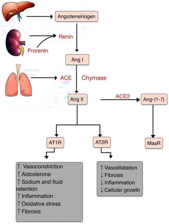
34. RAS and Inflammation

The interaction with AT1R promotes the classic effects of inflammation, vasoconstriction, OS and increased proliferation factors [15][16][15,16]. On the other hand, AT2R, ACE2, Ang1-7, Ang1-9 and Mas Receptor (MasR) have anti-inflammatory properties [17][18][17,18].
The binding Ang II-AT1R enhances vascular permeability and synthesis of vascular endothelial growth factor (VEGF), stimulates adhesion molecule expression by neutrophils (PMNs) and ECs, including selectins (P- and L- selectin), VCAM-1, and ICAM-1 [10][16][10,16]. Likewise, the activation of AT1R induces inflammatory responses, such as the migration of leukocytes and the release of pro-inflammatory cytokines (e.g., IL-1β, IL6, TNF, CXCL-1) [17]. Ang II contributes to endothelial dysfunction by activating COX-2, resulting in the production of ROS and prostaglandins [10]. Peroxisome proliferator-activated receptors (PPARs) expression has been found to be suppressed by Ang II. This suppression leads to a decrease in the PPARs’ capabilities to mitigate inflammation [19].
In the presence of inflammatory cytokines or a tissue injury, the mast cells degranulate to the ECM and the chymase is activated. Ang I is converted to Ang II by the active chymase as well as ACE [20]. Chymase also activates MMP-9 and TGF-β by converting their inactive precursors into active forms, which are associated with OS, inflammation and fibrosis [6][21][6,21].
Ang II, by stimulating NADPH oxidases (NOX), induces reactive oxygen species (ROS) production, an important messenger involved in intracellular signaling (Figure 2) [10][22][10,22]. ROS are small molecules derived from oxygen metabolism, including hydrogen peroxide, superoxide, singlet oxygen and hydroxyl radical. Low ROS levels can adjust biological activities, such as cellular growth, differentiation, proliferation, signaling and senescence. However, when the ROS production is increased (OS), Ang II signaling is disturbed, causing endothelial dysfunction, vascular remodeling and inflammation [23][24][25][26][23,24,25,26].
Figure 2. Ang II-mediated ROS activation. NF-kB, nuclear factor kappa B; JAK2, janus kinase 2; STAT3, signal transducer and activator of transcription 3; COX-2, cyclooxygenase-2; IL-6, interleukin-6 and its receptor, IL-6R; VCAM-1/ICAM-1, vascular cell/intercellular adhesion molecule 1; MMP-9, -2, matrix metalloproteinases -9, -2. (Adapted after [10][27][10,28]).

45. RAS and the Immune System

Inflammation is a vital mechanism for health. Through AT1R, the stimulation of ROS augments the inflammatory actions of the immune system [10][28][10,31].
The inflammatory response involves many cell types interacting with RAS: dendritic cells, T-cells, PMNs, mast cells and macrophages (Figure 3).
Figure 3. Interactions between Ang II and IIS (adapted after [10][14]).
Interactions between Ang II and IIS (adapted after [10,14]).

4.1. T Cells

5.1. T Cells

T-cells hold an intrinsic RAS that regulates their migration and proliferation. In the context of inflammation, Ang II acts through the AT1R to induce rearrangements in the cytoskeleton of T cells. This activation leads to the release of chemokines and cytokines that enhance the recruitment of T cells to inflammatory sites [14]. The endogenously produced Ang II in T cells contributes to their activation, increases the production of TNF-α and upregulates the expression of C-C chemokine receptor 5 (CCR5) [10]. CCR5 plays an important role in recruiting and activating inflammatory cells [29][32]. In addition, TNF-α triggers various events, including the production of pro-inflammatory cytokines (IL-1, -6 and -8), adhesion molecules, generation of NO and release of pro-coagulatory substances. TNF-α acts on two receptors: TNFR1 and TNFR2 [30][33]. TNFR1 is involved in promoting pro-inflammatory and cytotoxic responses, while TNFR2 is primarily associated with proliferation, tissue regeneration and cell survival [31][34]. Furthermore, TNF-α can reduce the viability of antigen-presenting cells (APCs) [10].
Upon activation, native CD4+ T cells have the capacity to differentiate into two major subsets of T helper cells known as Th1 and Th2 [32][35]. Th1 cells contribute to cellular immunity, promote the killing efficiency of macrophages and stimulate the proliferation of CD8+ T cells. On the contrary, Th2 cells play a role in humoral immunity by stimulating the proliferation of B-cells and facilitating Abs class switching in B-cells [10][33][10,36]. It has been proved that the RAS may be involved in promoting Th1-mediated AI diseases [34][37]. Ang II has been found responsible for disrupting the Th1/Th2 balance by promoting the production of Th1 cytokine IFN-γ, thereby exerting pro-inflammatory effects, while reducing the levels of the Th2 cytokine IL-4 [35][38]. Tregs are tissue resident memory cells (TRM), which constitute approximately 20–40% of the CD4 T-cells in both human and mice skin. Their primary function is to uphold the immune homeostasis of the skin, facilitate wound healing and participate in tissue repair. These cells play an important role in chronic inflammatory conditions affecting the skin, such as psoriasis and vitiligo.

4.2. Dendritic Cells

5.2. Dendritic Cells

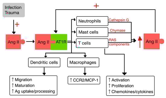
Dendritic cells (DCs) are specialized APCs that have a critical role in regulating the innate and also the adaptive immune responses [36][37][40,41]. In a study by Meng et al. [36][40], it was observed that Ang II exerts contrasting effects on DCs. On one hand, Ang II inhibits the phagocytic activity and proliferation of DCs. However, on the other hand, it promotes the maturation and the migration of DCs and also the expression of pro-inflammatory cytokines. Additionally, Ang II stimulates the T cell proliferation mediated by DCs [36][40].

4.3. Macrophages

5.3. Macrophages

Macrophages and their precursors, known as monocytes, are white blood cells specialized in clearing away cellular debris and pathogens through phagocytosis. Additionally, they possess the ability to trigger and activate other immune cells to respond to invading pathogens [14]. Aldosterone aims at monocytes/macrophages and promotes the activation/migration of these cells in the ECs by increasing the expression of VCAM-1 and ICAM-1 [37][41]. It was proven that the activity of AT1R in M1 macrophages promotes polarization, which accelerates the inflammation with progression of tissue damage [38][42]. Likewise, Ang II upregulates the expression of monocyte chemoattractant protein-1 (MCP-1) and one of its receptors, CCR2 [14][39][14,43].

4.4. Neutrophils

5.4. Neutrophils

PMNs, as the first responding cells to invading pathogens, play a crucial role in providing early immune protection. PMN bactericidal activity is increased by ACE present within them, regardless of the involvement of the Ang II/AT1R pathway. By interacting with AT1R, Ang II releases IL-8, which stimulates PMN recruitment and infiltration. Also, when stimulated by Ang II, PMNs produce oxidative bursts [10][40][10,44]. Cathepsin G (CatG), found in macrophages and PMNs, is a lysosomal protease that is upregulated in response to signals linked to infection and inflammation. CatG can elevate the local generation of Ang II by converting both angiotensinogen and Ang I to Ang II [10][41][42][10,45,46].

56. RAS in the Skin

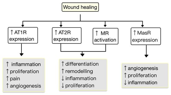
New research findings have unveiled the presence of a local RAS within the skin, where ACE has a role in the regulation of inflammation and autoimmunity [43][47]. The components of the RAS are situated in cutaneous and subcutaneous layers. They are crucial in various skin-related conditions, such as inflammation, fibrosis, scar formation and certain types of skin cancers [44][48]. Components of RAS are expressed in the human skin. Initial studies highlighting the presence of a local RAS in the skin revealed that skin cells, particularly keratinocytes, possess the ability to produce Ang II (as well as potentially other angiotensins) independently of the systemic circulation’s supply of RAS components. Keratinocytes are abundant in AT1R throughout all epidermal layers. AT1 receptors are also expressed in the hair follicles and sweat glands. In dermis, fibroblasts express AT1R, AT2R, MAS, angiotensinogen, renin, ACE, Ang II, and mast cells express chymase. In hypodermis, the subcutaneous fat expresses the same components of RAS as in dermis plus ACE2 [45][46][47][49,50,51]. RAS activity is involved in cell proliferation, differentiation, tissue remodeling and skin photoaging [48][49][52,53]. Stimulation of AT1R triggers cellular processes, including cell proliferation, migration, collagen synthesis and angiogenesis. Contrarily, the AT2R inhibit these actions by blocking the synthesis of certain pro-inflammatory molecules, like TGF-β, TNF-α and IL-6. Therefore, the interplay between AT1R and AT2R within RAS provides a fragile balance in regulating these cellular functions and inflammatory responses in the skin [50][54]. The expression of RAS components is upregulated in human wounded skin. Ang receptors have been implicated in wound healing and scar formation of the skin (Figure 4) [45][49]. Impaired wound healing has been associated with disruptions in the function of AT1R. In both aging and diabetes, RAS dysregulation occurs, characterized by high AT1R expression and low AT2R expression. This modification in the AT1R/AT2R ratio is linked with a reduction in epidermal thickness, collagen degeneration, dermal layer fractures and subcutaneous fat atrophy [51][55]. Research studies revealed that valsartan (an ARB) exhibits the highest level of skin penetration among other ARBs. Topical application of 1% valsartan gel has shown significant enhancement in wound healing. Researchers found that the rate of wound healing is superior while using topical valsartan compared to losartan. The beneficial effects of valsartan gel were mediated through the activation of AT2R, as the healing effect was absent in mice lacking AT2R. Conversely, the application of a 5% captopril gel resulted in a notable delay in the process of wound healing [44][48].
Figure 4. The role of RAS in the wound healing process. AT1R promotes inflammation, proliferation, angiogenesis and pain. AT2R and MR promote remodeling and differentiation and decrease inflammation and proliferation. MasR increases proliferation and angiogenesis and decreases inflammation (adapted after [45][49])
Hypertrophic and keloids scars are characterized by an aberrant wound healing process that results in excessive ECM production. There is evidence that both Ang II and AT1R concentrations are elevated in keloid and hypertrophic scars compared to normal skin. AT1R promotes scar formation. In hypertrophic and keloid scars, the AT1R activation leads to increased ECM production, transition of fibroblasts into myofibroblasts and contraction of granulation tissue. This process involves the activation of TGF-β signaling pathways. Elevated levels of Ang II, acting through AT1R, contribute to skin scar formation by upregulating the expression of inflammatory molecules (e.g., IL-6, VEGF, TGF-β1) [44][47][50][48,51,54]. ACEIs reduce scar formation, inhibit fibroblast proliferation, and suppress the expression of TGF-β1 and collagen. TGF-β1 has cytoprotective effects in mitigating tissue injury through promoting wound repair, tissue regeneration and exerting anti-inflammatory effects. Abnormal TGF-β1 signaling can cause pathological fibrosis in response to tissue injury [52][53][56,57]. Moreover, the dysregulation between pro-inflammatory (IL-6) and anti-inflammatory (IL-10) cytokines can lead to hypertrophic scarring. In a study by Hedayatyanfard et al. [50][54], a 5% topical ointment losartan was investigated as a treatment for hypertrophic and keloid scars. The results showed that the application of losartan ointment led to significant improvements, including vascularity, pigmentation, pliability and height; at the end of treatment period, scars were smaller.
Video Production Service