Delayed Cerebral Ischemia and Cisternal Drainage: Comparison
Please note this is a comparison between Version 2 by Fanny Huang and Version 3 by Fanny Huang.

Aneurysmal subarachnoid hemorrhage (aSAH) provokes a cascade reaction that is responsible for early and delayed brain injuries mediated by intracranial hypertension, hydrocephalus, cerebral vasospasm (CV), and delayed cerebral ischemia (DCI), which result in increased morbidity and mortality. During open microsurgical repair, cisternal access is achieved essentially to gain proximal vascular control and aneurysm exposition. Cisternostomy also allows brain relaxation, removal of cisternal clots, and restoration of the CSF dynamics through the communication between the anterior and posterior circulation cisterns and the ventricular system, with the opening of the Membrane of Liliequist and lamina terminalis, respectively. Continuous postoperative CSF drainage through a cisternal drain (CD) is a valuable option for treating acute hydrocephalus and intracranial hypertension. Moreover, it efficiently removes the blood and toxic degradation products, with a potential benefit on CV, DCI, and shunt-dependent hydrocephalus. Finally, the CD is an effective pathway to administer vasoactive, fibrinolytic, and anti-oxidant agents and shows promising results in decreasing CV and DCI rates while minimizing systemic effects.

  • cisternostomy
  • cisternal drain
  • delayed cerebral ischemia
  • cerebral vasospasm
  • hydrocephalus
  • aneurysmal subarachnoid hemorrhage

1. Pathophysiology of Delayed Cerebral Ischemia

DCI occurs in up to 30% of aSAH patients and results in poor functional outcomes in half of them despite adequate treatment [1][2][3]. CV is defined as a narrowing of the angiographically visible cerebral arteries that may occur in 30–70% of patients following aSAH [4]. It has been considered for decades the principal cause of DCI. Recent evidence demonstrated that CV in the major cerebral vessels is a contributing factor and not the determinant one [2]. Blood degradation products in the subarachnoid space trigger a molecular cascade that may lead to CV, microcirculatory dysfunction, excitotoxicity, oxidative stress, and inflammatory cascade [2][5][6]. The glymphatic system is responsible for clearing the CSF toxic products [7]. However, lymphatic vessel disruption and glymphatic system dysfunction following aSAH may exacerbate neuroinflammatory response and contribute to DCI [8][9][10]. Continuous CSF drainage seems a valuable option to increase clots evacuation, reduce DCI, and improve functional outcomes, but contrasting results are reported with different output pathways [11][12][13][14][15][16]. The studies reporting the impact of external CSF drains in CV and DCI are summarized in Table 1.
Table 1. The studies investigating the role of the lumbar drain, external ventricular drain, and cisternal drain for cerebral vasospasm and delayed cerebral ischemia management are summarized here. CD: cisternal drain; DCI: delayed cerebral ischemia; EVD: external ventricular drain; LD: lumbar drain; RCT: randomized controlled trial; STD: standard.
Author and Year Design Intervention Outcome
Wolf et al., 2023 [12] RCT LD (144) vs. STD care * (143) LD reduced DCI (p = 0.04) and unfavorable outcomes at 6 months (p = 0.04).
Al-Tamimi et al., 2012 [13] RCT LD (105) vs. STD care * (105) LD showed DCI reduction (p = 0.021) but no outcome improvement at 6 months.
Maeda et al., 2013 [11] Retrospective LD (34) vs. EVD (17) LD showed more rapid clot washout and a trend toward DCI reduction.
Klimo et al., 2004 [14] Retrospective LD (81) vs. STD care * (86) LD reduced CV (p < 0.001) and DCI (p = 0.05).
Ogura et al., 1988 [15] Retrospective CD (101) vs. no drain (31) The CD was associated with no significant reduction of CV, although positive trends were seen in patients with high Fisher grades.
Sakaki et al., 1987 [16] Retrospective CD (75) vs. STD care * (74) Early CD significantly reduced DCI (p < 0.05) and improved outcomes (p < 0.01).
* Standard care groups included patients treated with or without EVD, depending on the presence of acute hydrocephalus.
Moreover, increased intracranial pressure combined with micro and macrovascular changes may further reduce cerebral perfusion pressure and have been related to the risk of developing hypoperfusion and ischemia [17][18][19]. DCI is a complex process resulting from multiple pathological pathways secondary to aSAH and has been correlated with the extent of subarachnoid hemorrhage, as expressed by the modified Fisher grade [20][21][22]. The role of vasospasm has probably been overemphasized, and a therapeutic approach to all these factors should be preferred.

2. Cisternal Drainage, Cerebral Vasospasm and Delayed Cerebral Ischemia

Extensive and early evacuation of cisternal clots shortly after aSAH and before the process of blood degradation takes place could be a valid option to stop the pathological cascade and prevent DCI. Evacuation of a subarachnoid hemorrhage within 48h showed a reduction in CV and DCI in preclinical studies [23][24]. Moreover, the amount of postoperative clots and clot-clearance rate on serial brain CT seems to predict the risk of CV, consequent infarction, and unfavorable outcomes [25][26].
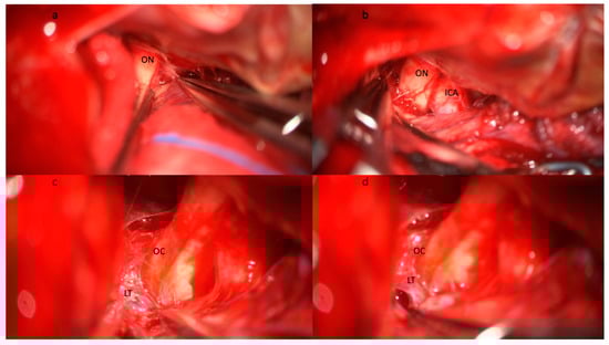
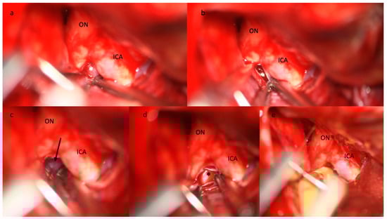
Yasargil pioneered the interest in understanding the anatomy of the basal cisterns. He was the first to report routine basal cistern opening, including LT (Figure 1) and MoL (Figure 2) fenestration to obtain brain relaxation, proximal exposure, extensive evacuation of the subarachnoid hemorrhage, and CSF flow restoration [27]. In the following years, the advent of microneurosurgery allowed widespread diffusion of cisternal procedures [27][28][29].
Figure 1. Step-by-step illustration of a cisternostomy performed during microsurgical clipping of an aneurysm of the right middle cerebral artery bifurcation. (a) Subfrontal approach and identification of the optic nerve (ON) with (b) progressive opening of the optico-carotid cistern and exposure of the internal carotid artery (ICA). (c) The dissection continues medially and posteriorly following the ON until the exposure of the optic chiasma (OC) and the lamina terminalis (LT). (d) The opening of the lamina terminalis gives access to the third ventricle and allows CSF drainage from the ventricular system. Brain relaxation is generally obtained at this stage after blood clot evacuation from the basal cisterns, the opening of the optico-carotid cisterns, and the LT. Abbreviations: CSF: cerebrospinal fluid; ICA: internal carotid artery; LT: lamina terminalis; OC: optic chiasma; ON: optic nerve.
Figure 2. Step-by-step illustration of a cisternostomy performed during microsurgical clipping of an aneurysm of the right middle cerebral artery bifurcation. (a) The diencephalic leaf of the Membrane of Liliequist (white asterisk) is seen through the optico-carotid triangle and (b) progressively opened, (c) enabling the exposure of the mesencephalic leaf (black arrow) that is finally opened. (d) The basilar artery (black asterisk) is visible in the pre-pontine cistern. (e) The tip of the cisternal drain is positioned between the ICA and the ON in the pre-pontine cistern. Abbreviations: ICA: internal carotid artery; ON: optic nerve.
Extensive subarachnoid clot removal was first described by Suzuki and Yoshimoto in 1976 [30]. Early surgery and clot evacuation within 24 h from the bleeding greatly lessen the risk of CV and improve functional outcomes, especially in patients with high Fisher grades aSAH [30][31]. A few reports published in Japan in the seventies and eighties confirmed the positive impact of early operation and clot evacuation on CV and delayed ischemia [25][32][33][34][35]. Moreover, the Japanese register showed an association between surgical clipping and a lower risk of cerebral infarction [36].
A few studies have been published dealing with the impact of cisternostomy on CV, DCI, and functional outcome, and the results are summarized in Table 2 [15][16][37][38][39][40][41][42].
Table 2. The studies investigating the role of the cisternal drain in CV and DCI management are summarized here. CD: cisternal drain; DCI: delayed cerebral ischemia; STD: standard.
Author and Year Design Intervention Outcomes
Inagawa T et al., 1991 [40] Retrospective CD (140) The total amount of CSF cisternal drainage was correlated with decreased CV and DCI.
Ogura et al., 1988 [15] Retrospective CD (101) vs. no drain (31) The CD was associated with no significant reduction of CV, although positive trends were seen in patients with high Fisher grades.
Sakaki et al., 1987 [16] Retrospective CD (75) vs. STD care * (74) Early CD significantly reduced DCI (p < 0.05) and improved outcomes (p < 0.01).
Kawakami et al., 1987 [39] Retrospective CD (22) Symptomatic CV occurred in 22% of cases and good functional outcome in 95% of cases.
Ito el al., 1986 [38] Retrospective CD (38) Effective cisternal drainage was correlated with reduced CV and improved outcomes.
* Standard care groups included patients treated with or without EVD, depending on the presence of acute hydrocephalus.
To researchers' knowledge, Ito et al. were the first to combine the insertion of cisternal drainage with cisternostomy to enhance their positive effects [38]. Cisternal drainage improves toxic substance clearance compared to EVD and may remove more than 1g of hemoglobin per day [16]. Its position in the basal cisterns creates a gravitational gradient to CSF circulation and clearance similar to lumbar drains but achieving it closer to the bleeding source [11][12][14][43]. Moreover, despite the emerging efficacy of LD in reducing DCI and unfavorable outcomes, obstructive hydrocephalus and compressed basal cisterns remain the main limitations in the use of LD, while both situations are efficiently treated with CD [12][43]. Indeed, LT and MoL fenestration combined with CD increase daily CSF drainage, resolve obstructive hydrocephalus, and were significantly associated with a reduction of symptomatic CV [38]. Kawakami et al. performed a continuous cisternal drainage for a minimum of fourteen days in 22 patients. Moderate CV occurred in 22% of cases, and good neurological outcome was observed in 95% of cases [39]. Inagawa et al. evaluated the efficacy of continuous cisternal drainage on CV for 140 consecutive patients. They classified patients according to the total amount of drainage regardless of the duration. Drainage of cisternal CSF of more than 500 mL was shown to be associated with decreased DCI, angiographic and symptomatic CV, and improved functional outcome [40]. Sakaki et al. reported that early clot evacuation combined with CD was associated with better neurological outcomes (p = 0.01). Moreover, they observed a reduction of 50% for symptomatic CV and a less severe angiographic spasm [16]. Ogura et al. assessed the efficacy of continuous postoperative cisternal drainage in a retrospective study and showed that it reduced CV and mortality incidence and improved outcomes in patients with severe clinical and radiological presentation [15]. Moreover, continuous postoperative CSF drainage decreases ICP, is directly related to cerebral perfusion, and contributes to DCI [15][42][44]. Cisternostomy may, thus, represent a valuable adjunct in ruptured aneurysms surgery [31][45][46][47][48], as it may allow clots evacuation and postoperative cisternal drainage [38], thus blocking the cascade responsible for DCI [15][16][38][39][40]. Therefore, cisternal drainage, as part of the surgical treatment, has several advantages that allow ICP control and the reduction of several processes that contribute to the development of DCI.

3. CD and Drugs Administration

Intrathecal drug administration presents many anatomic and pharmacodynamic advantages compared to systemic administration. A higher drug concentration can be reached with minimal systemic effects [49]. Cisternal injections, compared to ventricular and lumbar administration, allow drug delivery directly in the basal cisterns. It may enhance the pharmacologic effects on the large proximal arteries and improve the intraparenchymal diffusion through the perivascular Virchow spaces. Animal models confirmed a more effective and durable vasoactive response compared to intra-arterial administration [50][51]. Prophylactic injections of intrathecal nicardipine and milrinone have shown a reduction in angiographic CV and DCI and an increase in mean cerebral blood flow but without a significant improvement in functional outcomes [52][53][54][55][56][57]. Intraventricular nicardipine showed a significant reduction in DCI and improvement of functional outcomes in patients treated for significant CV [58]. Shibuya et al. administered 2 mg of cisternal nicardipine three times a day for 10 days in 50 patients treated for an aSAH. Prophylactic nicardipine reduced the incidence of radiographic and symptomatic CV by 26% and 20%, respectively, and increased early good clinical outcomes by 15%. However, no statistical significance was reached [55]. Similar results were obtained by Suzuki et al. [52].
Magnesium sulfate showed several potential beneficial effects in aSAH patients, such as vasodilatation and attenuation of neuronal death. However, intravenous administration failed to prevent DCI and improve functional outcomes [59], while continuous intracisternal administration significantly improved DCI and functional outcomes [60].
In researchers' institution, they introduced intrathecal nicardipine as a treatment for moderate (>50% arterial narrowing at angioCT) to severe (>75% arterial narrowing at angioCT) CV in patients with aSAH in 2019. In reseaourchers' experience, intrathecal nicardipine showed a significant reduction in DCI rate and improved functional outcome (unpublished data). Moreover, cisternal administration showed a positive trend toward a further reduction of DCI when compared to ventricular administration.

References

  1. Hoh, B.L.; Ko, N.U.; Amin-Hanjani, S.; Chou, S.H.-Y.; Cruz-Flores, S.; Dangayach, N.S.; Derdeyn, C.P.; Du, R.; Hänggi, D.; Hetts, S.W.; et al. 2023 Guideline for the Management of Patients with Aneurysmal Subarachnoid Hemorrhage: A Guideline From the American Heart Association/American Stroke Association. Stroke 2023, 54, E314–E370.
  2. Cossu, G.; Messerer, M.; Oddo, M.; Daniel, R.T. To Look beyond Vasospasm in Aneurysmal Subarachnoid Haemorrhage. Biomed. Res. Int. 2014, 2014, 628597.
  3. Li, K.; Barras, C.D.; Chandra, R.V.; Kok, H.K.; Maingard, J.T.; Carter, N.S.; Russell, J.H.; Lai, L.; Brooks, M.; Asadi, H. A Review of the Management of Cerebral Vasospasm after Aneurysmal Subarachnoid Hemorrhage. World Neurosurg. 2019, 126, 513–527.
  4. Mascia, L.; Del Sorbo, L. Diagnosis and Management of Vasospasm. F1000 Med. Rep. 2009, 1, 33.
  5. Rowland, M.J.; Hadjipavlou, G.; Kelly, M.; Westbrook, J.; Pattinson, K.T.S. Delayed Cerebral Ischaemia after Subarachnoid Haemorrhage: Looking beyond Vasospasm. Br. J. Anaesth. 2012, 109, 315–329.
  6. Macdonald, R.L.; Weir, B.K.; Grace, M.G.; Martin, T.P.; Doi, M.; Cook, D.A. Morphometric Analysis of Monkey Cerebral Arteries Exposed in Vivo to Whole Blood, Oxyhemoglobin, Methemoglobin, and Bilirubin. Blood Vessel. 1991, 28, 498–510.
  7. Louveau, A.; Smirnov, I.; Keyes, T.J.; Eccles, J.D.; Rouhani, S.J.; Peske, J.D.; Derecki, N.C.; Castle, D.; Mandell, J.W.; Lee, K.S.; et al. Structural and Functional Features of Central Nervous System Lymphatic Vessels. Nature 2015, 523, 337–341.
  8. Sun, B.; Xie, F.; Yang, M.; Cao, M.; Yuan, H.; Wang, H.; Wang, J.; Jia, L. Blocking Cerebral Lymphatic Drainage Deteriorates Cerebral Oxidative Injury in Rats with Subarachnoid Hemorrhage. In Early Brain Injury or Cerebral Vasospasm; Feng, H., Mao, Y., Zhang, J.H., Eds.; Springer: Vienna, Austria, 2011; pp. 49–53.
  9. Chen, J.; Wang, L.; Xu, H.; Xing, L.; Zhuang, Z.; Zheng, Y.; Li, X.; Wang, C.; Chen, S.; Guo, Z.; et al. Meningeal Lymphatics Clear Erythrocytes That Arise from Subarachnoid Hemorrhage. Nat. Commun. 2020, 11, 3159.
  10. Dodd, W.S.; Laurent, D.; Dumont, A.S.; Hasan, D.M.; Jabbour, P.M.; Starke, R.M.; Hosaka, K.; Polifka, A.J.; Hoh, B.L.; Chalouhi, N. Pathophysiology of Delayed Cerebral Ischemia After Subarachnoid Hemorrhage: A Review. J. Am. Heart Assoc. 2021, 10, e021845.
  11. Maeda, Y.; Shirao, S.; Yoneda, H.; Ishihara, H.; Shinoyama, M.; Oka, F.; Sadahiro, H.; Ueda, K.; Sano, Y.; Kudomi, S.; et al. Comparison of Lumbar Drainage and External Ventricular Drainage for Clearance of Subarachnoid Clots after Guglielmi Detachable Coil Embolization for Aneurysmal Subarachnoid Hemorrhage. Clin. Neurol. Neurosurg. 2013, 115, 965–970.
  12. Wolf, S.; Mielke, D.; Barner, C.; Malinova, V.; Kerz, T.; Wostrack, M.; Czorlich, P.; Salih, F.; Engel, D.C.; Ehlert, A.; et al. Effectiveness of Lumbar Cerebrospinal Fluid Drain Among Patients with Aneurysmal Subarachnoid Hemorrhage: A Randomized Clinical Trial. JAMA Neurol. 2023, 80, 833–842.
  13. Al-Tamimi, Y.Z.; Bhargava, D.; Feltbower, R.G.; Hall, G.; Goddard, A.J.P.; Quinn, A.C.; Ross, S.A. Lumbar Drainage of Cerebrospinal Fluid after Aneurysmal Subarachnoid Hemorrhage: A Prospective, Randomized, Controlled Trial (LUMAS). Stroke 2012, 43, 677–682.
  14. Klimo, P.; Kestle, J.R.W.; MacDonald, J.D.; Schmidt, R.H. Marked Reduction of Cerebral Vasospasm with Lumbar Drainage of Cerebrospinal Fluid after Subarachnoid Hemorrhage. J. Neurosurg. 2004, 100, 215–224.
  15. Ogura, K.; Hara, M.; Tosaki, F.; Hirai, N. Effect of Cisternal Drainage after Early Operation for Ruptured Intracranial Aneurysms. Surg. Neurol. 1988, 30, 441–444.
  16. Sakaki, S.; Ohta, S.; Kuwabara, H.; Shiraishi, M. The Role of Ventricular and Cisternal Drainage in the Early Operation for Ruptured Intracranial Aneurysms. Acta Neurochir. 1987, 88, 87–94.
  17. Lagares, A.; Cicuendez, M.; Ramos, A.; Salvador, E.; Alén, J.F.; Kaen, A.; Jiménez-Roldán, L.; Millán, J.M. Acute Perfusion Changes after Spontaneous SAH: A Perfusion CT Study. Acta Neurochir. 2012, 154, 402–405.
  18. Starnoni, D.; Maduri, R.; Hajdu, S.D.; Pierzchala, K.; Giammattei, L.; Rocca, A.; Grosfilley, S.B.; Saliou, G.; Messerer, M.; Daniel, R.T. Early Perfusion Computed Tomography Scan for Prediction of Vasospasm and Delayed Cerebral Ischemia After Aneurysmal Subarachnoid Hemorrhage. World Neurosurg. 2019, 130, e743–e752.
  19. Yang, J.; Lu, J.; Li, R.; Lin, F.; Chen, Y.; Han, H.; Yan, D.; Li, R.; Li, Z.; Zhang, H.; et al. Application of Intracranial Pressure-Directed Therapy on Delayed Cerebral Ischemia After Aneurysmal Subarachnoid Hemorrhage. Front. Aging Neurosci. 2022, 14, 831994.
  20. Frontera, J.A.; Claassen, J.; Schmidt, J.M.; Wartenberg, K.E.; Temes, R.; Connolly, E.S.; Macdonald, R.L.; Mayer, S.A. Prediction of Symptomatic Vasospasm after Subarachnoid Hemmorrage: The Modified Fisher Scale. Neurosurgery 2006, 59, 529–538.
  21. Friedrich, B.; Müller, F.; Feiler, S.; Schöller, K.; Plesnila, N. Experimental Subarachnoid Hemorrhage Causes Early and Long-Lasting Microarterial Constriction and Microthrombosis: An In-Vivo Microscopy Study. J. Cereb. Blood Flow. Metab. 2012, 32, 447–455.
  22. van der Steen, W.E.; Leemans, E.L.; van den Berg, R.; Roos, Y.B.W.E.M.; Marquering, H.A.; Verbaan, D.; Majoie, C.B.L.M. Radiological Scales Predicting Delayed Cerebral Ischemia in Subarachnoid Hemorrhage: Systematic Review and Meta-Analysis. Neuroradiology 2019, 61, 247–256.
  23. Nosko, M.; Weir, B.K.A.; Lunt, A.; Grace, M.; Allen, P.; Mielke, B. Effect of Clot Removal at 24 Hours on Chronic Vasospasm after SAH in the Primate Model. J. Neurosurg. 1987, 66, 416–422.
  24. Handa, Y.; Weir, B.K.A.; Nosko, M.; Mosewich, R.; Tsuji, T.; Grace, M. The Effect of Timing of Clot Removal on Chronic Vasospasm in a Primate Model. J. Neurosurg. 1987, 67, 558–564.
  25. Ota, N.; Matsukawa, H.; Kamiyama, H.; Tsuboi, T.; Noda, K.; Hashimoto, A.; Miyazaki, T.; Kinoshita, Y.; Saito, N.; Tokuda, S.; et al. Preventing Cerebral Vasospasm After Aneurysmal Subarachnoid Hemorrhage with Aggressive Cisternal Clot Removal and Nicardipine. World Neurosurg. 2017, 107, 630–640.
  26. Toyoda, T.; Yonekura, I.; Iijima, A.; Shinozaki, M.; Tanishima, T. Clot-Clearance Rate in the Sylvian Cistern Is Associated with the Severity of Cerebral Vasospasm After Subarachnoid. Acta Neurochir. Suppl. 2015, 120, 275–277.
  27. Yasargil, M. Microsurgical Anatomy of the Basal Cistern and Vessels of the Brain, Diagnostic Studies, General Operative Techniques and Pathological Considerations of the Intracranial Aneurysms; Thieme: New York, NY, USA, 1984; Volume 1.
  28. Sturiale, C.L.; Ricciardi, L.; Marchese, E.; Puca, A.; Olivi, A.; Albanese, A. Surgical Treatment of Anterior Communicating Artery Aneurysms: Hints and Precautions for Young Cerebrovascular Surgeons. J. Neurol. Surg. A Cent. Eur. Neurosurg. 2020, 81, 463–471.
  29. Sturiale, C.L.; Rapisarda, A.; Marchese, E.; Puca, A.; Olivi, A.; Albanese, A. Surgical Treatment of Middle Cerebral Artery Aneurysms: Hints and Precautions for Young Cerebrovascular Surgeons. J. Neurol. Surg. A Cent. Eur. Neurosurg. 2021, 83, 75–84.
  30. Suzuki, J.; Yoshimoto, T. Early Operation for the Ruptured Intracranial Aneurysms—Especially the Cases Operated within 48 Hours after the Last Subarachnoid Hemorrhage (Author’s Transl). No Shinkei Geka 1976, 4, 135–141.
  31. Cengiz, S.L.; Ilik, M.K.; Erdi, F.; Ustun, M.E. The Role of Fenestration of the Lamina Terminalis on Symptomatic Vasospasm After Aneurysmal Subarachnoid Hemorrhage: A Clinical Research. Turk. Neurosurg. 2016, 26, 714–719.
  32. Saito, I.; Ueda, Y.; Sano, K. Significance of Vasospasm in the Treatment of Ruptured Intracranial Aneurysms. J. Neurosurg. 1977, 47, 412–429.
  33. Taneda, M. Effect of Early Operation for Ruptured Aneurysms on Prevention of Delayed Ischemic Symptoms. J. Neurosurg. 1982, 57, 622–628.
  34. Mizukami, M.; Kawase, T.; Usami, T.; Tazawa, T. Prevention of Vasospasm by Early Operation with Removal of Subarachnoid Blood. Neurosurgery 1982, 10, 301–307.
  35. Hosoda, K.; Fujita, S.; Kawaguchi, T.; Shose, Y.; Hamano, S.; Iwakura, M. Effect of Clot Removal and Surgical Manipulation on Regional Cerebral Blood Flow and Delayed Vasospasm in Early Aneurysm Surgery for Subarachnoid Hemorrhage. Surg. Neurol. 1999, 51, 81–88.
  36. Kurogi, R.; Kada, A.; Nishimura, K.; Kamitani, S.; Nishimura, A.; Sayama, T.; Nakagawara, J.; Toyoda, K.; Ogasawara, K.; Ono, J.; et al. Effect of Treatment Modality on In-Hospital Outcome in Patients with Subarachnoid Hemorrhage: A Nationwide Study in Japan (J-ASPECT Study). J. Neurosurg. 2018, 128, 1318–1326.
  37. Garvayo, M.; Messerer, M.; Starnoni, D.; Puccinelli, F.; Vandenbulcke, A.; Daniel, R.T.; Cossu, G. The Positive Impact of Cisternostomy with Cisternal Drainage on Delayed Hydrocephalus after Aneurysmal Subarachnoid Hemorrhage. Acta Neurochir. 2023, 165, 187–195.
  38. Ito, U.; Tomita, H.; Yamazaki, S.; Takada, Y.; Inaba, Y. Enhanced Cisternal Drainage and Cerebral Vasospasm in Early Aneurysm Surgery. Acta Neurochir. 1986, 80, 18–23.
  39. Kawakami, Y.; Shimamura, Y. Cisternal Drainage after Early Operation of Ruptured Intracranial Aneurysm. Neurosurgery 1987, 20, 8–14.
  40. Inagawa, T.; Kamiya, K.; Matsuda, Y. Effect of Continuous Cisternal Drainage on Cerebral Vasospasm. Acta Neurochir. 1991, 112, 28–36.
  41. Yamamoto, I.; Shimoda, M.; Yamada, S.; Ikeda, A. Indications for Cisternal Drainage in Conjunction with Early Surgery for Ruptured Aneurysms and Timing of Its Discontinuation. Neurol. Med. Chir. 1989, 29, 407–413.
  42. Kasuya, H.; Shimizu, T.; Kagawa, M. The Effect of Continuous Drainage of Cerebrospinal Fluid in Patients with Subarachnoid Hemorrhage: A Retrospective Analysis of 108 Patients. Neurosurgery 1991, 28, 56–59.
  43. Hulou, M.M.; Essibayi, M.A.; Benet, A.; Lawton, M.T. Lumbar Drainage After Aneurysmal Subarachnoid Hemorrhage: A Systematic Review and Meta-Analysis. World Neurosurg. 2022, 166, 261–267.e9.
  44. Giammattei, L.; Starnoni, D.; Maduri, R.; Bernini, A.; Abed-Maillard, S.; Rocca, A.; Cossu, G.; Simonin, A.; Eckert, P.; Bloch, J.; et al. Implementation of Cisternostomy as Adjuvant to Decompressive Craniectomy for the Management of Severe Brain Trauma. Acta Neurochir. 2020, 162, 469–479.
  45. Mura, J.; Rojas-Zalazar, D.; Ruiz, A.; Vintimilla, L.C.; Marengo, J.J. Improved Outcome in High-Grade Aneurysmal Subarachnoid Hemorrhage by Enhancement of Endogenous Clearance of Cisternal Blood Clots: A Prospective Study That Demonstrates the Role of Lamina Terminalis Fenestration Combined with Modern Microsurgical Cistern. Minim. Invasive Neurosurg. 2007, 50, 355–362.
  46. Andaluz, N.; Zuccarello, M. Fenestration of the Lamina Terminalis as a Valuable Adjunct in Aneurysm Surgery. Neurosurgery 2004, 55, 1050–1059.
  47. Alaraj, A.; Charbel, F.T.; Amin-Hanjani, S. Peri-Operative Measures for Treatment and Prevention of Cerebral Vasospasm Following Subarachnoid Hemorrhage. Neurol. Res. 2009, 31, 651–659.
  48. Komotar, R.J.; Olivi, A.; Rigamonti, D.; Tamargo, R.J. Microsurgical Fenestration of the Lamina Terminalis Reduces the Incidence of Shunt-Dependent Hydrocephalus after Aneurysmal Subarachnoid Hemorrhage. Neurosurgery 2002, 51, 1403.
  49. Yamamoto, T.; Mori, K.; Esaki, T.; Nakao, Y.; Tokugawa, J.; Watanabe, M. Preventive Effect of Continuous Cisternal Irrigation with Magnesium Sulfate Solution on Angiographic Cerebral Vasospasms Associated with Aneurysmal Subarachnoid Hemorrhages: A Randomized Controlled Trial. J. Neurosurg. JNS 2016, 124, 18–26.
  50. Nishiguchi, M.; Ono, S.; Iseda, K.; Manabe, H.; Hishikawa, T.; Date, I. Effect of Vasodilation by Milrinone, a Phosphodiesterase III Inhibitor, on Vasospastic Arteries after a Subarachnoid Hemorrhage in Vitro and in Vivo: Effectiveness of Cisternal Injection of Milrinone. Neurosurgery 2010, 66, 158–164.
  51. Shibuya, M.; Suzuki, Y.; Takayasu, M.; Asano, T.; Ikegaki, I.; Sugita, K. Effects of Intrathecal Administration of Nicardipine and Nifedipine on Chronic Cerebral Vasospasm in Dogs. J. Clin. Neurosci. 1994, 1, 58–61.
  52. Suzuki, M.; Doi, M.; Otawara, Y.; Ogasawara, K.; Ogawa, A. Intrathecal Administration of Nicardipine Hydrochloride to Prevent Vasospasm in Patients with Subarachnoid Hemorrhage. Neurosurg. Rev. 2001, 24, 180–184.
  53. Hafeez, S.; Grandhi, R. Systematic Review of Intrathecal Nicardipine for the Treatment of Cerebral Vasospasm in Aneurysmal Subarachnoid Hemorrhage. Neurocrit. Care 2019, 31, 399–405.
  54. Koyanagi, M.; Fukuda, H.; Lo, B.; Uezato, M.; Kurosaki, Y.; Sadamasa, N.; Handa, A.; Chin, M.; Yamagata, S. Effect of Intrathecal Milrinone Injection via Lumbar Catheter on Delayed Cerebral Ischemia after Aneurysmal Subarachnoid Hemorrhage. J. Neurosurg. 2018, 128, 717–722.
  55. Shibuya, M.; Suzuki, Y.; Enomoto, H.; Okada, T.; Ogura, K.; Sugita, K. Effects of Prophylactic Intrathecal Administrations of Nicardipine on Vasospasm in Patients with Severe Aneurysmal Subarachnoid Haemorrhage. Acta Neurochir. 1994, 131, 19–25.
  56. Webb, A.; Kolenda, J.; Martin, K.; Wright, W.; Samuels, O. The Effect of Intraventricular Administration of Nicardipine on Mean Cerebral Blood Flow Velocity Measured by Transcranial Doppler in the Treatment of Vasospasm Following Aneurysmal Subarachnoid Hemorrhage. Neurocrit. Care 2010, 12, 159–164.
  57. Lu, N.; Jackson, D.; Luke, S.; Festic, E.; Hanel, R.A.; Freeman, W.D. Intraventricular Nicardipine for Aneurysmal Subarachnoid Hemorrhage Related Vasospasm: Assessment of 90 Days Outcome. Neurocrit. Care 2012, 16, 368–375.
  58. Sadan, O.; Waddel, H.; Moore, R.; Feng, C.; Mei, Y.; Pearce, D.; Kraft, J.; Pimentel, C.; Mathew, S.; Akbik, F.; et al. Does Intrathecal Nicardipine for Cerebral Vasospasm Following Subarachnoid Hemorrhage Correlate with Reduced Delayed Cerebral Ischemia? A Retrospective Propensity Score-Based Analysis. J. Neurosurg. 2022, 136, 115–124.
  59. Dorhout Mees, S.M.; Algra, A.; Wong, G.K.C.; Poon, W.S.; Bradford, C.M.; Saver, J.L.; Starkman, S.; Rinkel, G.J.E.; van den Bergh, W.M.; van Kooten, F.; et al. Early Magnesium Treatment After Aneurysmal Subarachnoid Hemorrhage. Stroke 2015, 46, 3190–3193.
  60. Takeuchi, S.; Kumagai, K.; Toyooka, T.; Otani, N.; Wada, K.; Mori, K. Intravenous Hydrogen Therapy with Intracisternal Magnesium Sulfate Infusion in Severe Aneurysmal Subarachnoid Hemorrhage. Stroke 2021, 52, 20–27.
More
Video Production Service