Physiological and Anti-Tumor Activities of Vitamin C: Comparison
Please note this is a comparison between Version 2 by Camila Xu and Version 1 by YOOL LEE.

Vitamin C (L-ascorbic acid, ascorbate, VC) is an essential nutrient for the normal maintenance of cellular functions, such as neural pathways, molecule biosynthesis (e.g., collagen, norepinephrine), immune signaling, chromatin remodeling, and cell division. This versatile nutrient has the potential as an anti-tumor agent. At high doses, it acts as a pro-oxidant, selectively targeting cancer cells. At low doses, it acts as an antioxidant, enhancing anti-tumor immunity. VC's potential lies in its ability to target cancer stem cells (CSCs), the self-renewing cells responsible for tumor recurrence and chemoresistance. Recent studies suggest that VC can selectively target CSCs via epigenetic and metabolic pathways. Understanding how VC exerts anti-tumor activity by targeting CSCs provides a rationale for its use in cancer treatment.

  • vitamin C
  • cancer stem cells
  • epithelial–mesenchymal transition

1. Introduction

Vitamin C (L-ascorbic acid, ascorbate, VC) is an essential nutrient for the normal maintenance of cellular functions, such as neural pathways, molecule biosynthesis (e.g., collagen, norepinephrine), immune signaling, chromatin remodeling, and cell division [1,2,3,4][1][2][3][4]. At physiological concentrations (40–80 µM in human plasma), VC acts as an antioxidant by serving as an electron donor and effectively scavenges reactive oxygen species (ROS). In contrast, at high doses (10–20 mM), VC acts as a pro-oxidant that induces oxidative stress and suppresses tumor growth, without notable damage to normal cells and tissues [5,6][5][6]. Recently, multiple studies have uncovered that VC has multifaceted anti-tumor effects [7]. For example, it acts as a cofactor for enzymes that regulate gene expression and suppresses oncogenes while reactivating tumor suppressor genes [8,9,10][8][9][10]. It has also been reported that VC induces various forms of canonical cell death and may be involved in non-canonical mechanisms relating to energy crises resulting from ATP depletion [11]. Aligned with its pleiotropic effects as a cancer-specific, pro-oxidative cytotoxic agent, anti-cancer epigenetic regulator, and immune modulator, high-dose VC has been proposed as a potent adjuvant treatment for cancer, acting synergistically with numerous standard (chemo-) therapies and alleviating the toxic side effects of chemotherapy (refer to for comprehensive reviews [12,13][12][13] of this). Furthermore, emerging animal model studies suggest that VC enhances anti-tumor effects when used in combination with dietary intervention (intermittent fasting, IF) [14] or cancer immunotherapy that facilitates anti-tumor immune environments [15[15][16],16], suggesting that VC mediates beneficial anti-cancer effects by targeting both tumor-intrinsic and -extrinsic pathways [17].
One of the most critical issues in cancer biology, as well as cancer diagnosis and treatment, is cancer cell plasticity, the adaptive and reversible capacity of diverse cancer cell populations to shift between cancer stem cell (CSC) and non-CSC/differentiated cell states in response to the tumor microenvironment [18]. At the top of the heterogeneous tumor hierarchy, the CSC, a self-renewing and multi-potent cancer cell type, is responsible for tumor recurrence, metastasis, chemoresistance, and mortality [18]. Phenotypically, CSCs are associated with the epithelial–mesenchymal transition (EMT), which confers cancer cells with increased motility and invasion ability that is characteristic of malignant and drug-resistant cells [19]. A growing number of studies suggest that CSC heterogeneity and plasticity are influenced not just by genetic factors but also by non-genetic factors, including epigenetic pathways [20], metabolic processes [21], and tumor-microimmune environments [22,23,24][22][23][24]. CSCs develop resistance to traditional chemotherapy due to their adaptable phenotype, which enables them to withstand therapy by overexpressing anti-apoptotic factors, defending against oxidative stress, and effectively repairing DNA damage [25]. Unfortunately, the vast majority of anti-cancer drugs have been designed to target rapidly dividing non-CSCs, rather than dormant CSCs, frequently leading to tumor relapse and treatment ineffectiveness [26,27][26][27]. Furthermore, many chemotherapy drugs trigger diverse mechanisms of plasticity in cancer cells, including EMT, autophagy, and metabolic reprogramming, contributing to the evolution of therapy-resistant tumors [28]. While extensive pharmacological efforts have been made to specifically target CSCs, with the aim of eradicating this malignant and drug-resistant cell population [29[29][30][31],30,31], many synthetic drugs have toxic effects on normal tissues and their use can be accompanied by several detrimental side effects on physiology and behavior [32]. This underscores the need for safer and more targeted therapeutic approaches that can effectively eliminate CSCs without causing significant harm to normal tissues, thus minimizing adverse effects. In this context, there is a growing recognition that less toxic natural products, possessing anti-cancer stem cell (CSC) activities, such as flavonoids, FDA-approved drugs derived from natural sources, and nutritional herbs commonly employed in traditional Chinese medicine, hold promise as potential alternatives for addressing therapy-resistant cancers [33,34][33][34].
In recent years, an increasing body of research has indicated that VC has a preferential ability to target CSC populations by modulating epigenetic and metabolic pathways in various cancer types, including leukemia [35[35][36],36], liver cancer [37[37][38],38], and breast cancer [39,40][39][40]. Moreover, a more recent study has shown that pharmacological VC enhances the effectiveness of combination nanomedicines and reduces cancer cell stemness, thus preventing post-surgery recurrence and systemic metastasis [41].

2. Physiological and Anti-Tumor Activities of Vitamin C

Physiologically, VC exists largely in its reduced (ascorbic acid [AA]) or oxidized (dehydroascorbic acid [DHA]) forms, which, depending on its redox state, involves the loss or gain of two electrons [7] (Figure 1A).
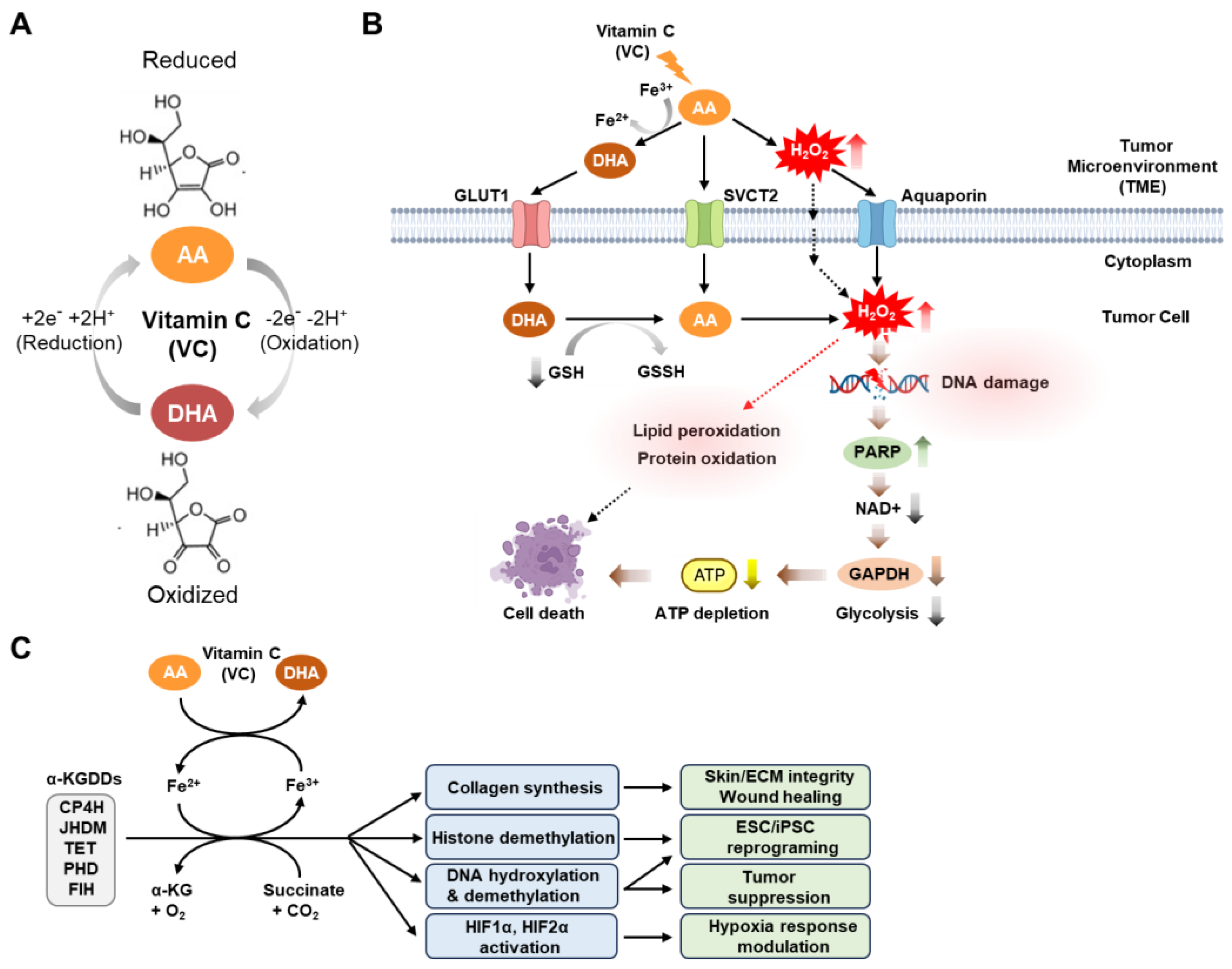
Figure 1. Physiological and anti-cancer mechanisms of vitamin C activity. (A) Physiological vitamin C (VC) exists largely in its reduced (ascorbic acid (AA)) or oxidized (dehydroascorbic acid (DHA)) forms, determined by either the gain or loss of two electrons and two hydrogens (reduction: +2e +2H+; oxidation: −2e −2H+). (B) Pharmacological VC can induce cancer cell death through two complementary mechanisms that elevate oxidative stress. Following VC treatment, hydrogen peroxide (H2O2) is produced in the extracellular environment by AA oxidation via Fenton chemistry that is facilitated by the presence of labile ferric iron (Fe3+) that enters cancer cells from the tumor microenvironment through either aquaporins or passive diffusion. VC enters cells through sodium-dependent vitamin C transporters (mainly SVCT2) when it is in its reduced form (AA), or via glucose transporters (mainly GLUT1) when it is in its oxidized form (DHA). Once inside the cell, dehydroascorbic acid (DHA) is rapidly converted to ascorbic acid (AA) through the action of the reducing agent glutathione (GSH). This process depletes the intracellular glutathione, resulting in elevated levels of intracellular H2O2 and several detrimental effects, including DNA damage, lipid peroxidation, and protein oxidation. In particular, DNA damage triggers the activation of the DNA repair enzyme poly (ADP-ribose) polymerase (PARP), which depletes cellular NAD+ levels. This depletion, in turn, inhibits the activity of glyceraldehyde 3-phosphate dehydrogenase (GAPDH) and glycolysis in cancer cells, resulting in decreased ATP production and cell death. (C) VC plays a pivotal role in numerous biological processes by serving as a cofactor for Fe2+ and alpha-ketoglutarate-dependent dioxygenases (Fe2+/α-KGDDs). These enzymes encompass a range of proteins, including collagen prolyl hydroxylases (CP4H), JmjC histone demethylases (JHDMs), ten–eleven translocation (TET) DNA hydroxylases, and hypoxia-inducible factor (HIF) hydroxylases (such as proline hydroxylase domain proteins (PHDs), and asparagine hydroxylase (factor-inhibiting HIF [FIH])). These enzymes have diverse functions, such as regulating collagen synthesis to maintain skin tissue and extracellular matrix (ECM) integrity as well as to facilitate efficient wound healing. They can also promote histone and DNA demethylation, thereby enhancing induced pluripotent stem cell (iPSC) reprogramming and suppressing leukemia progression. Furthermore, they can modulate various responses under low-oxygen conditions (hypoxia). This figure was created using BioRender, with modifications inspired by [42,43,44][42][43][44].
Notably, VC exhibits several drug-like properties that make it a promising therapeutic agent. VC has a low molecular weight (<500 Da, 176.12 g/mol), is water-soluble, and has a high oral bioavailability. The bioavailability of VC in foods is generally considered equivalent to the purified form within the recommended nutritional range of 15–200 mg [45]. Yet, this bioavailability diminishes by over 50% with higher amounts, such as doses exceeding 1000 mg. Since VC was first chemically synthesized in 1933, the bioavailability of synthetic and natural VC has been a subject of extensive research [46]. Animal studies indicate varying bioavailability between synthetic and natural VC, depending on the study design and animal model. In contrast, human studies have consistently shown no significant differences in their bioavailability [47]. While synthetic and natural VC share the same molecular and physicochemical properties, it has been reported that fruits and vegetables offer a wealth of micronutrients, dietary fiber, and phytochemicals that can modulate the absorption and utilization of VC [47,48][47][48]. Furthermore, VC is highly susceptible to oxidation and degradation, particularly in biological fluids such as plasma and blood [47,48][47][48]. The stability of VC in these fluids is influenced by several factors that can occur during processing (e.g., heat and light, pH, metal ions) and storage (e.g., temperature, oxygen exposure). Particularly, in vivo VC levels are determined by a balance between uptake, metabolism, and excretion (refer to [49] for a comprehensive review of these processes). Notably, the conversion of ascorbic acid to DHA in foods or the gastrointestinal tract can diminish the bioactivity of VC [49]. To enhance the chemical stability and bioavailability of VC, various chemically synthesized ascorbic analogs, such as ascorbate 2-sulfate, ascorbate 2-monophosphate, and ascorbate 2-triphosphate, have been developed [45]. Additionally, encapsulating VC in specific nanoparticles has been shown to improve stability during storage and delivery [45]. The transport of the reduced form of VC (AA) occurs through specialized transporters known as sodium-dependent vitamin C transporters (SVCT) 1 and 2, which are conserved across mammalian species, including humans [50,51,52,53][50][51][52][53] (Figure 1B). Notably, it has been reported that SVCT2 is a key protein for VC uptake in both normal [54,55][54][55] and cancer cells [37,56][37][56]. Moreover, VC has been shown to inhibit breast cancer cell growth [56] or preferentially kill CSC populations in live cancer in an SVCT2-dependent manner [37]. In contrast, transport of the oxidized form of VC (DHA) into cells is primarily facilitated by glucose transporters known as GLUTs (GLUTs 1–4 and 8) [57,58,59,60][57][58][59][60]. According to the prevailing model, DHA is the most active anti-cancer form of VC in tumors, as it generates cytotoxic reactive oxygen species (ROS) upon its intracellular conversion to AA following its entry into cells [61,62][61][62]. However, recent studies involving direct treatment of various cancer cell lines, including human breast cancer and neuroblastoma cells, with DHA have consistently shown that DHA has minimal or no significant impact on cell death [42,43,63][42][43][63]. This suggests the possibility that the cytotoxic responses to DHA can vary depending on the specific cancer cell type or experimental conditions. Taken together, these results underscore the need for further studies in a wide range of cancer cell types to explore how the distinct redox forms of VC contribute to VC-induced cell death. Numerous preclinical investigations of various human cancer models have indicated that the extracellular generation of hydrogen peroxide (H2O2) is a pivotal factor in the anti-cancer efficacy of high-dose VC [13,63][13][63]. AA readily oxidizes to DHA through a two-electron oxidation process in the presence of catalytic metals, like copper (Cu+/Cu2+) and iron (Fe3+/Fe2+), leading to elevated H2O2 concentrations in the extracellular space of tumors [7,64,65][7][64][65]. The H2O2 can subsequently permeate cells by utilizing peroxiporins within the plasma membrane to exert its influence on redox-dependent signaling and metabolic pathways pertinent to the viability of cancer cells, including the pathways regulating processes such as cell-cycle arrest, DNA damage, and apoptosis [66] (Figure 1B). However, the role of iron in the anti-cancer action of VC has recently been debated, with varying findings in different in vitro studies. Some investigations have observed that reducing or depleting intracellular iron levels enhances the growth inhibition and apoptosis induced by VC in neuroblastoma and K562 leukemic cells [67[67][68],68], while others have reported that extracellular iron diminishes the anti-cancer effects of VC in PC-3 and LNCaP prostate cancer cell lines [69]. More recently, it has been documented that exogenous iron impairs the anti-cancer effects of VC in specific cancer cell lines, both in vitro and in vivo [70]. These findings suggest that the impact of iron on VC-induced cytotoxicity may vary depending on the cell type or experimental method employed, such as inhibiting intracellular iron using iron chelators or exogenous iron treatment, necessitating further exploration in other cancer types. Notably, the mechanisms of cell death underlying the anti-cancer effects of VC have undergone extensive investigation. Previous research has suggested that pharmacological VC can trigger various forms of cell death, including apoptosis, necroptosis, and autophagy, with the specific outcome contingent upon the concentration and cell type employed in the experiment [43]. Earlier studies have also indicated that VC-induced cytotoxicity is primarily mediated through caspase-dependent apoptosis or necrosis, based on assessments of changes in the protein levels of key cell death effectors, such as caspases, BAX, BID, and receptor-interacting protein kinase (RIPK1), in response to VC exposure [71,72,73][71][72][73]. However, emerging evidence indicates that classical inhibitors of apoptosis or necrosis, such as the pan-caspase inhibitor Z-VAD-FMK and the RIP1-targeted necroptosis inhibitor Nec-1, do not prevent the cell death induced by pharmacological VC, suggesting that there are non-canonical cell death mechanisms at play [74,75][74][75]. Recently, non-apoptotic forms of cell death, such as ferroptosis, parthanatos, and pyroptosis, have garnered attention as promising targets for cancer therapy with natural or synthetic compounds that induce ROS [76]. These findings suggest that the cytotoxic effects induced by high-dose VC may entail multiple cell death pathways operating synergistically, rather than a single pathway. In addition to cell death mechanisms, metabolic crises are a recurring phenomenon in cancer cell death triggered by pharmacological VC treatment [18]. Consistent with the Warburg hypothesis, which indicates cancer cells’ preference for glycolysis over oxidative phosphorylation for energy production, ATP depletion and cell demise in response to VC are primarily attributed to the hindered glycolysis caused by the VC-induced H2O2-mediated inhibition of glyceraldehyde 3-phosphate dehydrogenase (GAPDH) activity [43,62][43][62]. In this context, recent studies have proposed a model suggesting that VC-induced H2O2 inflicts DNA damage, consequently promoting poly (ADP-ribose) polymerase (PARP) activation, which, in turn, consumes NAD and depletes ATP through the reduction of GAPDH activity and glycolysis [43,61,77,78][43][61][77][78]. However, subsequent investigations have reported that treatment with the PARP inhibitor Olaparib maintains NAD+ and ATP levels but results in increased DNA double-strand breaks and does not prevent ascorbate-induced cell death [43]. This suggests that the PARP-associated DNA damage response may not be the exclusive cause of this cytotoxicity, implying that supplementary mechanisms contribute to the NAD+- and ATP depletion-dependent cytotoxicity of VC treatment [43] (Figure 1B). In addition to its pro-oxidant properties, several studies have uncovered additional VC-mediated anti-tumor mechanisms involving epigenetic and post-translational pathways (Figure 1C). VC has been found to serve as a key cofactor that catalyzes the activity of various iron-containing dioxygenase enzymes, such as ferrous iron Fe2+ and α-ketoglutarate (αKG)-dependent dioxygenases (Fe2+/α-KGDDs), that play diverse roles in many biological processes, including the regulation of metabolic adaptations to hypoxia, the epigenetic regulation of gene transcription, and the reprogramming of cellular metabolism [7]. These enzymes include hypoxia-inducible factor (HIF) hydroxylases (e.g., prolyl-hydroxylase domain-containing proteins (PHDs 1–3) and factor inhibiting HIF (FIH)), as well as DNA demethylases (e.g., TET1–3) [8,9,10][8][9][10]. The activities of these enzymes contribute to the suppression of oncogenes and the re-expression of tumor suppressor genes, resulting in post-translational and/or epigenetic anti-tumor effects in both hematological and solid tumors [36,79,80][36][79][80]. Indeed, a growing number of studies report the involvement of these enzymes in VC-induced tumor suppression across multiple cancer types, including leukemia, melanoma, and renal cell carcinoma [4,79,80,81,82][4][79][80][81][82]. Harnessing its multifaceted effects in cancer, high-dose VC has emerged as a promising therapeutic strategy, either as a standalone treatment or in combination with various standard (chemo-) therapies, potentially alleviating the toxic side effects associated with chemotherapy [12,13,83][12][13][83]. Notably, even intravenous administration of very high doses of VC, ranging from 1 to 200 g and administered repeatedly, was reported to be well tolerated in the majority of patients [84]. However, caution has been noted regarding the administration of high doses, as they may lead to overt side effects in certain susceptible patients, such as the formation of oxalate renal stones [85]. Moreover, it is important to note that some patients may experience side effects, including diarrhea, nausea, abdominal cramps, and other gastrointestinal issues [85]. In light of these considerations, national clinical trials (NCT) have been actively investigating the effects of vitamin C as a standalone treatment or in combined therapies across various cancers, including EGFR mutant non-small cell lung cancer (NSCLC) (NCT04033107), recurrent high-grade glioma (NCT01891747), metastatic colorectal cancer (NCT04516681, NCT02969681), KRAS and BRAF mutant colon cancer (NCT04035096), hepatocellular carcinoma, pancreatic cancer, gastric cancer, colorectal cancer (NCT04033107), and acute myeloid leukemia (AML) (NCT02877277).

References

  1. Figueroa-Méndez, R.; Rivas-Arancibia, S. Vitamin C in Health and Disease: Its Role in the Metabolism of Cells and Redox State in the Brain. Front. Physiol. 2015, 6, 397.
  2. Pullar, J.M.; Carr, A.C.; Vissers, M.C.M. The Roles of Vitamin C in Skin Health. Nutrients 2017, 9, 866.
  3. Carr, A.C.; Maggini, S. Vitamin C and Immune Function. Nutrients 2017, 9, 1211.
  4. Chen, H.Y.; Almonte-Loya, A.; Lay, F.Y.; Hsu, M.; Johnson, E.; González-Avalos, E.; Yin, J.; Bruno, R.S.; Ma, Q.; Ghoneim, H.E.; et al. Epigenetic remodeling by vitamin C potentiates plasma cell differentiation. Elife 2022, 11, e73754.
  5. Kaźmierczak-Barańska, J.; Boguszewska, K.; Adamus-Grabicka, A.; Karwowski, B.T. Two Faces of Vitamin C—Antioxidative and Pro-Oxidative Agent. Nutrients 2020, 12, 1501.
  6. Chen, Q.; Espey, M.G.; Sun, A.Y.; Pooput, C.; Kirk, K.L.; Krishna, M.C.; Khosh, D.B.; Drisko, J.; Levine, M. Pharmacologic doses of ascorbate act as a prooxidant and decrease growth of aggressive tumor xenografts in mice. Proc. Natl. Acad. Sci. USA 2008, 105, 11105–11109.
  7. Villagran, M.; Ferreira, J.; Martorell, M.; Mardones, L. The Role of Vitamin C in Cancer Prevention and Therapy: A Literature Review. Antioxidants 2021, 10, 1894.
  8. Zhitkovich, A. Nuclear and Cytoplasmic Functions of Vitamin C. Chem. Res. Toxicol. 2020, 33, 2515–2526.
  9. Brabson, J.P.; Leesang, T.; Mohammad, S.; Cimmino, L. Epigenetic Regulation of Genomic Stability by Vitamin C. Front. Genet. 2021, 12, 675780.
  10. Cenigaonandia-Campillo, A.; Serna-Blasco, R.; Gómez-Ocabo, L.; Solanes-Casado, S.; Baños-Herraiz, N.; Puerto-Nevado, L.D.; Cañas, J.A.; Aceñero, M.J.; García-Foncillas, J.; Aguilera, Ó. Vitamin C activates pyruvate dehydrogenase (PDH) targeting the mitochondrial tricarboxylic acid (TCA) cycle in hypoxic. Theranostics 2021, 11, 3595–3606.
  11. Szarka, A.; Kapuy, O.; Lőrincz, T.; Bánhegyi, G. Vitamin C and Cell Death. Antioxid. Redox Signal 2021, 34, 831–844.
  12. Böttger, F.; Vallés-Martí, A.; Cahn, L.; Jimenez, C.R. High-dose intravenous vitamin C, a promising multi-targeting agent in the treatment of cancer. J. Exp. Clin. Cancer Res. 2021, 40, 343.
  13. Mussa, A.; Idris, R.A.M.; Ahmed, N.; Ahmad, S.; Murtadha, A.H.; Din, T.A.D.A.A.T.; Yean, C.Y.; Rahman, W.F.W.A.; Lazim, N.M.; Uskoković, V.; et al. High-Dose Vitamin C for Cancer Therapy. Pharmaceuticals 2022, 15, 711.
  14. Di Tano, M.; Raucci, F.; Vernieri, C.; Caffa, I.; Buono, R.; Fanti, M.; Brandhorst, S.; Curigliano, G.; Nencioni, A.; de Braud, F.; et al. Synergistic effect of fasting-mimicking diet and vitamin C against KRAS mutated cancers. Nat. Commun. 2020, 11, 2332.
  15. Magrì, A.; Germano, G.; Lorenzato, A.; Lamba, S.; Chilà, R.; Montone, M.; Amodio, V.; Ceruti, T.; Sassi, F.; Arena, S.; et al. High-dose vitamin C enhances cancer immunotherapy. Sci. Transl. Med. 2020, 12, eaay8707.
  16. Bedhiafi, T.; Inchakalody, V.P.; Fernandes, Q.; Mestiri, S.; Billa, N.; Uddin, S.; Merhi, M.; Dermime, S. The potential role of vitamin C in empowering cancer immunotherapy. Biomed. Pharmacother. 2021, 146, 112553.
  17. Li, W.-N.; Zhang, S.-J.; Feng, J.-Q.; Jin, W.-L. Repurposing Vitamin C for Cancer Treatment: Focus on Targeting the Tumor Microenvironment. Cancers 2022, 14, 2608.
  18. Das, P.K.; Pillai, S.; Rakib, M.A.; Khanam, J.A.; Gopalan, V.; Lam, A.K.Y.; Islam, F. Plasticity of Cancer Stem Cell: Origin and Role in Disease Progression and Therapy Resistance. Stem Cell Rev. Rep. 2020, 16, 397–412.
  19. Tanabe, S.; Quader, S.; Cabral, H.; Ono, R. Interplay of EMT and CSC in Cancer and the Potential Therapeutic Strategies. Front. Pharmacol. 2020, 11, 904.
  20. Kumar, V.E.; Nambiar, R.; De Souza, C.; Nguyen, A.; Chien, J.; Lam, K.S. Targeting Epigenetic Modifiers of Tumor Plasticity and Cancer Stem Cell Behavior. Cells 2022, 11, 1403.
  21. Yadav, U.P.; Singh, T.; Kumar, P.; Sharma, P.; Kaur, H.; Sharma, S.; Singh, S.; Kumar, S.; Mehta, K. Metabolic Adaptations in Cancer Stem Cells. Front. Oncol. 2020, 10, 1010.
  22. Albini, A.; Bruno, A.; Gallo, C.; Pajardi, G.; Noonan, D.M.; Dallaglio, K. Cancer stem cells and the tumor microenvironment: Interplay in tumor heterogeneity. Connect. Tissue Res. 2015, 56, 414–425.
  23. Wu, B.; Shi, X.; Jiang, M.; Liu, H. Cross-talk between cancer stem cells and immune cells: Potential therapeutic targets in the tumor immune microenvironment. Mol. Cancer 2023, 22, 38.
  24. Clara, J.A.; Monge, C.; Yang, Y.; Takebe, N. Targeting signalling pathways and the immune microenvironment of cancer stem cells—A clinical update. Nat. Rev. Clin. Oncol. 2020, 17, 204–232.
  25. Chae, Y.C.; Kim, J.H. Cancer stem cell metabolism: Target for cancer therapy. BMB Rep. 2018, 51, 319–326.
  26. Batlle, E.; Clevers, H. Cancer stem cells revisited. Nat. Med. 2017, 23, 1124–1134.
  27. Shibue, T.; Weinberg, R.A. EMT, CSCs, and drug resistance: The mechanistic link and clinical implications. Nat. Rev. Clin. Oncol. 2017, 14, 611–629 .
  28. Qin, S.; Jiang, J.; Lu, Y.; Nice, E.C.; Huang, C.; Zhang, J.; He, W. Emerging role of tumor cell plasticity in modifying therapeutic response. Signal Transduct. Target. Ther. 2020, 5, 228.
  29. Gupta, P.B.; Onder, T.T.; Jiang, G.; Tao, K.; Kuperwasser, C.; Weinberg, R.A.; Lander, E.S. Identification of selective inhibitors of cancer stem cells by high-throughput screening. Cell 2009, 138, 645–659.
  30. Du, F.Y.; Zhou, Q.F.; Sun, W.J.; Chen, G.L. Targeting cancer stem cells in drug discovery: Current state and future perspectives. World J. Stem Cells 2019, 11, 398–420.
  31. Lee, Y.; Tanggono, A.S. Potential Role of the Circadian Clock in the Regulation of Cancer Stem Cells and Cancer Therapy. Int. J. Mol. Sci. 2022, 23, 14181.
  32. Basak, D.; Arrighi, S.; Darwiche, Y.; Deb, S. Comparison of Anticancer Drug Toxicities: Paradigm Shift in Adverse Effect Profile. Life 2021, 12, 48.
  33. Meerson, A.; Khatib, S.; Mahajna, J. Natural Products Targeting Cancer Stem Cells for Augmenting Cancer Therapeutics. Int. J. Mol. Sci. 2021, 22, 13044.
  34. Telang, N.T.; Nair, H.B.; Wong, G.Y.C. Growth Inhibitory Efficacy of Chinese Herbs in a Cellular Model for Triple-Negative Breast Cancer. Pharmaceuticals 2021, 14, 1318.
  35. Cimmino, L.; Dolgalev, I.; Wang, Y.; Yoshimi, A.; Martin, G.H.; Wang, J.; Ng, V.; Xia, B.; Witkowski, M.T.; Mitchell-Flack, M.; et al. Restoration of TET2 Function Blocks Aberrant Self-Renewal and Leukemia Progression. Cell 2017, 170, 1079–1095.e1020.
  36. Agathocleous, M.; Meacham, C.E.; Burgess, R.J.; Piskounova, E.; Zhao, Z.; Crane, G.M.; Cowin, B.L.; Bruner, E.; Murphy, M.M.; Chen, W.; et al. Ascorbate regulates haematopoietic stem cell function and leukaemogenesis. Nature 2017, 549, 476–481.
  37. Lv, H.; Wang, C.; Fang, T.; Li, T.; Lv, G.; Han, Q.; Yang, W.; Wang, H. Vitamin C preferentially kills cancer stem cells in hepatocellular carcinoma via SVCT-2. NPJ Precis. Oncol. 2018, 2, 1.
  38. Wan, J.; Zhou, J.; Fu, L.; Li, Y.; Zeng, H.; Xu, X.; Lv, C.; Jin, H. Ascorbic Acid Inhibits Liver Cancer Growth and Metastasis. Front. Pharmacol. 2021, 12, 726015.
  39. Bonuccelli, G.; De Francesco, E.M.; de Boer, R.; Tanowitz, H.B.; Lisanti, M.P. NADH autofluorescence, a new metabolic biomarker for cancer stem cells: Identification of Vitamin C and CAPE as natural products targeting “stemness”. Oncotarget 2017, 8, 20667–20678.
  40. Sen, U.; Chaudhury, D.; Shenoy, P.S.; Bose, B. Differential sensitivities of triple-negative breast cancer stem cell towards various doses of vitamin C: An insight into the internal antioxidant systems. J. Cell Biochem. 2021, 122, 349–366.
  41. Jiang, X.; Liu, J.; Mao, J.; Han, W.; Fan, Y.; Luo, T.; Xia, J.; Lee, M.J.; Lin, W. Pharmacological ascorbate potentiates combination nanomedicines and reduces cancer cell stemness to prevent post-surgery recurrence and systemic metastasis. Biomaterials 2023, 295, 122037.
  42. El Banna, N.; Hatem, E.; Heneman-Masurel, A.; Léger, T.; Baïlle, D.; Vernis, L.; Garcia, C.; Martineau, S.; Dupuy, C.; Vagner, S.; et al. Redox modifications of cysteine-containing proteins, cell cycle arrest and translation inhibition: Involvement in vitamin C-induced breast cancer cell death. Redox Biol. 2019, 26, 101290.
  43. Ma, E.; Chen, P.; Wilkins, H.M.; Wang, T.; Swerdlow, R.H.; Chen, Q. Pharmacologic ascorbate induces neuroblastoma cell death by hydrogen peroxide mediated DNA damage and reduction in cancer cell glycolysis. Free Radic. Biol. Med. 2017, 113, 36–47.
  44. Cimmino, L.; Neel, B.G.; Aifantis, I. Vitamin C in Stem Cell Reprogramming and Cancer. Trends Cell Biol. 2018, 28, 698–708.
  45. Maurya, V.K.; Shakya, A.; McClements, D.J.; Srinivasan, R.; Bashir, K.; Ramesh, T.; Lee, J.; Sathiyamoorthi, E. Vitamin C fortification: Need and recent trends in encapsulation technologies. Front. Nutr. 2023, 10, 1229243.
  46. Pelletier, O.; Keith, M.O. Bioavailability of synthetic and natural ascorbic acid. J. Am. Diet. Assoc. 1974, 64, 271–275.
  47. Carr, A.C.; Vissers, M.C. Synthetic or food-derived vitamin C—Are they equally bioavailable? Nutrients 2013, 5, 4284–4304.
  48. Carr, A.C.; Bozonet, S.M.; Vissers, M.C. A randomised cross-over pharmacokinetic bioavailability study of synthetic versus kiwifruit-derived vitamin C. Nutrients 2013, 5, 4451–4461.
  49. Michels, A.J.; Hagen, T.M.; Frei, B. Human genetic variation influences vitamin C homeostasis by altering vitamin C transport and antioxidant enzyme function. Annu. Rev. Nutr. 2013, 33, 45–70.
  50. Tsukaguchi, H.; Tokui, T.; Mackenzie, B.; Berger, U.V.; Chen, X.Z.; Wang, Y.; Brubaker, R.F.; Hediger, M.A. A family of mammalian Na+-dependent L-ascorbic acid transporters. Nature 1999, 399, 70–75.
  51. Wang, H.; Dutta, B.; Huang, W.; Devoe, L.D.; Leibach, F.H.; Ganapathy, V.; Prasad, P.D. Human Na(+)-dependent vitamin C transporter 1 (hSVCT1): Primary structure, functional characteristics and evidence for a non-functional splice variant. Biochim. Biophys. Acta 1999, 1461, 1–9.
  52. Daruwala, R.; Song, J.; Koh, W.S.; Rumsey, S.C.; Levine, M. Cloning and functional characterization of the human sodium-dependent vitamin C transporters hSVCT1 and hSVCT2. FEBS Lett. 1999, 460, 480–484.
  53. Wang, Y.; Mackenzie, B.; Tsukaguchi, H.; Weremowicz, S.; Morton, C.C.; Hediger, M.A. Human vitamin C (L-ascorbic acid) transporter SVCT1. Biochem. Biophys. Res. Commun. 2000, 267, 488–494.
  54. Pierce, M.R.; Raj, A.; Betke, K.M.; Zeidan, L.N.; Matthies, H.J.; May, J.M. Sodium-dependent vitamin C transporter-2 mediates vitamin C transport at the cortical nerve terminal. J. Neurosci. Res. 2015, 93, 1881–1890.
  55. Salazar, K.; Espinoza, F.; Cerda-Gallardo, G.; Ferrada, L.; Magdalena, R.; Ramírez, E.; Ulloa, V.; Saldivia, N.; Troncoso, N.; Oviedo, M.J.; et al. SVCT2 Overexpression and Ascorbic Acid Uptake Increase Cortical Neuron Differentiation, Which Is Dependent on Vitamin C Recycling between Neurons and Astrocytes. Antioxidants 2021, 10, 1413.
  56. Hong, S.W.; Lee, S.H.; Moon, J.H.; Hwang, J.J.; Kim, D.E.; Ko, E.; Kim, H.S.; Cho, I.J.; Kang, J.S.; Kim, D.J.; et al. SVCT-2 in breast cancer acts as an indicator for L-ascorbate treatment. Oncogene 2013, 32, 1508–1517.
  57. Rumsey, S.C.; Kwon, O.; Xu, G.W.; Burant, C.F.; Simpson, I.; Levine, M. Glucose transporter isoforms GLUT1 and GLUT3 transport dehydroascorbic acid. J. Biol. Chem. 1997, 272, 18982–18989.
  58. Rumsey, S.C.; Daruwala, R.; Al-Hasani, H.; Zarnowski, M.J.; Simpson, I.A.; Levine, M. Dehydroascorbic acid transport by GLUT4 in Xenopus oocytes and isolated rat adipocytes. J. Biol. Chem. 2000, 275, 28246–28253.
  59. Corpe, C.P.; Eck, P.; Wang, J.; Al-Hasani, H.; Levine, M. Intestinal dehydroascorbic acid (DHA) transport mediated by the facilitative sugar transporters, GLUT2 and GLUT8. J. Biol. Chem. 2013, 288, 9092–9101.
  60. Vera, J.C.; Rivas, C.I.; Fischbarg, J.; Golde, D.W. Mammalian facilitative hexose transporters mediate the transport of dehydroascorbic acid. Nature 1993, 364, 79–82.
  61. Yun, J.; Mullarky, E.; Lu, C.; Bosch, K.N.; Kavalier, A.; Rivera, K.; Roper, J.; Chio, I.I.; Giannopoulou, E.G.; Rago, C.; et al. Vitamin C selectively kills KRAS and BRAF mutant colorectal cancer cells by targeting GAPDH. Science 2015, 350, 1391–1396.
  62. Ngo, B.; Van Riper, J.M.; Cantley, L.C.; Yun, J. Targeting cancer vulnerabilities with high-dose vitamin C. Nat. Rev. Cancer 2019, 19, 271–282.
  63. Chen, Q.; Espey, M.G.; Krishna, M.C.; Mitchell, J.B.; Corpe, C.P.; Buettner, G.R.; Shacter, E.; Levine, M. Pharmacologic ascorbic acid concentrations selectively kill cancer cells: Action as a pro-drug to deliver hydrogen peroxide to tissues. Proc. Natl. Acad. Sci. USA 2005, 102, 13604–13609.
  64. Shen, J.; Griffiths, P.T.; Campbell, S.J.; Utinger, B.; Kalberer, M.; Paulson, S.E. Ascorbate oxidation by iron, copper and reactive oxygen species: Review, model development, and derivation of key rate constants. Sci. Rep. 2021, 11, 7417.
  65. Du, J.; Wagner, B.A.; Buettner, G.R.; Cullen, J.J. Role of labile iron in the toxicity of pharmacological ascorbate. Free Radic. Biol. Med. 2015, 84, 289–295.
  66. Lennicke, C.; Rahn, J.; Lichtenfels, R.; Wessjohann, L.A.; Seliger, B. Hydrogen peroxide—Production, fate and role in redox signaling of tumor cells. Cell Commun. Signal. 2015, 13, 39.
  67. Carosio, R.; Zuccari, G.; Orienti, I.; Mangraviti, S.; Montaldo, P.G. Sodium ascorbate induces apoptosis in neuroblastoma cell lines by interfering with iron uptake. Mol. Cancer 2007, 6, 55.
  68. Tsuma-Kaneko, M.; Sawanobori, M.; Kawakami, S.; Uno, T.; Nakamura, Y.; Onizuka, M.; Ando, K.; Kawada, H. Iron removal enhances vitamin C-induced apoptosis and growth inhibition of K-562 leukemic cells. Sci. Rep. 2018, 8, 17377.
  69. Mojić, M.; Bogdanović Pristov, J.; Maksimović-Ivanić, D.; Jones, D.R.; Stanić, M.; Mijatović, S.; Spasojević, I. Extracellular iron diminishes anticancer effects of vitamin C: An in vitro study. Sci. Rep. 2014, 4, 5955.
  70. Zhong, B.; Zhao, L.; Yu, J.; Hou, Y.; Ai, N.; Lu, J.J.; Ge, W.; Chen, X. Exogenous iron impairs the anti-cancer effect of ascorbic acid both in vitro and in vivo. J. Adv. Res. 2023, 46, 149–158.
  71. Kim, J.E.; Kang, J.S.; Lee, W.J. Vitamin C Induces Apoptosis in Human Colon Cancer Cell Line, HCT-8 Via the Modulation of Calcium Influx in Endoplasmic Reticulum and the Dissociation of Bad from 14-3-3β. Immune Netw. 2012, 12, 189–195.
  72. Shin, H.; Nam, A.; Song, K.H.; Lee, K.; Rebhun, R.B.; Seo, K.W. Anticancer effects of high-dose ascorbate on canine melanoma cell lines. Vet. Comp. Oncol. 2018, 16, 616–621.
  73. Chen, X.Y.; Chen, Y.; Qu, C.J.; Pan, Z.H.; Qin, Y.; Zhang, X.; Liu, W.J.; Li, D.F.; Zheng, Q. Vitamin C induces human melanoma A375 cell apoptosis via Bax- and Bcl-2-mediated mitochondrial pathways. Oncol. Lett. 2019, 18, 3880–3886.
  74. Baek, M.W.; Cho, H.S.; Kim, S.H.; Kim, W.J.; Jung, J.Y. Ascorbic Acid Induces Necrosis in Human Laryngeal Squamous Cell Carcinoma via ROS, PKC, and Calcium Signaling. J. Cell Physiol. 2017, 232, 417–425.
  75. Kim, J.H.; Hwang, S.; Lee, J.H.; Im, S.S.; Son, J. Vitamin C Suppresses Pancreatic Carcinogenesis through the Inhibition of Both Glucose Metabolism and Wnt Signaling. Int. J. Mol. Sci. 2022, 23, 12249.
  76. Greco, G.; Catanzaro, E.; Fimognari, C. Natural Products as Inducers of Non-Canonical Cell Death: A Weapon against Cancer. Cancers 2021, 13, 304.
  77. Chen, P.; Yu, J.; Chalmers, B.; Drisko, J.; Yang, J.; Li, B.; Chen, Q. Pharmacological ascorbate induces cytotoxicity in prostate cancer cells through ATP depletion and induction of autophagy. Anticancer Drugs 2012, 23, 437–444.
  78. Buranasudja, V.; Doskey, C.M.; Gibson, A.R.; Wagner, B.A.; Du, J.; Gordon, D.J.; Koppenhafer, S.L.; Cullen, J.J.; Buettner, G.R. Pharmacologic Ascorbate Primes Pancreatic Cancer Cells for Death by Rewiring Cellular Energetics and Inducing DNA Damage. Mol. Cancer Res. 2019, 17, 2102–2114.
  79. Zhang, X.; Li, S.; He, J.; Jin, Y.; Zhang, R.; Dong, W.; Lin, M.; Yang, Y.; Tian, T.; Zhou, Y.; et al. TET2 Suppresses VHL Deficiency-Driven Clear Cell Renal Cell Carcinoma by Inhibiting HIF Signaling. Cancer Res. 2022, 82, 2097–2109.
  80. Gustafson, C.B.; Yang, C.; Dickson, K.M.; Shao, H.; Van Booven, D.; Harbour, J.W.; Liu, Z.J.; Wang, G. Epigenetic reprogramming of melanoma cells by vitamin C treatment. Clin. Epigenet. 2015, 7, 51.
  81. Miles, S.L.; Fischer, A.P.; Joshi, S.J.; Niles, R.M. Ascorbic acid and ascorbate-2-phosphate decrease HIF activity and malignant properties of human melanoma cells. BMC Cancer 2015, 15, 867.
  82. Brabson, J.P.; Leesang, T.; Yap, Y.S.; Wang, J.; Lam, M.Q.; Fang, B.; Dolgalev, I.; Barbieri, D.A.; Strippoli, V.; Bañuelos, C.P.; et al. Oxidized mC modulates synthetic lethality to PARP inhibitors for the treatment of leukemia. Cell Rep. 2023, 42, 112027.
  83. Zasowska-Nowak, A.; Nowak, P.J.; Ciałkowska-Rysz, A. High-Dose Vitamin C in Advanced-Stage Cancer Patients. Nutrients 2021, 13, 735.
  84. Padayatty, S.J.; Sun, A.Y.; Chen, Q.; Espey, M.G.; Drisko, J.; Levine, M. Vitamin C: Intravenous use by complementary and alternative medicine practitioners and adverse effects. PLoS ONE 2010, 5, e11414.
  85. Doseděl, M.; Jirkovský, E.; Macáková, K.; Krčmová, L.K.; Javorská, L.; Pourová, J.; Mercolini, L.; Remião, F.; Nováková, L.; Mladěnka, P.; et al. Vitamin C-Sources, Physiological Role, Kinetics, Deficiency, Use, Toxicity, and Determination. Nutrients 2021, 13, 615.
More
ScholarVision Creations