Your browser does not fully support modern features. Please upgrade for a smoother experience.
Please note this is a comparison between Version 1 by Lucia Kottferová and Version 2 by Rita Xu.
Pet hedgehogs, which are increasingly favoured companions, have garnered attention due to their potential as carriers of zoonotic diseases. These small insectivorous mammals, native to Europe, Asia, and Africa, are commonly kept as pets.
- hedgehog
- infections
- Trichophyton erinacei
- zoonoses
1. Introduction
In recent years, the upsurge in exotic pet ownership has introduced a charming yet intricate dimension to the human‒animal bond. Among unconventional companions, pet hedgehogs have garnered considerable attention. However, sharing our lives with these animals has not been without challenges, particularly concerning the transmission of fungal infections. Trichophyton erinacei, a dermatophytic fungal pathogen, has garnered scientific interest in recent years. Its potential to serve as a zoonotic bridge between these unconventional pets and their human caretakers has raised significant concerns within the veterinary and public health communities. A thorough scientific investigation is necessary to fully understand the complex landscape of zoonotic potential and public health consequences surrounding T. erinacei transmission from pet hedgehogs. Hedgehogs, small nocturnal insectivorous mammals widespread across Europe, Asia, and Africa, comprise sixteen recognised species organised into five genera: Hemiechinus (two species), Atelerix (four species), Erinaceus (four species), Paraechinus (four species), and Mesechinus (two species) [1][2][3][4][1,2,3,4].
Dermatomycosis infections from T. erinacei primarily originate from imported, unprotected white-bellied or African pygmy hedgehogs (Atelerix albiventris) available for purchase in pet shops, as well as Egyptian long-eared hedgehogs (Hemiechinus auritus). Understanding and addressing the potential health implications associated with these hedgehog species in the context of exotic pet ownership are crucial [5].
2. Classification and Taxonomy of the Dermatophytes
Dermatophytes, a group of highly successful pathogenic fungi, are responsible for causing superficial mycoses, commonly referred to as dermatophytosis or ringworm, in both humans and animals. These fungi are taxonomically and ecologically related, belonging to the family Arthrodermataceae within the order Onygenales. What sets them apart is their unique capability to metabolise keratin as their exclusive nutrient source [6][7][6,7]. Traditionally, dermatophytes were classified into three genera: Trichophyton, Epidermophyton, and Microsporum, with their sexual states grouped under Arthroderma [8][9][8,9]. However, recent taxonomic revisions, the elimination of dual nomenclature, and the utilisation of multi-gene phylogenies have expanded the number of genera within the dermatophyte group. Despite these changes, many of the most clinically significant primary pathogenic species still fall within the original three genera [10]. In the newly proposed taxonomy, Trichophyton contains 16 species, namely Epidermophyton (1 species), Nannizzia (9 species), Microsporum (3 species), Lophophyton (1 species), Arthroderma (21 species), and Ctenomyces (1 species), but more detailed studies remain needed to establish species borderlines. Each species now has a single valid name. Two new genera have been introduced: Guarromyces and Paraphyton. The number of genera has increased, but species that are relevant to routine diagnostics now belong to smaller groups, which enhances their identification [8]. One notable taxonomic refinement involves the former entity known as T. mentagrophytes var. erinacei. Smith and Marples first described it in 1963, but it has since undergone a reclassification and is now officially known as Trichophyton erinacei [9]. This reclassification aligns with earlier observations by mycologists who referred to it as the “hedgehog mushroom” [11][12][11,12]. The recognition of T. erinacei as a distinct dermatophyte species dates back to 1966 when Quaife emphasised that it is not a mere variant or subspecies but indeed constitutes a separate species [13]. In the last few decades, dermatophytes have come a long way in their modern systematic classification, which is mostly based on molecular species identification from ITS sequences [13]. The molecular investigations suggest a close genetic or evolutionary relationship between T. erinacei and the Trichophyton anamorph of A. benhamiae. This relationship extends to the teleomorph A. benhamiae. The findings indicate that this close relationship is observed in strains of these fungi found in both the American‒European and African regions [14][15][14,15]. Throughout the years, the aetiological agent of ringworm in hedgehogs has been reported under different names, reflecting evolving species concepts. These names include T. erinacei var. erinacei, T. erinacei, T. mentagrophytes var. erinacei, Arthroderma benhamiae var. erinacei, and A. benhamiae. These taxonomic refinements reflect a comprehensive understanding of the fungal species and their intricate relationships [12][16][12,16].3. Clinical Signs
3.1. Clinical Signs in Hedgehogs
T. erinacei infections in hedgehogs are characterised by several clinical signs. These signs are essential for recognising and understanding the disease in these animals. Hedgehogs infected with T. erinacei often exhibit crusty lesions on their skin [17]. These lesions can appear as scaly patches or areas of skin with an abnormal texture. Additionally, infected hedgehogs may experience alopecia, which is the loss of fur or spines, as you can see in Figure 1 and Figure 2. These clinical signs are particularly prominent on the head area of the hedgehog or on their back [8][18][8,18].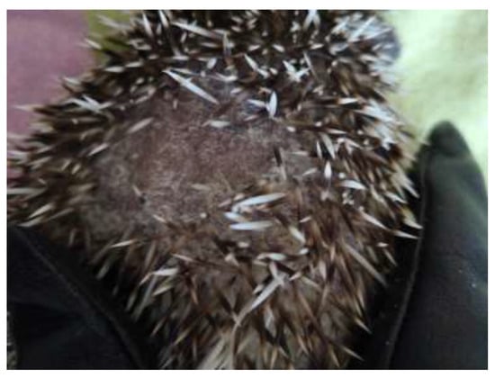
Figure 1. Crusty lesions, alopecia, and loss of spines in pet hedgehog caused by mycotic infection.
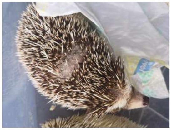
Figure 2. Lesion on the back of an African pygmy hedgehog due to infection by T. erinaceus.
3.2. Clinical Signs in Humans
Trichophyton infections in humans often present with distinctive clinical signs, primarily affecting the skin. One of the most common initial signs is the emergence of itchy and inflamed skin eruptions, often displaying a reddish base at the point where the skin comes into contact with an infected animal. While these symptoms can start off as mild, they tend to intensify over time. Unfortunately, there are instances where the initial rash resembles other inflammatory skin conditions, such as eczema, leading to the improper use of topical or oral corticosteroids. Regrettably, this misdiagnosis can expedite the progression of the infection, making it more severe [21]. Usually, there are single lesions in humans, but several cases have had two or three separate body areas affected. When evaluating skin lesions, particularly on the hands, obtaining a thorough and accurate medical history is crucial. This history should include any recent contact with animals, particularly hedgehogs. In cases where recent animal contact is reported, especially with hedgehogs, maintaining a high level of suspicion is vital for initiating prompt and appropriate treatment. Early recognition of Trichophyton infection and the commencement of antifungal therapy can lead to the timely relief of symptoms and clearance of the infection [22][23][24][22,23,24]. Furthermore, T. erinacei infections in humans can manifest in various ways (Table 1), primarily affecting the skin. Recognising these clinical signs is pivotal for accurate diagnosis and tailored treatment [2]. In human cases, T. erinacei infections frequently result in inflammatory dermatological conditions, with two common presentations being tinea manus and tinea corporis. These infections can cause discomfort and skin abnormalities, including redness, itching, and, in some instances, blister formation [18]. However, T. erinacei does not stop there; it has been identified as the causative agent in a range of other dermatophyte infections, including Tinea faciei, Tinea capitis, and Tinea barbae [21][22][21,22]. These diverse clinical manifestations highlight the critical need for precise species identification and treatments tailored to the specific infection [24][25][26][24,25,26].Table 1. Clinical presentation in humans after contact with hedgehog infected by T. erinaceus.
| Clinical Signs in Humans | Region | Authors |
|---|---|---|
| Tinea corporis | Asia | [22][27][28][22,27,28] |
| Europe | [29][29[30],30] | |
| Tinea faciei | America | [31] |
| Asia | [27][32][27,32] | |
| Europe | [28][33][34][28,33,34] | |
| Tinea barbae | Europe | [25] |
| Tinea manus | America | [21] |
| Asia | [26][28][35][36][37][26,28,35,36,37] | |
| Europe | [30][38][[41][3039][40],38,39,40,41] | |
| Onychomycosis | Asia | [42] |