Your browser does not fully support modern features. Please upgrade for a smoother experience.
Please note this is a comparison between Version 2 by Camila Xu and Version 1 by Doaa Sami Khafaga.
Bio-signals are records of biological events inside the human body, such as a heartbeat or muscle contraction. These signals are used to detect whether there is a problem or disorder in a human organ.
- COVIDOA
- compression
- Haar wavelet
- reconstruction
- optimization
- ECG
1. Introduction
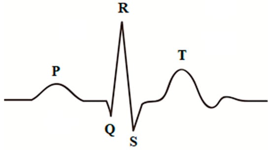
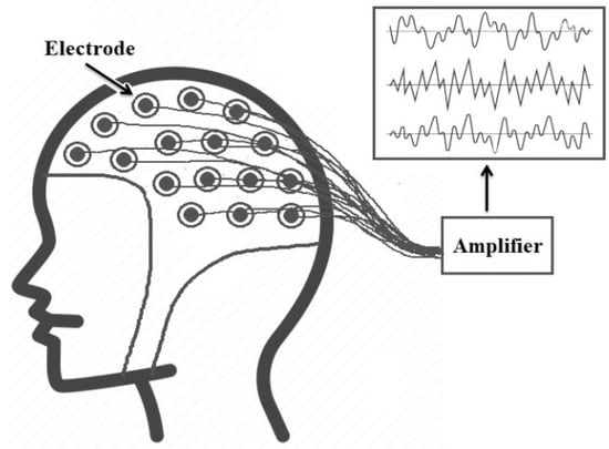
Smart healthcare systems deal with massive amounts of medical data daily for healthcare monitoring and early detection and diagnosis of diseases [1]. Bio-signals are records of biological events inside the human body, such as a heartbeat or muscle contraction. These signals are used to detect whether there is a problem or disorder in a human organ. There are many kinds of bio-signals used for various clinical purposes, such as ECG, which is used for recording human heart activity, EEG for recording the electrical activity of the brain, EMG for evaluating the electrical activity of skeletal muscles, ERG for measuring the electrical responses of various cell types in the retina, and EGG for recording the myoelectrical signal generated by the movement of the smooth muscle of the stomach [2]. ECG and EEG are the most widely used bio-signals for diagnoses of cardiac and brain disturbances [3]. Three main elements represent a typical heartbeat signal: the P wave, which indicates depolarization of the atria, the QRS complex, which shows depolarization of the ventricles, and the T wave, which represents repolarization of the ventricles, as shown in Figure 1. On the other hand, EEG signals record the brain’s electrical activity. Several sensors are positioned on various parts of the scalp to record EEG signals, as shown in Figure 2. EEG signals help to identify various common diseases, such as epilepsy and autism spectrum disorder [4].

Figure 1.
Single heartbeat.
In smart healthcare systems, the bio-signals are recorded by sensors attached to the patient’s body and then digitized. The digital bio-signals are processed using digital computers or computer-based medical devices [5]. An effective compression method is a basic need in such systems to minimize the volume of medical data and enhance the transmission’s efficiency [6]. However, obtaining high compression ratios is insufficient for an efficient compression algorithm, and data quality must also be maintained since the loss of medical data could lead to misdiagnosis problems.
2. Compression of Bio-Signals Using Block-Based Haar Wavelet Transform and COVIDOA for IoMT Systems
Over the last few decades, various algorithms have been proposed to compress medical data. These algorithms are either lossless or lossy compression. The lossless compression methods can achieve small compression ratios with no data loss, while lossy algorithms achieve much higher compression ratios but some information will be lost [8][7]. Data quality is crucial in the medical field, and losing some features may significantly impact the diagnosis process. However, suppose the data loss is within an acceptable limit and does not affect the data’s visual appearance. In that case, lossy compression techniques will be a good choice due to the high compression ratio they can achieve [9][8]. In [10][9], an ASCII character-encoding-based lossless compression method was proposed. In [11][10], Chen and Wang used two Huffman coding tables to develop a useful lossless compression method to reduce the storage and transmission demands for ECG signals. This algorithm has the advantages of low cost and power consumption. Rzepka [12][11] used selective linear prediction to compress multi-channel ECG. Most lossy compression algorithms are based on transform coding, where a specific transform is applied to the input signal, and some information is used to be discarded. In contrast, the others are used in the reconstruction process. The result of this process will not be identical to the original input, but it should be close enough according to the application’s purpose. The most popular transform-based compression techniques involve the DCT [13][12], DWT [7][13], and moment-based transform [14]. For bio-signal compression, Batista et al. [15] utilized Golomb–Rice coding with optimum DCT coefficients to compress ECG signals. They used the well-known MIT-BIH Arrhythmia database to evaluate their algorithm, where CR of 10.4:1 and PRD ≅≅ 2.5% were achieved. Jha and Kolekar [16] proposed another DCT-based algorithm to compress ECG signals. They employed DOST and dead-zone quantization to transform coefficients. Recent work includes the technique proposed in [17] for assessing compressed and decompressed ECG databases. The proposed algorithm used DCT, 16-bit quantization, run-length encoding for compression, and convolution neural network for classification. The obtained CR was 2.56, and the classification accuracies were 0.966 and 0.990 for the compressed and decompressed databases, respectively. Further, Pal et al. [18] proposed a compression algorithm for 2D ECG signals based on the combination of DCT and embedded zero-tree wavelet. The results showed that the suggested approach could raise the sparsity of the transform domain, which boosts compression effectiveness with a small degradation in reconstruction quality. Other DCT-based signal compression algorithms are proposed in [19,20][19][20]. The WT is a powerful tool for signal analysis because of its compact representation of signals and images, and its most popular applications are denoising and compression of signals [21]. Recently, OMs, such as Tchebichef and Hahn moments, have been used in signal reconstruction and compression due to their ability to represent signals [22]. Signal compression techniques based on orthogonal moments are presented in [23,24][23][24]. It is observed from the state of the art that the wavelet-based algorithms provide superior performance compared to the other compression methods [21]. Jha and Kolekar [25] used the DWT to select an appropriate mother wavelet to compress the ECG signal while guaranteeing quality. The same authors employed EMD and DWT to develop another ECG compression algorithm [26]. Based on the obtained results, it is noticed that the suggested method performs better than several current ECG compressors. Singhai et al. [27] used DWT and PSO to design a compression algorithm where the PSO selects threshold values and the optimal wavelet parameters. The compression ratio obtained using this algorithm was 28.43 at PRD = 2.63. Kolekar et al. [28] proposed an ECG compression technique based on the modified run-length encoding of wavelet coefficients. The proposed approach used dead-zone quantization for WT coefficients, and the obtained coefficients were encoded using modified run-length encoding. Shi et al. [29] proposed a new ECG compression method based on a binary convolutional auto-encoder (BCAE) equipped with residual error compensation (REC). The proposed method aimed to achieve efficient ECG compression through deep learning while ensuring high signal quality. The performance is tested using several measures, such as PRD, QS, SNR, and CR. The average performance in CR, PRD, NPRD, and SNR is 17.18, 3.92, 6.36, and 28.27 dB, respectively, for 48 ECG records. The achieved results in CR and PRD are 117.33 and 7.76, respectively. Recently, Singhai et al. [30] presented an algorithm for ECG compression based on DWT and nature-inspired optimization algorithms. The algorithm used the optimization algorithm to find the optimal wavelet design parameter values and optimal threshold levels. The results show the capability of this technique to provide high compression ratios with high signal quality. Lossy compression depends on using only some features and omitting others in return for reduced size. The question is, therefore, which features should be selected and which should not? The answer should be that the features that contain the most important clinical features and lead to the highest reconstruction quality should be selected, and the remaining features should be neglected. An optimization algorithm would be very helpful in selecting the most important feature subset. Motivated by the simplicity and efficiency of the HWT in signal and image processing and the efficiency of COVIDOA in solving various optimization tasks, researchers utilized the HWT in combination with COVIDOA to develop an efficient compression algorithm for bio-signals. In this approach, the signal is transformed using the HWT and then the best feature subset from the wavelet coefficients that should be used for reconstructing the signal will be selected with the help of COVIDOA.References
- Tian, S.; Yang, W.; Le Grange, J.M.; Wang, P.; Huang, W.; Ye, Z. Smart healthcare: Making medical care more intelligent. Glob. Health J. 2019, 3, 62–65.
- Reaz, M.B.I.; Hussain, M.S.; Mohd-Yasin, F. Techniques of EMG signal analysis: Detection, processing, classification, and applications. Biol. Proced. Online 2006, 8, 11–35.
- Houssein, E.H.; Kilany, M.; Hassanien, A.E. ECG signals classification: A review. Int. J. Intell. Eng. Inform. 2017, 5, 376–396.
- Nagel, S. Towards a Home-Use BCI: Fast Asynchronous Control and Robust Non-Control State Detection. Ph.D. Thesis, Universität Tübingen, Tübingen, Germany, 2019.
- Jeong, J.S.; Han, O.; You, Y.Y. A design characteristics of smart healthcare system as the IoT application. Indian J. Sci. Technol. 2016, 9, 52.
- Abdellatif, A.A.; Emam, A.; Chiasserini, C.F.; Mohamed, A.; Jaoua, A.; Ward, R. Edge-based compression and classification for smart healthcare systems: Concept, implementation, and evaluation. Expert Syst. Appl. 2019, 117, 1–14.
- Mentzer, F.; Gool, L.V.; Tschannen, M. Learning better lossless compression using lossy compression. In Proceedings of the IEEE/CVF Conference on Computer Vision and Pattern Recognition, Seattle, WA, USA, 14–19 June 2020; pp. 6638–6647.
- Hosny, K.M.; Khalid, A.M.; Mohamed, E.R. Efficient compression of volumetric medical images using Legendre moments and differential evolution. Soft Comput. 2020, 24, 409–427.
- Mukhopadhyay, S.K.; Mitra, S.; Mitra, M. A lossless ECG data compression technique using ASCII character encoding. Comput. Electr. Eng. 2011, 37, 486–497.
- Chen, S.L.; Wang, J.G. VLSI implementation of low-power cost-efficient lossless ECG encoder design for wireless healthcare monitoring application. Electron. Lett. 2013, 49, 91–93.
- Rzepka, D. Low-complexity lossless multichannel ECG compression based on selective linear prediction. Biomed. Signal Process. Control 2020, 57, 101705.
- Zhou, X.; Bai, Y.; Wang, C. Image compression based on discrete cosine transform and multistage vector quantization. Int. J. Multimed. Ubiquitous Eng. 2015, 10, 347–356.
- Makbol, N.M.; Khoo, B.E.; Rassem, T.H. Block-based discrete wavelet transform-singular value decomposition image watermarking scheme using human visual system characteristics. IET Image Process. 2016, 10, 34–52.
- Mahmmod, B.M.; Ramli, A.R.B.; Abdulhussain, S.H.; Al-Haddad, S.A.R.; Jassim, W.A. Signal compression and enhancement using a new orthogonal-polynomial-based discrete transform. IET Signal Process. 2018, 12, 129–142.
- Batista, L.V.; Carvalho, L.C.; Melcher, E.U.K. Compression of ECG signals based on optimum quantization of discrete cosine transform coefficients and Golomb-Rice coding. In Proceedings of the 25th Annual International Conference of the IEEE Engineering in Medicine and Biology Society (IEEE Cat. No. 03CH37439), Cancún, Mexico, 17–21 September 2003; Volume 3, pp. 2647–2650.
- Jha, C.K.; Kolekar, M.H. Electrocardiogram data compression using DCT-based discrete orthogonal Stockwell transform. Biomed. Signal Process. Control 2018, 46, 174–181.
- Soni, E.; Nagpal, A.; Garg, P.; Pinheiro, P.R. Assessment of Compressed and Decompressed ECG Databases for Telecardiology Applying a Convolution Neural Network. Electronics 2022, 11, 2708.
- Pal, H.S.; Kumar, A.; Vishwakarma, A.; Balyan, L.K. A Hybrid 2D ECG Compression Algorithm using DCT and Embedded Zero Tree Wavelet. In Proceedings of the 2022 IEEE 6th Conference on Information and Communication Technology (CICT), Gwalior, India, 18–20 November 2022; pp. 1–5.
- Su, Y.; Lu, X.; Huang, L.; Du, X.; Guizani, M. A novel DCT-based compression scheme for 5G vehicular networks. IEEE Trans. Veh. Technol. 2019, 68, 10872–10881.
- Pandey, A.; Saini, B.S.; Singh, B.; Sood, N. Quality-controlled ECG data compression based on 2D discrete cosine coefficient filtering and iterative JPEG2000 encoding. Measurement 2020, 152, 107252.
- Rajankar, S.O.; Talbar, S.N. An electrocardiogram signal compression techniques: A comprehensive review. Analog Integr. Circuits Signal Process. 2019, 98, 59–74.
- Bencherqui, A.; Daoui, A.; Karmouni, H.; Qjidaa, H.; Alfidi, M.; Sayyouri, M. Optimal reconstruction and compression of signals and images by Hahn moments and artificial bee Colony (ABC) algorithm. Multimed. Tools Appl. 2022, 81, 29753–29783.
- Akkar, H.A.; Hadi, W.A.; Al-Dosari, I.H. A Squared-Chebyshev wavelet thresholding based 1D signal compression. Def. Technol. 2019, 15, 426–431.
- Hosny, K.M.; Khalid, A.M.; Mohamed, E.R. Efficient compression of bio-signals by using Tchebichef moments and Artificial Bee Colony. Biocybern. Biomed. Eng. 2018, 38, 385–398.
- Kumar Jha, C.; Kolekar, M.H. Diagnostic quality assured ECG signal compression with selection of appropriate mother wavelet for minimal distortion. IET Sci. Meas. Technol. 2019, 13, 500–508.
- Jha, C.K.; Kolekar, M.H. Empirical mode decomposition and wavelet transform-based ECG data compression scheme. IRBM 2021, 42, 65–72.
- Singhai, P.; Ateek, A.; Kumar, A.; Ansari, I.A.; Bhalerao, S. ECG Signal Compression based on Wavelet Parameterization and Thresholding using PSO. In Proceedings of the 2020 International Conference on Communication and Signal Processing (ICCSP), Chennai, India, 28–30 July 2020.
- Kolekar, M.H.; Jha, C.K.; Kumar, P. ECG Data Compression Using Modified Run Length Encoding of Wavelet Coefficients for Holter Monitoring. IRBM 2022, 43, 325–332.
- Shi, J.; Wang, F.; Qin, M.; Chen, A.; Liu, W.; He, J.; Wang, H.; Chang, S.; Huang, Q. New ECG Compression Method for Portable ECG Monitoring System Merged with Binary Convolutional Auto-Encoder and Residual Error Compensation. Biosensors 2022, 12, 524.
- Singhai, P.; Kumar, A.; Ateek, A.; Ansari, I.A.; Singh, G.K.; Lee, H.N. ECG Signal Compression Based on Optimization of Wavelet Parameters and Threshold Levels Using Evolutionary Techniques. Circuits Syst. Signal Process. 2023, 1–29.
More

