Monooxygenases and Antibiotic Resistance: Comparison
Please note this is a comparison between Version 1 by Daniela Minerdi and Version 2 by Peter Tang.

Carbapenems are a group of broad-spectrum beta-lactam antibiotics that in many cases are the last effective defense against infections caused by multidrug-resistant bacteria, such as some strains of Klebsiella pneumoniae, Escherichia coli, Pseudomonas aeruginosa, and Acinetobacter baumannii. Resistance to carbapenems has emerged and is beginning to spread, becoming an ongoing public-health problem of global dimensions, causing serious outbreaks, and dramatically limiting treatment options. 

  • antibiotic resistance
  • carbapenem resistance
  • flavin monooxygenases
  • Baeyer–Villiger monooxygenases

1. Introduction

Antibiotics, discovered approximately a century ago, are often regarded as miraculous drugs that have revolutionized the treatment of infectious diseases. They have been a cornerstone of modern medicine, and their availability has significantly contributed to public health improvements. However, their effectiveness and widespread use have sometimes been taken for granted. Lately, the efficacy of administered antibiotics has changed dramatically, giving rise to the phenomenon of antibiotic resistance, through which more and more human pathogenic bacteria have acquired the capability to withstand or tolerate the effects of an attack by one or multiple antibacterial molecules. Conventional treatments are becoming ineffective, and the loss of effectiveness in even last-resort antibiotics has become a grave concern in recent years [1]. This development has had significant consequences, including the persistence of infections and an increased risk of their spread to others. Every year, 700,000 people die globally from antibiotic resistance [2] and, by 2050, the United Nations estimates that the superbugs and associated forms of multidrug resistance are projected to cause the deaths of up to 10 million people annually, which is comparable to the number of deaths caused by cancer. Additionally, it is estimated that this global health threat will result in a staggering economic cost of approximately USD 100 trillion [2]. Bacteria causing common blood-stream infections, pneumonia, urinary tract infections are developing antibiotic resistance all over the world. A high percentage of nosocomial infections are caused by multidrug-resistant bacteria (i.e., Gram-negative bacteria resistant to all β-lactam antibiotics, methicillin-resistant Staphylococcus aureus, vancomycin-resistant enterococci). Patients affected by these types of infections not only have a high risk of death but also consume a substantial amount of healthcare resources, leading to significant health and macroeconomic consequences, particularly for emerging economies. The reliance on antibiotics for the treatment of infections places a strain on healthcare systems, resulting in increased healthcare costs and resource allocation challenges. In emerging countries, the economic burden of antibiotic use is particularly profound. Limited healthcare budgets and infrastructure strain are exacerbated by the high demand for antibiotics due to infectious disease prevalence. The cost of acquiring antibiotics, administering treatments, and managing potential complications adds to the economic burden, diverting resources that could be allocated to other critical healthcare needs. About 100 years after the first antibiotic treatment was given to the first infected patient, bacterial infections such as gonorrhea, pneumonia, and tuberculosis have become a threat once again, making the world face the possibility of a post-antibiotic era in which common infections could kill again unless immediate and counteractions are taken. About 70% of all pathogenic bacteria are resistant to at least one commercially available antibiotic [3] and antibiotic resistance may have worsened due to COVID-19 because of the overuse of antibiotics in humans [3].
Effluent from hospitals, agricultural runoff, and wastewater treatment facilities pose potential pathways for the dissemination of antibiotic-resistant bacteria and their associated resistance genes within soil and the surrounding ecosystems. Factors contributing to antibiotic resistance include the excessive and unregulated use of antibiotics, inadequate treatment processes, and the recycling of wastewater. This issue is further exacerbated by the misuse of antimicrobials within the medical, agricultural, and veterinary sectors. Additionally, the widespread presence of antimicrobial resistance within the food supply chain can be attributed to the excessive or inappropriate use of antimicrobials to combat infections in animals, plants, and humans. Moreover, the routine administration of antibiotics for growth promotion and disease prevention in healthy animals is eroding the effectiveness of these medications. Antimicrobial resistance is pervasive at the animal–human–environment interface, with the farm-to-fork continuum emerging as a potential reservoir for resistance development and dissemination. These environments may serve as repositories for genes associated with antibiotic resistance, which could potentially be acquired by pathogens, beneficial microorganisms related to food production, or other entities. Recognizing the food chain as a significant source of antimicrobial-resistant microorganisms highlights the potential for their dispersion at various stages of food production [4]. Until antibiotics had been synthetized, bacterial antibiotic resistance had been a natural phenomenon of adaptation of bacteria to toxic molecules in the environment for more than 2 billion years.
The anthropogenic application of a great amount of antibiotics in medicine and agriculture for many years has led to a significant evolution of the mechanism of resistance in a relatively short period of time.

2. Antibiotic Resistance Mechanisms

The ability of bacteria to withstand the effects of antibiotic molecules can be attributed to several factors, some of which are inherent to the bacteria themselves. This intrinsic resistance arises from specific characteristics within the bacterial structure or physiology. Specifically, the ability to withstand the effects of antibiotic agents may originate from inherent bacterial mechanisms. These mechanisms could result from the absence of a specific target, variations in the composition of the cytoplasmic membrane, or the antibiotic molecule’s incapability to traverse the outer membrane. In parallel with intrinsic resistance, bacteria possess the capability to acquire resistance through diverse mechanisms, which usually encompass a minimum of four distinct methods. These methods not only underscore the adaptability of bacterial species but also emphasize the multifaceted nature of antibiotic resistance development.

2.1. Modification of Antibiotic Target

Modification of the target sites of antibiotics represents a frequently observed mechanism of antibiotic resistance. Clinical strains displaying resistance can be identified across all classes of antibiotics, irrespective of their mechanisms of action. This resistance phenomenon arises from a series of genetic mutations that can affect various components, including the gene responsible for encoding the target protein, proteins involved in drug transport, and those associated with drug activation when exposed to the antibiotic [5][6].

2.2. Antibiotic Resistance Mediated by Bacterial Enzymes

Bacteria during evolution have adopted several mechanisms of resistance to antibiotics; in some, the cell uses its own genes to survive antibiotic exposure; in others, the bacteria are able to survive thanks to new capacities attained by the acquisition of new genetic material that allow survival. Excellent reviews deal with this subject [6][13]. The aim of this resviearchw is to focus on the mechanisms of resistance mediated by bacterial proteins, specifically on the enzymes that modify drugs in detail. This researchpaper aims to specifically highlight the role of monooxygenases in antibiotic resistance, both in a general context and with a specific focus on the involvement of Baeyer–Villiger monooxygenases in carbapenem resistance. The role of bacterial proteins in the development of antibiotic resistance is versatile and it includes (i) drug alteration (a) enzymes modify the antibacterial drug by destroying its antibacterial activity; (ii) drug alteration (b) that can occur through an enzymatic process where an enzyme covalently transfers various chemical groups to the drug, thereby preventing its binding to its intended target; (iii) target protection (a) proteins bind to the target of the antibiotic molecules, leading to allosteric dissociation of the drug from its intended target; (iii) target protection (b) proteins can bind to the antibiotic target causing a conformational change that allows the functioning of the target protein even in the presence of the drug; and (iv) target bypass where the enzyme target of the antibiotic molecule becomes redundant thanks to the acquisition of a gene that encodes an alternative enzyme that fulfils the function of the drug target [7][14] (Figure 1).
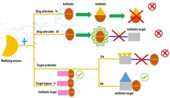
Figure 1.
Antibiotic resistance mediated by enzyme modifications.
Oxidoreductases, transferases, and hydrolases (Figure 1) are the main classes of enzyme responsible for the antibiotic resistance. Among hydrolases, the most common enzymes that catalyze antibiotic hydrolysis are β-actamases and macrolide esterases that destroy β-lactams, macrolides, chloramphenicol, and phosphomycin. β-lactamases hydrolyze the amide bond in the β-lactam ring, the common structural element of all β-lactam antibiotics like penicillins, cephalosporins, carbapenems, and monobactams [8][15]. The emergence of high rates of new mutations in the genes coding for β-lactamases that produce enzymes with a different structure and their location on mobile genetic elements contributes to the rapid spread of resistant bacteria [9][16]. Macrolide esterases are implicated in macrolide detoxification. Macrolides are a class of antibiotics extensively used in both agriculture and medicine. Erythromycin and azithromycin have been used as substitutes for β-lactam antibiotics in patients with penicillin allergies [10][17]. Transferases are a class of enzymes with a different substrate specificity, type of modification, and mechanism of action that modify the antibiotic molecules by covalently binding different chemical groups [10][17].

2.3. FMOs and Antibiotic Resistance

2.3.1. Tetracyclines

Microbial FMOs are able to modify antibiotics through oxidation, leading to resistance. The class A flavin-dependent monooxygenase tetracycline destructase TetX confers resistance to all clinically relevant tetracyclines, including the broad-spectrum antibiotic tigecycline, which is successfully used against multidrug-resistant pathogens. TetX was isolated from transposons Tn4351 and Tn4400 present in anaerobic bacteria of the genus Bacteroides [11][28]. It is able to regioselectively hydroxylate tetracycline antibiotics to 11a-hydroxy-tetracyclines (Figure 2A). Overexpression in E. coli and protein purification followed by mass spectral and NMR characterization demonstrated that TetX requires NADPH, Mg2+, and molecular oxygen to hydroxylate tetracyclines at C11a. This hydroxylation weakens the binding to magnesium, altering the physical properties of tigecycline and decreasing its affinity to ribosomes. Intramolecular cyclization and non-enzymatic breakdown to non-defined products follow the hydroxylation step [12][29]. The crystallographic structure of TetX from Bacteroides thetaiotaomicron and its complexes with tetracyclines showed the extremely versatile substrate diversity of the enzyme [13][30]. The environmental bacterium Mycobacterium abscessus has emerged as an important human pathogen causing bronchopulmonary infections. It possesses a WhiB7-independent tetracycline-inactivating monooxygenase, MabTetX, that confers a high level of resistance to tetracycline and doxycycline. Both antibiotic molecules are monooxygenated by the purified MabTetX [14][31].
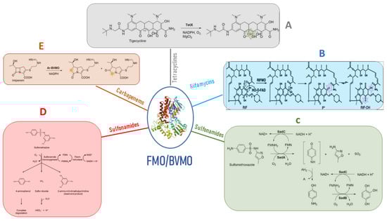
Figure 2. Antibiotic resistance mediated by FMOs’ modifications of antibiotic molecules. (A) the class A flavin-dependent monooxygenase tetracycline destructase TetX hydroxylates tetracycline antibiotics to 11a-hydroxy-tetracyclines inactivating the antibiotic molecule. (B) Rifampicin monooxygenase (RIFMO) catalyzes the hydroxylation of rifampicin (RIF) at the C2 atom inactivating the antibiotic activity of the molecule by preventing key contacts with the RNA polymerase target. (C) the monooxygenases SadA and SadC attack the sulfonamide molecules and release 4-aminophenol, which is converted by the monooxygenase SadB to 1,2,4-trihydroxybenzene that lacks antibiotic activity. (D) sulfonamide monooxygenase and flavin reductase are required for the cleavage of sulfamethazine producing the not active dead-end products and 4-aminophenol. (E) Baeyer–Villiger asymmetric oxygen insertion within the carbapenem ring of imipenem inactivates the antibiotic property of the molecule.
Tet (56), which shares similarity with TetX, has been identified in Legionella longbeachae, the pathogen responsible for causing Legionnaires’ disease. Detailed analysis using X-ray crystal structure revealed that Tet 56 possesses a structural configuration comprising a flavin adenine dinucleotide (FAD)-binding Rossmann-type fold domain, a domain responsible for binding tetracycline, and a C-terminal α-helix that serves as a connecting link between these two domains [15][32]. Additionally, genes encoding TetX3 and TetX4 have been detected in a multitude of Enterobacteriaceae and Acinetobacter strains originating from both animals and humans [15][32].

2.3.2. Rifamycins

The rifamycins represent a group of naturally occurring compounds, as well as their semi-synthetic derivatives, characterized by their activity against a wide spectrum of bacteria, encompassing both Gram-positive and Gram-negative species. These compounds possess a structural framework that incorporates a naphthalene core, connected via a polyketide “handle” and linked to the naphthalene segment through a cyclic amide bond. Their three-dimensional configuration resembles that of a basket, a crucial feature enabling them to bind effectively to the RNA exit tunnel located on the bacterial RNA polymerase’s β subunit, which serves as their target site [16][33]. The enzyme rifampicin monooxygenase (RIFMO) is another class A FMO that modifies rifampicin (RIF), an antibiotic that acts by inhibiting DNA-dependent RNA polymerase, used in combination therapy for the treatment of tuberculosis and mycobacterial and non-mycobacterial infections [17][34]. Nocardia farcinica [18][35] as well as Streptomyces and Rhodococcus species present in the environment possess the rifmo gene [18][35]. RIF was proposed to be hydroxylated by RIFMO to produce 2′-N-hydroxy-4-oxo-RIF, leading to subsequent RIF decomposition [19][36] (Figure 2B), but it was unclear the mechanism by which RIF was modified and degraded. Years later, it was described in Streptomyces venezuelae, with the monooxygenation of position 2 of the naphthyl group with a consequent ring opening and linearization of the antibiotic, leading to an antibiotic that cannot adopt the basket-like structure that is essential for binding to the RNA exit tunnel of the target ribosome site [20][37]. Liu and colleagues [21][38] successfully elucidated the crystal structure of RIFMO in association with the hydroxylated product of RIF. Their structural analysis revealed a notable disruption in the ansa aliphatic chain of RIF, positioned precisely between the naphthoquinone C2 and amide N1. This observation strongly implies that RIFMO hydroxylates RIF at the C2 atom, subsequently leading to the cleavage of the ansa linkage. This cleavage event ultimately results in the deactivation of the antibiotic properties of RIF by preventing critical interactions with its target, the RNA polymerase.

2.3.3. Sulfonamides

Sulfonamides were synthesized and introduced into the environment approximately 90 years ago. This relatively short timeframe has limited the opportunity for bacteria to evolve resistance to these compounds [22][39]. Nevertheless, sulfonamide-degrading bacteria have been found in seawater, agricultural soil, activated sludge, and acclimated membrane reactors, suggesting the existence of resistance mechanisms. The bacterial breakdown of sulfonamides has dual significance in environmental cleanup by eliminating pollutants and in the context of antibiotic resistance, where the enzymes responsible for degradation can be viewed as potential mechanisms for resistance development. SadA and SadB are two monooxygenases using the FMN reductase SadC that enable Microbacterium sp. strain BR1 and other Actinomycetes to inactivate sulfonamide antibiotics, using them as a carbon source. SadA and SadC attack the sulfonamide molecules and release 4-aminophenol, which is converted by SadB to 1,2,4-trihydroxybenzene that lacks antibiotic activity [23][40] (Figure 2C). Microbacterium sp. CJ77 possesses a two-component class D FMO system encoded by the monooxygenase gene sulX and the flavin reductase gene sulR [24][41] and are similar to the Microbacterium sp. strain BR1 SadA/SadB system. The presence of sulfonamides upregulates the expression of the sulX and sulR genes. Sulfonamide monooxygenase and flavin reductase were expressed and purified in E. coli, and biochemical analysis showed that sulfamethazine is cleaved by the two-component monooxygenase system to produce 4-aminophenol and the dead-end products (Figure 2D) [24][41]. SulR and sulX genes were co-expressed in E. coli, conferring decreased susceptibility to sulfamethoxazole, suggesting that the two genes are potential resistance determinants and encode drug-inactivating enzymes. All sulfonamide-degrading actinobacteria possess the gene cluster sulX-sulR in a genomic island also carrying class 1 integrons. This implies that the ability to metabolize sulfonamides may have been obtained by sulfonamide-resistant bacteria that had previously acquired the class 1 integron due to selective pressures from sulfonamide exposure [24][41].

3. Carbapenems

Carbapenems belong to a class of semi-synthetic β-lactam antibiotics derived from thienamycin, a natural compound produced by the soil microorganism Streptomyces cattleya, which was discovered in 1970 [25][42]. They are characterized by their wide spectrum of activity and exceptional potency against both Gram-negative and Gram-positive bacteria. Consequently, they are reserved as a last-line treatment for patients suffering from severe infections or suspected of having multidrug-resistant bacterial infections [26][43]. Carbapenems exhibit relative resistance to hydrolysis by most β-lactamases. Their structural composition consists of a β-lactam ring, which is similar to penicillins (“penams”) fused with an unsaturated five-membered ring. The key structural difference lies in the presence of a double bond between C-2 and C-3 (“-penem”) and the presence of carbon (“carba-”) at position 1 [27][44]. The presence of this carbon is fundamental for their spectrum of activity, stability against β-lactamases, and potency. They resist most β-lactamases due to the presence of a trans hydroxyethyl side chain, which replaces the acylamino substituent found on the β-lactam ring in penicillins and cephalosporins. Although carbapenems acylate the serine residue on β-lactamases rapidly, the subsequent hydrolysis of the acylated enzyme is very slow because the trans-1-hydroxyethyl moiety displaces the water necessary for hydrolysis at the active site. As a result, the enzyme becomes acylated and loses its activity [28][45]. Carbapenems are hence considered highly reliable as last-resort drugs for treating bacterial infections. Carbapenems exhibit varying antibacterial activities. Some, like panipenem, imipenem, and doripenem, are effective against Gram-positive bacteria, while others, including ertapenem, biapenem, meropenem, and doripenem, have limited effectiveness against Gram-negative bacteria [29][46]. They are preferred over other antimicrobial agents for treating invasive or life-threatening infections due to their concentration-independent bactericidal effect and minimal adverse effects [30][47]. Carbapenems like doripenem, tebipenem, meropenem, imipenem, ertapenem, biapenem, and panipenem are used worldwide, especially in response to the increasing resistance to cephalosporin antibiotics within the Enterobacteriaceae group.

4. BVMO and β-Lactams Resistance

4.1. Carbapenems

Minerdi and colleagues [31][79] made a pioneering observation, shedding light on a novel aspect of carbapenem resistance. Their study revealed that carbapenemases are not the sole culprits in carbapenem inactivation; Baeyer–Villiger monooxygenases (BVMOs) also possess this capability through oxygenation. This researchtudy focused on an Acinetobacter radioresistens strain isolated from the environment. This particular strain was found to harbor a Baeyer–Villiger monooxygenase known as Ar-BVMO. Remarkably, Ar-BVMO exhibited a complete amino acid sequence identity with the ethionamide monooxygenase found in multidrug-resistant A. baumannii. The significance of this finding is underscored by the fact that carbapenem antibiotics are considered the last-resort treatment for multidrug-resistant (MDR) A. baumannii infections. Unfortunately, there has been a global surge in the prevalence of A. baumannii strains that have developed resistance to these vital antimicrobial drugs [32][80]. Carbapenem resistance in A. baumannii mainly arises from acquired carbapenem-hydrolyzing oxacillinases such as OXA-23 that are classified under the Ambler class D β-lactamases [32][80]. Interestingly, the source of the blaOXA-23 gene is A. radioresistens, a commensal bacterial species commonly found on the skin of hospitalized and healthy patients [33][81]. Furthermore, two kinase inhibitors, tozasertib (VX-680) and danusertib (PHA-739358), previously identified as potential targets in anticancer therapy, were demonstrated to be metabolized by Ar-BVMO. To assess whether the expression of Ar-BVMO in imipenem-sensitive E. coli BL21 cells could confer resistance to imipenem, a disk diffusion assay was performed following EUCAST guidelines. According to the EUCAST clinical breakpoint table for Enterobacteriaceae, imipenem zone diameter breakpoints for sensitivity and resistance are ≥22 mm and <16 mm, respectively. Notably, E. coli BL21 cells transformed with pT7-Ar-BVMO but not induced, as well as E. coli BL21 cells transformed with an empty expression plasmid and induced with IPTG, displayed inhibition zones with average diameters of 25.5 mm and 28.0 mm, respectively. Conversely, E. coli BL21, when expressed in Ar-BVMO, showed resistance to imipenem (zone diameter breakpoint, <16 mm). These findings offered preliminary evidence that imipenem-sensitive E. coli expressing Ar-BVMO may become resistant to the antibiotic molecule [34][82]. The functionality of the purified Ar-BVMO enzyme in the presence of imipenem was evaluated by monitoring the utilization of NADPH. The derived kinetic parameters, Km and Vmax strongly suggested that imipenem could indeed serve as a genuine substrate for Ar-BVMO. Furthermore, liquid chromatography–mass spectrometry analysis revealed a classical Baeyer–Villiger asymmetric oxygen insertion within the carbapenem ring of imipenem (as depicted in Figure 2E). This compelling evidence confirms that imipenem qualifies as a substrate for Ar-BVMO. Remarkably, this marks the first instance of an antibiotic inactivating BVMO enzyme that, in the course of its typical BV oxidation activity, concurrently employs an unprecedented mechanism for carbapenem resistance. It is hypothesized that genetic exchange may have occurred between A. radioresistens and A. baumannii within the human body [35][83]. This outcome may have facilitated the transfer of the BVMO gene to the latter species.

4.2. β-Lactams

Methicillin and/or vancomycin-resistant Staphylococcus aureus is one of the most common opportunistic human pathogens. It causes skin infections but also sepsis and pneumonia. Hwang and colleagues [36][84] described the crystal structure of a BVMO originating from Methicillin-resistant Staphylococcus aureus (SAFMO). This enzyme could potentially contribute to vancomycin and/or methicillin resistance by breaking down these molecules. Molecular docking simulations showed that vancomycin and methicillin can be accommodated in the SAFMO active site. If confirmed, this hypothesis could lead to a novel drug target against S. aureus.
ScholarVision Creations