Flavones in Hypertensive Disease: Comparison
Please note this is a comparison between Version 2 by Rita Xu and Version 1 by Nathaniel Bloodworth.

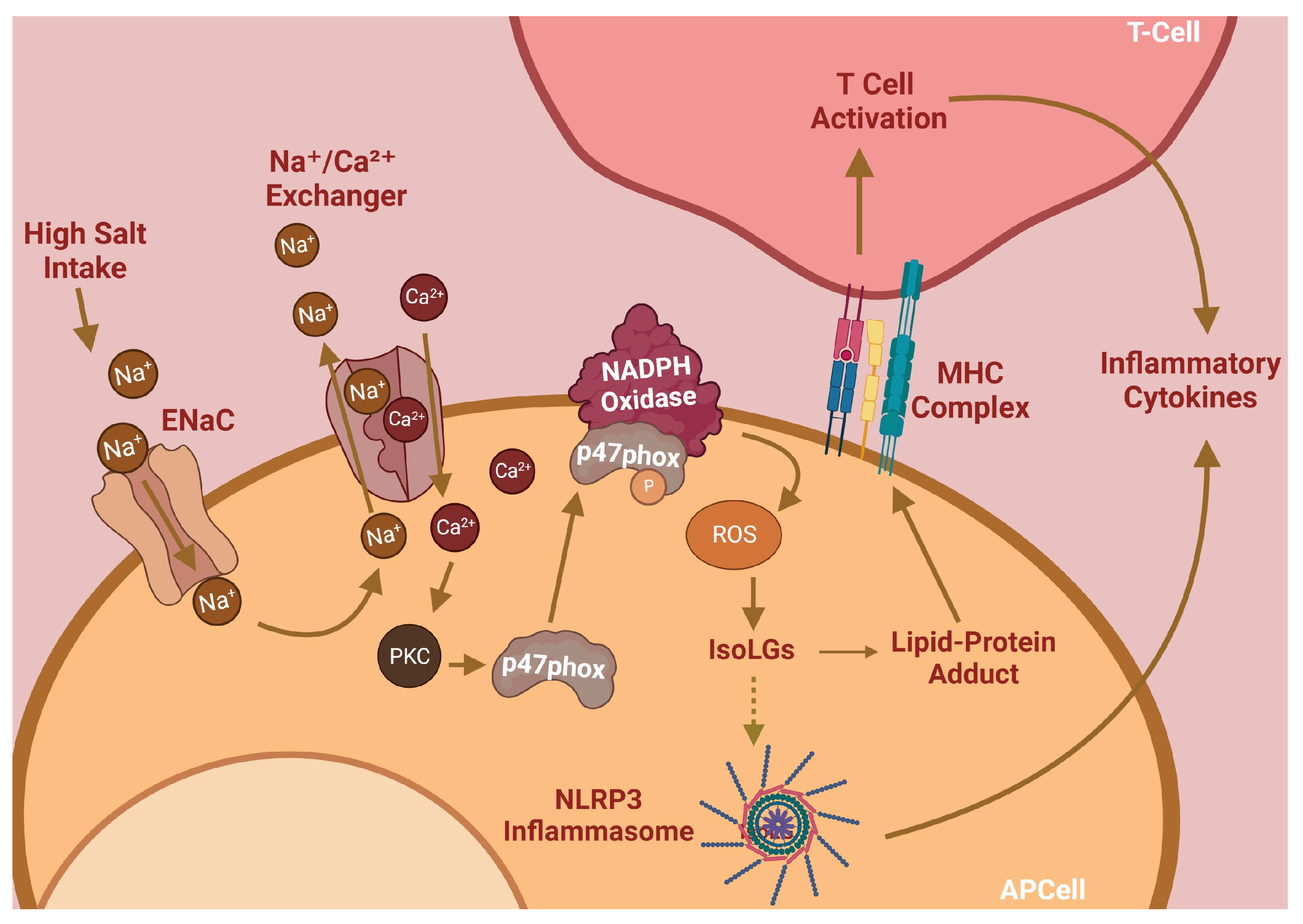
Hypertension is the leading remediable risk factor for cardiovascular morbidity and mortality in the United States. Excess dietary salt consumption, which is a catalyst of hypertension, initiates an inflammatory cascade via activation of antigen-presenting cells (APCs). This pro-inflammatory response is driven primarily by sodium ions (Na

+

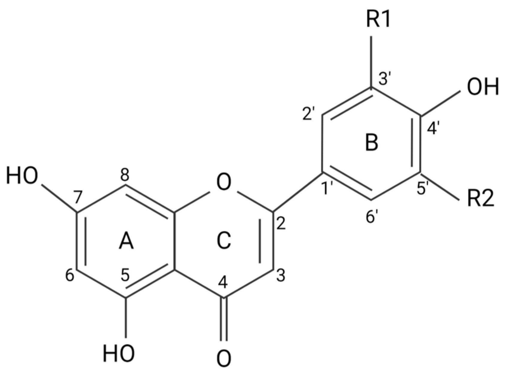
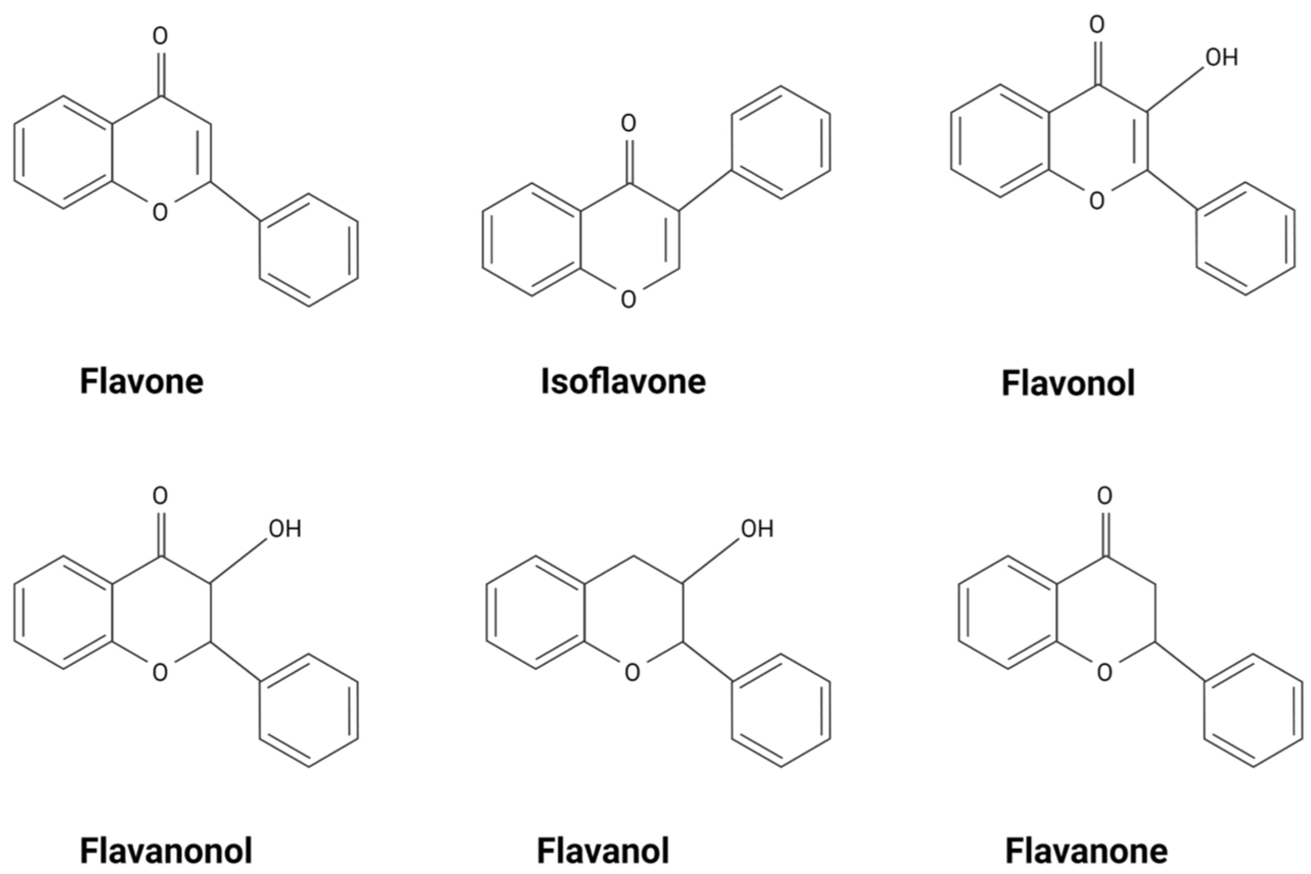
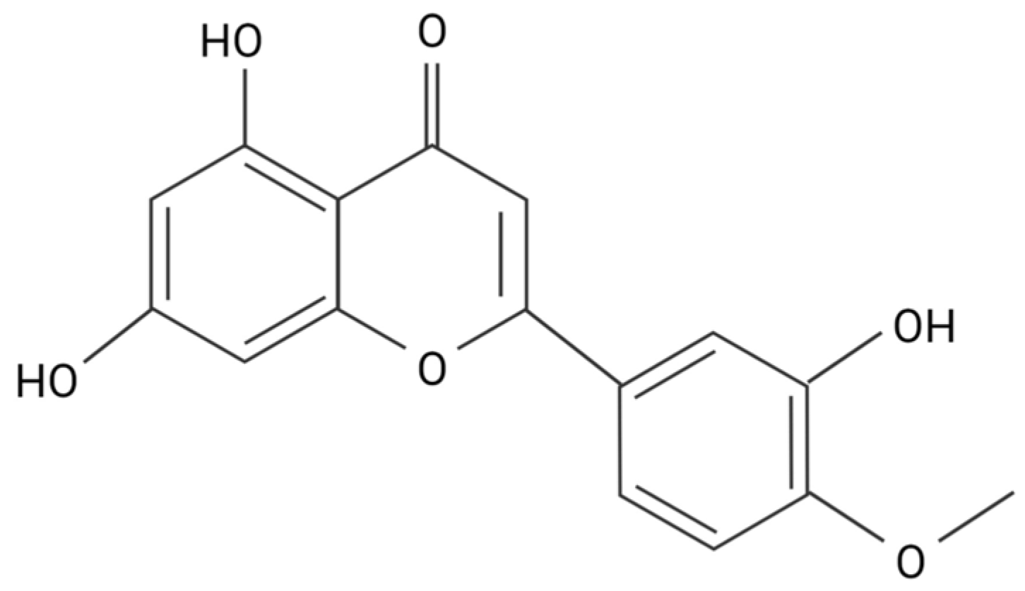
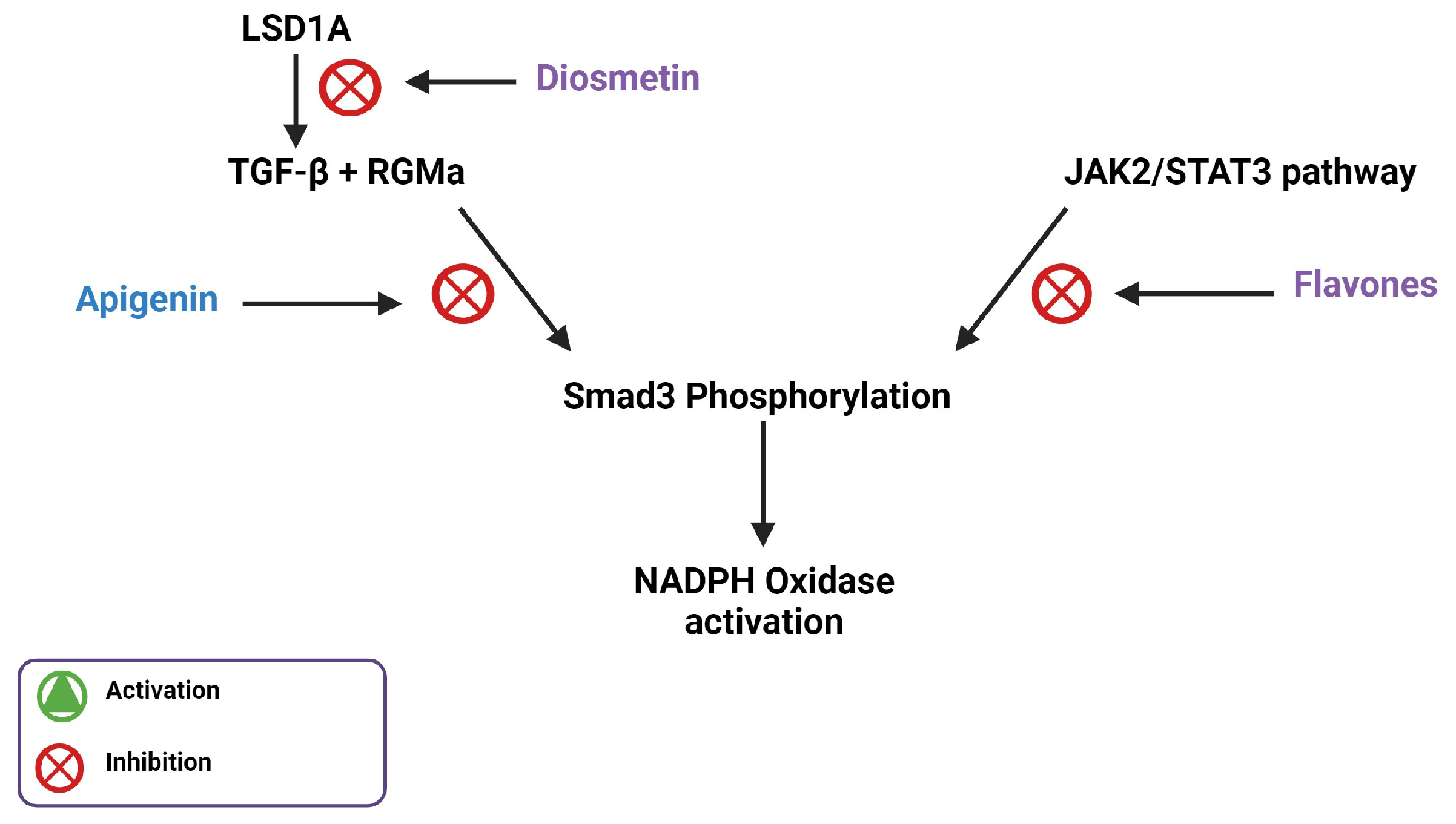
) transporting into APCs by the epithelial sodium channel (ENaC) and subsequent NADPH oxidase activation, leading to high levels of oxidative stress. Flavonoids, a natural phenolic compound, have these therapeutic benefits and can potentially serve as anti-hypertensives. Flavones are a type of flavonoid that have increased anti-inflammatory effects that may allow them to act as therapeutic agents for hypertension, including diosmetin, which is able to induce significant arterial vasodilation in several different animal models.

  • diosmetin
  • flavone
  • flavonoid
  • hypertension
  • inflammation

1. Introduction

Hypertension is the single most important cause of cardiovascular disease and premature death in the world, affecting 1.4 billion people and incurring an estimated annual cost of $46 billion [1]. Nearly half of the adults in the United States (47%) develop hypertension, defined as a systolic blood pressure of 130 mmHg or higher [2,3][2][3]. A major risk factor for hypertension is excess dietary salt intake. The American Heart Association (AHA) recommends for individuals to consume a maximum of 2300 mg of salt per day; however, less than 10% of the United States population observes this recommendation [4]. The mechanisms describing the pathogenesis of hypertension are variable, but emerging data in mice and humans implicate immune cell activation as a key contributor to salt-sensitivity in blood pressure.
The initiation and maintenance of hypertension are dependent on inflammation caused by activated immune cells [5]. OurThe lab has previously found that salt induces immune cell activation via the induction of reactive oxygen species (ROS) and lipid peroxides, a mechanism summarized in Figure 1. Sodium ions (Na+) enter antigen-presenting cells (APCs) through the epithelial sodium channel (ENaC) and incite a further influx of calcium via the Na⁺/Ca2⁺ ion exchanger. Elevated intracellular Ca2+ levels activate PKC to induce phosphorylation of p47phox, a critical component of the NADPH oxidase complex responsible for its assembly and activation [6]. NADPH oxidase generates ROS, which in turn activates the NLRP3 inflammasome and stimulates the subsequent production of pro-inflammatory cytokines [7]. Elevated NADPH oxidase activity also induces the formation of a class of lipid peroxides through ROS-induced peroxidation of arachidonic acid. These lipid peroxides, called Isolevuglandins (IsoLGs), are nucleophiles that preferentially react with lysine residues on proteins to form lipid-protein adducts. These adducts can be proteolyzed and presented to patrolling T-cells [6,7][6][7]. These important mechanisms, while critical for hypertension pathogenesis, are currently unaddressed by conventional therapies; thus, they represent a unique opportunity for impactful pharmacologic interventions.
Figure 1. Salt-sensitive hypertension pathway.
Flavonoids and flavonoid-derived compounds represent one such potential intervention. Flavonoids are natural phenolic compounds found in fruits, vegetables, wines, flowers, tea, and other natural products [8]. They are known for their anti-inflammatory, anti-oxidative, and anti-mutagenic effects. The structure of flavonoids consists of a phenyl ring, B, a benzene ring, A, and a heterocyclic C ring (Figure 2). Flavonoids are divided into different subgroups based on their structure, including flavonols, flavones, flavanones, flavanols, flavanonols, and isoflavones (Figure 3) [8]. Flavonols and flavones play a crucial role against ROS as a source of external antioxidants due to their anti-radical capabilities, which occur in a structurally dependent manner, with C4 and C3 hydroxyl groups associated with stronger scavenging potential [9]. Flavones are the most hydrophobic of the flavonoid subgroups, one of the qualities that indicate the membrane permeability of the flavonoid, which is important to the exertion of therapeutic effects [10]. Many flavonoids are thought to lower blood pressure and aid in hypertension. Diosmetin (5,7-dihydroxy-2-(3-hydroxy-4-methoxyphenyl)chromen-4-one), a flavone, induces significant arterial vasodilation in hypertensive rats [11]. In this review, we discuss the pharmacologic properties of flavones and their antioxidative effects on pro-inflammatory pathways implicated in hypertension.
Figure 2. Flavonoid carbon numbers and ring names. (A) benzene ring; (B) phenyl ring; (C) heterocyclic ring.
Figure 3. Types of flavonoids.

2. Flavonoids Exert Their Effects by Targeting Intracellular Proteins

Although flavonols are more active antioxidants than flavones, the metabolic stability and membrane permeability are much higher in flavones [9,12][9][12]. This and the ability to produce a physiologic response depends primarily on its hydrophobicity [13]. A partition coefficient (PC) test using an n-octanol/HEPES solution determined the hydrophobicity of four flavonoids, eriodyctiol, quercetin, luteolin, and taxifolin. Of the four flavonoids tested, luteolin had the highest affinity for octanol, making it the most hydrophobic, followed by quercetin, eriodyctiol, and then taxifolin [14]. The hydrophobicity of flavonoids is determined by the number of hydroxyl functional groups attached to the rings, as well as the number and placement of pi bonds in the rings. Flavonols, like quercetin, and flavanonols, like taxifolin, have similar structures. Of these two types of flavonoids, flavanonols have fewer pi bonds, making them less hydrophobic. Flavones, like luteolin, have fewer hydroxyl groups, causing them to be more hydrophobic. Methoxy flavones, like diosmetin, are considered the most hydrophobic of the flavonoids, with correspondingly higher metabolic stability and membrane transport ability when compared with other flavonoids (Figure 4) [12]. Although flavonoid hydrophobicity correlates with its ability to pass through the cell membrane, whether this correlates with physiological activity has yet to be fully discovered. Diosmetin has vasorelaxation effects that are over 10 times greater than verapamil, a potent vasodilatory agent used to treat hypertension, due to its greater inhibitory effect on Ca2⁺ release from intracellular Ca2⁺ stores [11].
Figure 4. Structure of diosmetin.

3. Flavones Target NADPH Oxidase Transcription by Inhibiting Smad3 Phosphorylation

NADPH oxidase expression is required for the initiation and maintenance of hypertension, making the pathways regulating NADPH oxidase expression important therapeutic targets. Smad3 is an important transcriptional regulator of NADPH oxidase gene expression in APCs. Its phosphorylation and subsequent activation are regulated by a variety of pathways upstream [15]. IL-6 signaling, via the JAK2/STAT3 pathway, results in Smad3 phosphorylation, which leads to the upregulation of NADPH oxidase components [15,16][15][16]. The canonical TGF-β1 signaling pathway, which is also involved in the activation of the pro-inflammatory transcription factor NF-κB, is dependent on Smad3 phosphorylation [17]. A study done by Li et al. used an MTT assay and western blot to reveal the effect of apigenin on TGF-β1 and Smad3 protein levels, respectively [18]. Apigenin, a flavone, prevented TGF-β1-induced Smad3 phosphorylation, possibly by inhibiting JAK2/STAT3 signaling [18]. A different study conducted by Ning et al. used western blot to reveal that the application of diosmetin in µM amounts decreased the expression of phosphorylated STAT3 in human osteosarcoma cells [19].
Pectolinarigenin treatment, another dimethoxy flavone, is implicated in the suppression of the TGF-β/Smad3 and the JAK2/STAT3 pathways in mice with hyperuricemic nephropathy [20]. In a study by Ren et al., western blot analysis showed that pectolinarigenin treatment decreased TGF-β expression and Smad3 phosphorylation. Pectolinarigenin treatment was also shown by this study to significantly decrease IL-6 protein levels and STAT3 phosphorylation, which would decrease JAK2/STAT3 signaling [20]. Together, this indicates that certain flavones can alter NADPH oxidase activity, and the associated inhibition of hypertension, through Smad3 inhibition.
Flavones potentially interact with other genes or proteins involved in TGF-β1 activation to inhibit Smad3 phosphorylation. A study carried out by Zhang et al. hypothesized that RGMa may play an integral role in TGF-β1-mediated phosphorylation of Smad3 [21] This study used western blot and immunofluorescence staining to find that as the expression of TGF-β1 increases, its surface receptor is activated, which enhances RGMa expression. RGMa forms a complex with Smad3 and the TGF-β1 receptor, facilitating Smad3 phosphorylation. This study further showed that RGMa inhibition was correlated with a reduction in Smad3 phosphorylation [21]. A study by Arango et al. utilized second-generation (PD) sequencing, a method that discovers small molecule–protein interactions, to identify RGMa as a target gene for apigenin [22]. In another study, Gao et al. found that apigenin improves hypertension in spontaneously hypertensive rats by down-regulating NADPH oxidase-dependent ROS expression [23]. These findings suggest that apigenin inhibits NADPH oxidase-mediated ROS production, potentially by interacting with RGMa to regulate Smad3 phosphorylation, showing how cardiac hypertrophy may be modulated on the basis of apigenin-dependent inflammation and ROS.
Resveratrol is a polyphenol found in grapes and peanuts that is known for its biological activities and pharmacological effects, including inhibition of TGF-β1-mediated epithelial-mesenchymal transition (EMT) in breast cancer, which is mediated by Smad3 phosphorylation [24,25][24][25]. Since the structure of resveratrol is similar to the structure of flavones, this inhibition suggests the importance of the hydroxyl group on the 4′ carbon of the phenyl ring, as well as the 5 carbon and 7 carbon of the benzene ring because both compounds share these groups.
Diosmetin can also inhibit protein lysine-specific histone demethylase 1A (LSD1A), expressed by KDM1A, which in turn regulates TGF-β1 production [26]. LSD1A is an epigenetic regulator that promotes EMT in various kinds of cancer [27]. EMT allows solid cancer cells to migrate by repressing epithelial markers and activating mesenchymal markers. LSD1A aids in this process by facilitating heterochromatin demethylation and euchromatin methylation, which activates the transcription of oncogenes and suppresses the expression of tumor suppressor genes, respectively [28], thereby leading to TGF-β-mediated EMT [29]. This shows that LSD1A is involved in the activation of TGF-β-mediated EMT, which is dependent on Smad3 phosphorylation [30]. LSD1A is a target for diosmetin, which potentially elucidates the inhibitory effects of flavones on TGF-β1-mediated Smad3 phosphorylation (Figure 5).
Figure 5. Illustration of Smad3 phosphorylation inhibition by flavones through the inhibition of LSD1A and the JAK2/STAT3 pathway.

4. Flavones Affect Mitochondrial Biogenesis, Dynamics, and Energetics

Given the roles of flavones in NADPH oxidase expression, there are also potential roles in the mitochondrial formation of signaling factors. Mitochondria are known to play a role in hypertension, with their dysfunction being linked to inflammation in cardiac tissue [31]. Flavones can alter mitochondrial metabolism through action sites that are located between complexes I and III, conducive to changes in mitochondrial membrane properties [32]. Novel anti-tumor drugs have incorporated flavone side chains in the structure, which can selectively generate ROS in hepatoma cells through preferential targeting of mitochondria [33]. In other conditions, flavones can protect against oxidative stress and inhibit myocardial ischemia, suggesting that flavone derivatives mechanistically target mitochondria with disease-dependent responses [34]. This underscores the importance of further understanding the pluralistic roles of flavones in ROS generation, which may be dependent on how flavones are implicated in the mitochondrial structure or the type of flavone.
Salvigenin, a trimethoxylated flavone, not only stimulated mitochondria but also decreased lipid levels to protect against metabolic syndrome [35]. Given the antimetastatic nature of flavonoid compounds, through induction of mitochondrial-mediated apoptosis [33], the roles of flavones in mitochondria-mediated hypertension need to be better elucidated. Flavones have been implicated in the metabolic process through mitochondrial alteration. Specifically, sudachitin, fruit-derived flavone, reduced weight gain in high-fat diet mice through mitochondrial biogenesis, resulting in improved insulin resistance [36]. In skeletal muscle, it is possible that flavones interact with Sirt1 and PGC-1α to mediate mitochondrial dynamics to protect against metabolic disorders [36]. Similarly, epigallocatechin-3-gallate, a green-tea-derived flavone, increases mitochondrial biogenesis through the activation of AMPK [37]. As previously reviewed, AMPK can both upregulate PGC-1α as well as NADH-mediated Sirt1 [38]. This suggests a pathway in which flavones can modulate mitochondrial biogenesis through the upregulation of PGC-1α, leading to NRF1 and NRF2, and subsequently TFAM activation to result in mtDNA-mediated mitochondrial biogenesis [38]. However, as previously reviewed, flavones are unique from other categories of flavonoids, including flavonols, flavanones, isoflavone, anthocyanins, and chalcones, which may play unique roles in mitophagy [38]. One key aspect in which flavones may uniquely modulate mitochondrial-dependent hypertension is through mitochondria fusion.
It is important to consider how flavones may impact mitochondria through pro-fission or pro-fusion factors, which can alter the relative mitochondrial dynamics that govern mitochondria biogenesis and subsequent energetics. Xanthohumol, another flavone, has been reported to upregulate mitofusin 2, a mitochondrial profusion factor, to alleviate murine neuronal death in nervous system diseases [39]. Similarly, while hypertension can be spawned by reduced mitochondria fusion, flavonoids have been shown to reverse Angiotensin II-dependent increases in dynamin-related protein 1 (profission) and decreases in optic atrophy 1 (profusion) [40]. These mitochondrial structural changes may have functional impacts on flavone-mediated hypertension.
Specifically, diosmetin treatment in colitis shows interaction with Sirt1 along with decreased inflammation [41], suggesting mitochondria biogenesis-linked resistance to hypertension. Similarly, in a cancer model, through the upregulation of p53 and similar pathways, Diosmetin results in potential mitochondrial membrane alteration to cause apoptosis [42]. Similarly, Diosmetin also has cytoprotective effects in myocardial ischemia injury through decreasing oxidative stress [43]. The modulation of oxidative stress and membrane potential by diosmetin suggests that it also causes structural changes that may impact bioenergetic-dependent hypertension development; however, it is unclear if diosmetin specifically modulates mitochondria biogenesis and structure.

References

  1. GBD 2013 Risk Factors Collaborators; Forouzanfar, M.H.; Alexander, L.; Anderson, H.R.; Bachman, V.F.; Biryukov, S.; Brauer, M.; Burnett, R.; Casey, D.; Coates, M.; et al. Global, regional, and national comparative risk assessment of 79 behavioural, environmental and occupational, and metabolic risks or clusters of risks in 188 countries, 1990–2013: A systematic analysis for the Global Burden of Disease Study 2013. Lancet 2015, 386, 2287–2323.
  2. Whelton, P.K.; Carey, R.M.; Aronow, W.S.; Casey, D.E., Jr.; Collins, K.J.; Himmelfarb, C.D.; DePalma, S.M.; Gidding, S.; Jamerson, K.A.; Jones, D.W.; et al. 2017 ACC/AHA/AAPA/ABC/ACPM/AGS/APhA/ASH/ASPC/NMA/PCNA Guideline for the Prevention, Detection, Evaluation, and Management of High Blood Pressure in Adults: A Report of the American College of Cardiology/American Heart Association Task Force on Clinical Practice Guidelines. Hypertension 2018, 71, e13–e115.
  3. Unger, T.; Borghi, C.; Charchar, F.; Khan, N.A.; Poulter, N.R.; Prabhakaran, D.; Ramirez, A.; Schlaich, M.; Stergiou, G.S.; Tomaszewski, M.; et al. 2020 International Society of Hypertension Global Hypertension Practice Guidelines. Hypertension 2020, 75, 1334–1357.
  4. Elijovich, F.; Laffer, C.L.; Sahinoz, M.; Pitzer, A.; Ferguson, J.F.; Kirabo, A. The Gut Microbiome, Inflammation, and Salt-Sensitive Hypertension. Curr. Hypertens. Rep. 2020, 22, 79.
  5. Patrick, D.M.; Van Beusecum, J.P.; Kirabo, A. The role of inflammation in hypertension: Novel concepts. Curr. Opin. Physiol. 2021, 19, 92–98.
  6. Barbaro, N.R.; Foss, J.D.; Kryshtal, D.O.; Tsyba, N.; Kumaresan, S.; Xiao, L.; Mernaugh, R.L.; Itani, H.A.; Loperena, R.; Chen, W.; et al. Dendritic Cell Amiloride-Sensitive Channels Mediate Sodium-Induced Inflammation and Hypertension. Cell Rep. 2017, 21, 1009–1020.
  7. Pitzer, A.; Elijovich, F.; Laffer, C.L.; Ertuglu, L.A.; Sahinoz, M.; Saleem, M.; Krishnan, J.; Dola, T.; Aden, L.A.; Sheng, Q.; et al. DC ENaC-Dependent Inflammasome Activation Contributes to Salt-Sensitive Hypertension. Circ. Res. 2022, 131, 328–344.
  8. Panche, A.N.; Diwan, A.D.; Chandra, S.R. Flavonoids: An overview. J. Nutr. Sci. 2016, 5, e47.
  9. Spiegel, M.; Andruniów, T.; Sroka, Z. Flavones’ and Flavonols’ Antiradical Structure–Activity Relationship—A Quantum Chemical Study. Antioxidants 2020, 9, 461.
  10. Premathilaka, R.; Rashidinejad, A.; Golding, M.; Singh, J. Oral delivery of hydrophobic flavonoids and their in-corporation into functional foods: Opportunities and challenges. Food Hydrocolloid 2022, 128, 107567.
  11. Ahmad, T.; Javed, A.; Khan, T.; Althobaiti, Y.S.; Ullah, A.; Almutairi, F.M.; Shah, A.J. Investigation into the Antihypertensive Effects of Diosmetin and Its Underlying Vascular Mechanisms Using Rat Model. Pharmaceuticals 2022, 15, 951.
  12. Zuiter, A. Proanthocyanidin: Chemistry and Biology: From Phenolic Compounds to Proanthocyanidins. Chem. Mol. Sci. Chem. Eng. 2014, 1, 29.
  13. Wang, T.-Y.; Li, Q.; Bi, K.-S. Bioactive flavonoids in medicinal plants: Structure, activity and biological fate. Asian J. Pharm. Sci. 2018, 13, 12–23.
  14. Areias, F.M.; Rego, A.C.; Oliveira, C.R.; Seabra, R.M. Antioxidant effect of flavonoids after ascorbate/Fe2+-induced oxidative stress in cultured retinal cells. Biochem. Pharmacol. 2001, 62, 111–118.
  15. Bondi, C.D.; Manickam, N.; Lee, D.Y.; Block, K.; Gorin, Y.; Abboud, H.E.; Barnes, J.L. NAD(P)H Oxidase Mediates TGF-beta1–Induced Activation of Kidney Myofibroblasts. J. Am. Soc. Nephrol. 2010, 21, 93–102.
  16. Saleem, M.; Aden, L.; Pitzer, A.; Ishimwe, J.; Ertuglu, L.; Laffer, C.; Wanjalla, C.; Pakala, S.; Kastner, P.; Park, J.; et al. Dendritic cell-specific SMAD3, downstream of JAK2, contributes to inflammation and salt-sensitivity of blood pressure in humans and mice. Physiology 2023, 38, 5734894.
  17. Qiu, X.; Zhu, J.; Sun, Y.; Fan, K.; Yang, D.-R.; Li, G.; Yang, G.; Chang, C. TR4 nuclear receptor increases prostate cancer invasion via decreasing the miR-373-3p expression to alter TGFbetaR2/p-Smad3 signals. Oncotarget 2015, 6, 15397–15409.
  18. Li, L.H.; Lu, B.; Wu, H.K.; Zhang, H.; Yao, F.F. Apigenin inhibits TGF-beta1-induced proliferation and migration of airway smooth muscle cells. Int. J. Clin. Exp. Pathol. 2015, 8, 12557–12563.
  19. Ning, R.; Chen, G.; Fang, R.; Zhang, Y.; Zhao, W.; Qian, F. Diosmetin inhibits cell proliferation and promotes apoptosis through STAT3/c-Myc signaling pathway in human osteosarcoma cells. Biol. Res. 2021, 54, 40.
  20. Ren, Q.; Wang, B.; Guo, F.; Huang, R.; Tan, Z.; Ma, L.; Fu, P. Natural Flavonoid Pectolinarigenin Alleviated Hyperuricemic Nephropathy via Suppressing TGFbeta/SMAD3 and JAK2/STAT3 Signaling Pathways. Front. Pharmacol. 2021, 12, 792139.
  21. Zhang, R.; Wu, Y.; Xie, F.; Zhong, Y.; Wang, Y.; Xu, M.; Feng, J.; Charish, J.; Monnier, P.P.; Qin, X. RGMa mediates reactive astrogliosis and glial scar formation through TGFbeta1/Smad2/3 signaling after stroke. Cell Death Differ. 2018, 25, 1503–1516.
  22. Arango, D.; Morohashi, K.; Yilmaz, A.; Kuramochi, K.; Parihar, A.; Brahimaj, B.; Grotewold, E.; Doseff, A.I. Molecular basis for the action of a dietary flavonoid revealed by the comprehensive identification of apigenin human targets. Proc. Natl. Acad. Sci. USA 2013, 110, E2153–E2162.
  23. Gao, H.-L.; Yu, X.-J.; Hu, H.-B.; Yang, Q.-W.; Liu, K.-L.; Chen, Y.-M.; Zhang, Y.; Zhang, D.-D.; Tian, H.; Zhu, G.-Q.; et al. Apigenin Improves Hypertension and Cardiac Hypertrophy Through Modulating NADPH Oxidase-Dependent ROS Generation and Cytokines in Hypothalamic Paraventricular Nucleus. Cardiovasc. Toxicol. 2021, 21, 721–736.
  24. Sun, Y.; Zhou, Q.-M.; Lu, Y.-Y.; Zhang, H.; Chen, Q.-L.; Zhao, M.; Su, S.-B. Resveratrol Inhibits the Migration and Metastasis of MDA-MB-231 Human Breast Cancer by Reversing TGF-beta1-Induced Epithelial-Mesenchymal Transition. Molecules 2019, 24, 1131.
  25. Ashrafizadeh, M.; Najafi, M.; Orouei, S.; Zabolian, A.; Saleki, H.; Azami, N.; Sharifi, N.; Hushmandi, K.; Zarrabi, A.; Ahn, K.S. Resveratrol Modulates Transforming Growth Factor-Beta (TGF-beta) Signaling Pathway for Disease Therapy: A New Insight into Its Pharmacological Activities. Biomedicines 2020, 8, 261.
  26. Gilson, M.K.; Liu, T.; Baitaluk, M.; Nicola, G.; Hwang, L.; Chong, J. BindingDB in 2015: A public database for medicinal chemistry, computational chemistry and systems pharmacology. Nucleic Acids Res. 2016, 44, D1045–D1053.
  27. Lin, T.; Ponn, A.; Hu, X.; Law, B.K.; Lu, J. Requirement of the histone demethylase LSD1 in Snai1-mediated transcriptional repression during epithelial-mesenchymal transition. Oncogene 2010, 29, 4896–4904.
  28. McDonald, O.G.; Wu, H.; Timp, W.; Doi, A.; Feinberg, A.P. Genome-scale epigenetic reprogramming during epithelial-to-mesenchymal transition. Nat. Struct. Mol. Biol. 2011, 18, 867–874.
  29. Kim, D.; Kim, K.I.; Baek, S.H. Roles of lysine-specific demethylase 1 (LSD1) in homeostasis and diseases. J. Biomed. Sci. 2021, 28, 41.
  30. Muthusamy, B.P.; Budi, E.H.; Katsuno, Y.; Lee, M.K.; Smith, S.M.; Mirza, A.M.; Akhurst, R.J.; Derynck, R. ShcA Protects against Epithelial–Mesenchymal Transition through Compartmentalized Inhibition of TGF-beta-Induced Smad Activation. PLoS Biol. 2015, 13, e1002325.
  31. Dikalov, S.I.; Dikalova, A.E. Crosstalk Between Mitochondrial Hyperacetylation and Oxidative Stress in Vascular Dysfunction and Hypertension. Antioxid. Redox Signal. 2019, 31, 710–721.
  32. Valdameri, G.; Herrerias, T.; Carnieri, E.G.S.; Cadena, S.M.S.C.; Martinez, G.R.; Rocha, M.E.M. Importance of the core structure of flavones in promoting inhibition of the mitochondrial respiratory chain. Chem. Interact. 2010, 188, 52–58.
  33. Dai, F.; Li, Q.; Wang, Y.; Ge, C.; Feng, C.; Xie, S.; He, H.; Xu, X.; Wang, C. Design, Synthesis, and Biological Evaluation of Mitochondria-Targeted Flavone–Naphthalimide–Polyamine Conjugates with Antimetastatic Activity. J. Med. Chem. 2017, 60, 2071–2083.
  34. Zheng, R.-F.; Du, Y.-W.; Zeng, C.; Wang, H.-F.; Xing, J.-G.; Xu, M. Total flavones of Dracocephalum moldavica L. protect astrocytes against H(2)O(2)-induced apoptosis through a mitochondria-dependent pathway. BMC Complement. Med. Ther. 2020, 20, 78.
  35. Serino, E.; Chahardoli, A.; Badolati, N.; Sirignano, C.; Jalilian, F.; Mojarrab, M.; Farhangi, Z.; Rigano, D.; Stornaiuolo, M.; Shokoohinia, Y.; et al. Salvigenin, a Trimethoxylated Flavone from Achillea wilhelmsii C. Koch, Exerts Combined Lipid-Lowering and Mitochondrial Stimulatory Effects. Antioxidants 2021, 10, 1042.
  36. Tsutsumi, R.; Yoshida, T.; Nii, Y.; Okahisa, N.; Iwata, S.; Tsukayama, M.; Hashimoto, R.; Taniguchi, Y.; Sakaue, H.; Hosaka, T.; et al. Sudachitin, a polymethoxylated flavone, improves glucose and lipid metabolism by increasing mitochondrial biogenesis in skeletal muscle. Nutr. Metab. 2014, 11, 32.
  37. Xiong, L.-G.; Chen, Y.-J.; Tong, J.-W.; Gong, Y.-S.; Huang, J.-A.; Liu, Z.-H. Epigallocatechin-3-gallate promotes healthy lifespan through mitohormesis during early-to-mid adulthood in Caenorhabditis elegans. Redox Biol. 2018, 14, 305–315.
  38. Chen, Q.; Ruan, D.; Shi, J.; Du, D.; Bian, C. The multifaceted roles of natural products in mitochondrial dysfunction. Front. Pharmacol. 2023, 14, 1093038.
  39. Wang, C.C.; Ho, Y.H.; Hung, C.F.; Kuo, J.R.; Wang, S.J. Xanthohumol, an active constituent from hope, affords protection against kainic acid-induced excitotoxicity in rats. Neurochem. Int. 2020, 133, 104629.
  40. Li, Y.; Dang, Q.; Li, Z.; Han, C.; Yang, Y.; Li, M.; Li, P. Restoration of Mitochondrial Function Is Essential in the Endothelium-Dependent Vasodilation Induced by Acacetin in Hypertensive Rats. Int. J. Mol. Sci. 2022, 23, 11350.
  41. Li, H.-L.; Wei, Y.-Y.; Li, X.-H.; Zhang, S.-S.; Zhang, R.-T.; Li, J.-H.; Ma, B.-W.; Shao, S.-B.; Lv, Z.-W.; Ruan, H.; et al. Diosmetin has therapeutic efficacy in colitis regulating gut microbiota, inflammation, and oxidative stress via the circ-Sirt1/Sirt1 axis. Acta Pharmacol. Sin. 2022, 43, 919–932.
  42. Wang, C.; Li, S.; Ren, H.; Sheng, Y.; Wang, T.; Li, M.; Zhou, Q.; He, H.; Liu, C. Anti-Proliferation and Pro-Apoptotic Effects of Diosmetin via Modulating Cell Cycle Arrest and Mitochondria-Mediated Intrinsic Apoptotic Pathway in MDA-MB-231 Cells. Med. Sci. Monit. 2019, 25, 4639–4647.
  43. Mo, G.L.; He, Y.; Zhang, X.Q.; Lei, X.; Luo, Q. Diosmetin exerts cardioprotective effect on myocardial ischaemia injury in neonatal rats by decreasing oxidative stress and myocardial apoptosis. Clin. Exp. Pharmacol. Physiol. 2020, 47, 1713–1722.
More
Video Production Service