Vascular Endothelial Cell Senescence and Death: Comparison
Please note this is a comparison between Version 3 by Jessie Wu and Version 4 by Lan-Lan Bu.

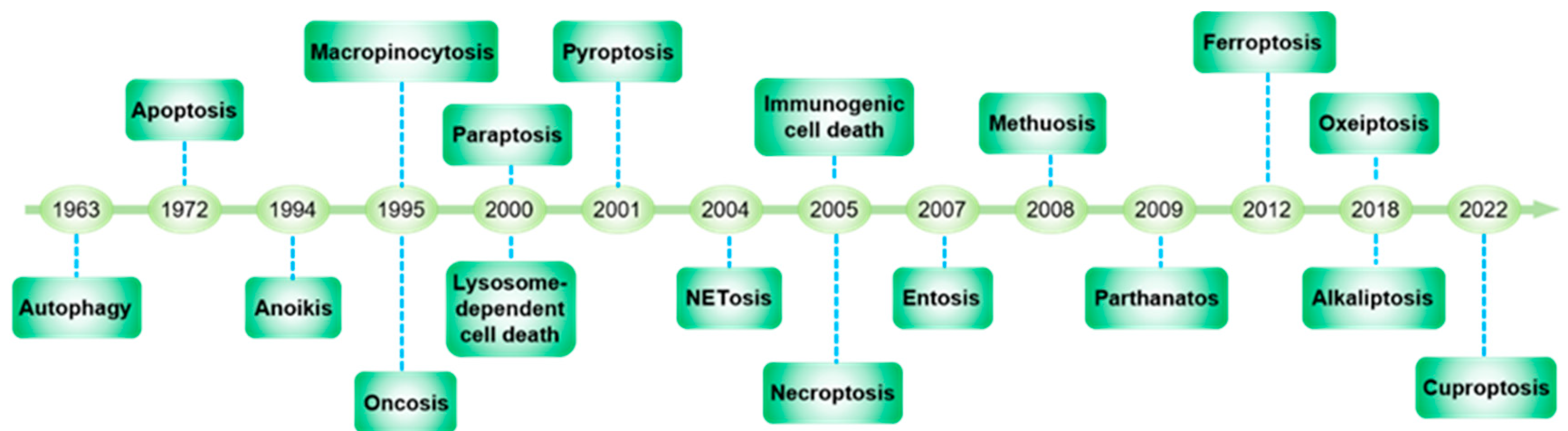
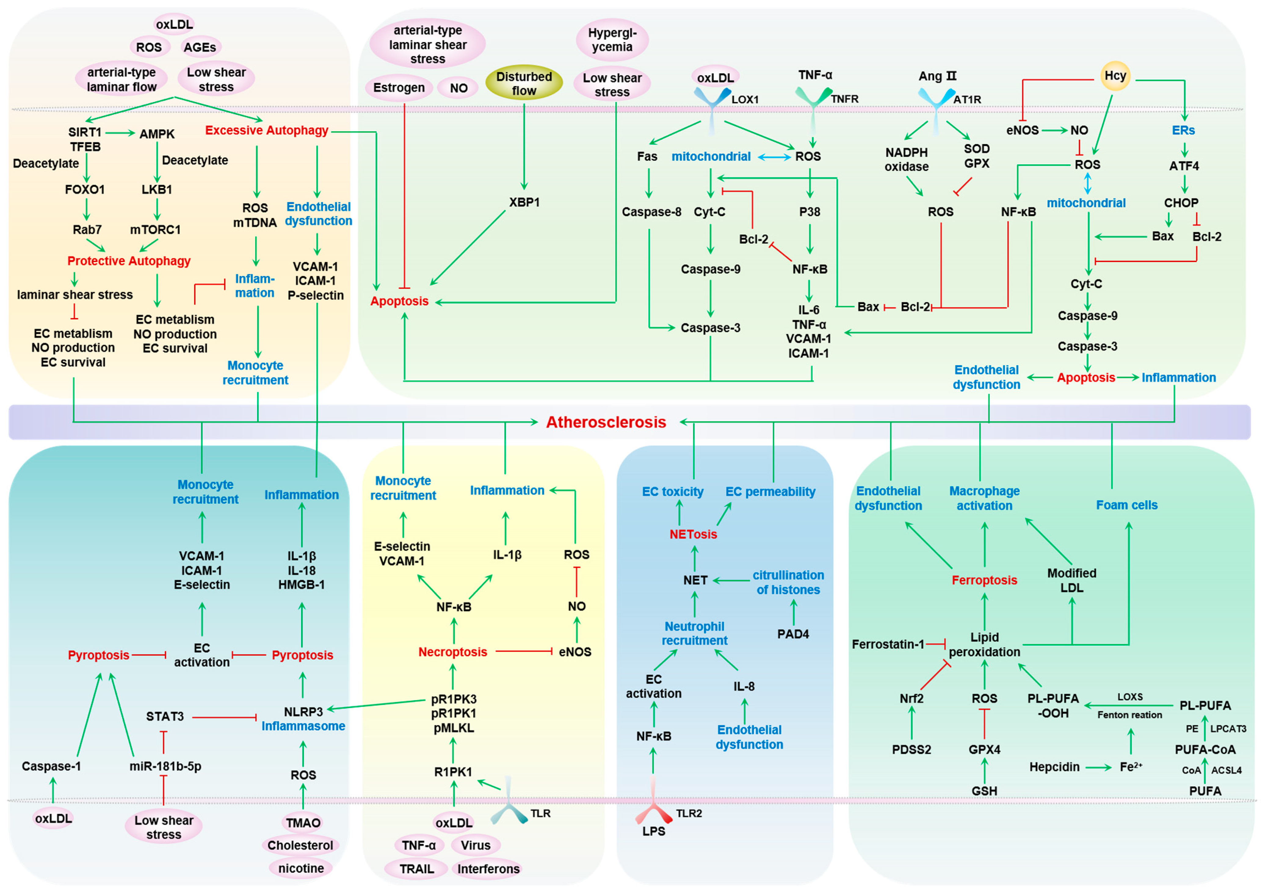
Endothelial cells (ECs) form the inner linings of blood vessels, and are directly exposed to endogenous hazard signals and metabolites in the circulatory system. The senescence and death of ECs are not only adverse outcomes, but also causal contributors to endothelial dysfunction, an early risk marker of atherosclerosis. The pathophysiological process of EC senescence involves both structural and functional changes and has been linked to various factors, including oxidative stress, dysregulated cell cycle, hyperuricemia, vascular inflammation, and aberrant metabolite sensing and signaling. Multiple forms of EC death have been documented in atherosclerosis, including autophagic cell death, apoptosis, pyroptosis, NETosis, necroptosis, and ferroptosis. Despite this, the molecular mechanisms underlying EC senescence or death in atherogenesis are not fully understood.

  • atherosclerosis
  • endothelial cell
  • endothelial cell senescence
  • endothelial cell death

1. Endothelial Cell Death and Atherosclerosis

The roles of various forms of regulated endothelial cell (EC) death, including apoptosis, autophagic cell death, NETosis-related cell death, ferroptosis, pyroptosis, and necroptosis, in the development of atherosclerosis have been investigated extensively, as summarized in Figure 1 and Table 1. Various proatherogenic signals, such as low-density lipoprotein (LDL), hyperglycemia, oxidative stress, and low shear stress, have been shown to induce EC death. Conversely, several athero-protective factors, like estrogen and NO, have been demonstrated to prevent EC death. The long-term influence of death-inducing factors causes a substantial loss of EC, leading to not only systemic endothelial dysfunction, but also plaque instability or surface erosion in atherosclerotic lesions locally. However, each mode of cell death is associated with the specific cellular signaling cascades and unique impacts on atherosclerosis, constituting a plethora of potential therapeutic targets. Before focusing on the death of ECs, researchers must emphasize the fact that multiple cell types have been reported to undergo cell death in atherosclerotic lesions, and the deaths of different cell types may yield various outcomes. For example, apoptosis of macrophages and foam cells precipitates cholesterol crystals, which enlarge the necrotic core and destabilize plaques [1]. Apoptosis of smooth muscle cells (SMCs) is frequently associated with the weakening of collagen fibrils, thinning of the fibrous cap, and calcification of lesions [2]. Non-apoptotic death has also been extensively characterized for macrophages and SMCs in atherosclerosis [3]. Herein, researchers will specifically discuss several well-studied forms of EC death in the context of atherosclerosis.
Figure 1. Timeline of cell death research.
Table 1. The main features of cell death.

2. Autophagy—Protective and Detrimental

Autophagy is an essential cytoprotective process that degrades intracellular organelles and recycles the protein and lipids [18]. Genetic activation of autophagy by overexpression of ATG5, a key regulator of autophagosome formation, can even extend the lifespan of mice, partially through increasing cellular resistance to oxidative stress [19]. Notably, autophagic flux is critical to maintaining vascular cell homeostasis, and the decline in EC autophagy contributes to arterial aging [20]. Autophagy-enhancing agents, such as trehalose, spermidine, and rapamycin, revert the phenotypes of aged arteries in mice, including reduced vascular NO levels, elevated endothelial inflammation, and impaired endothelium-dependent vasodilation [20][21][22].
At homeostasis or in response to mild-to-moderate vascular stress, EC autophagy is generally considered to be vasoprotective [23][24]. This is attributed to its ability to balance the status of cellular redox and bioenergetics, which is central to proper EC functions and survival [25]. Multiple triggering signals for autophagy are present during aging, such as ROS, oxLDL, advanced glycation end-products (AGEs), and shear stress [26][27][28][29][30]. Specifically, autophagy can protect EC metabolism, NO production, and survival to restrict chronic vascular inflammation and atherogenesis, with control of the level of cellular ROS as a main mechanism [30][31][32]. In an inflammatory environment, intense autophagy in ECs has been found to reduce the expression of endothelial adhesion molecules, such as CD31 and VE-cadherin [33]. This process limits the migration of neutrophils across the endothelium, ultimately disrupting the cycle of inflammation. Moreover, EC autophagy mediates the vasoprotective effects of laminar shear stress, suppresses EC inflammation, apoptosis, and senescence, and alleviates the atherosclerotic burden in hyperlipidemic mice [34].
At the molecular level, SIRT1 has been recognized as a main signaling mediator of protective autophagy in ECs. Physiological, arterial-type laminar flow induces ROS in ECs, which stimulates EC autophagy through flow-activated SIRT1 and TFEB, a master transcriptional regulator of lysosomal biogenesis [35]. SIRT1 takes part in different stages of autophagic flux, ranging from autophagosome initiation to lysosomal degradation, which has been extensively reviewed elsewhere [36][37]. Herein, researchers briefly focus on the steps that have been experimentally validated in ECs. In HUVECs, SIRT1-dependent AMPK activation, likely via deacetylating/activating LKB1, inhibits mTORC1, in turn stimulating autophagy [38]. As mTOR is a master negative regulator of all main stages of autophagy [39][40], further studies are warranted to dissect the specific signaling role of the SIRT1-mTOR axis in EC autophagy. In addition to regulating the LKB1-AMPK-mTOR pathway, the deacetylase activity of SIRT1 works on a diverse range of pro-autophagic proteins. For example, upon nutrient starvation, SIRT1 deacetylates ATG5, ATG7, and ATG12 to facilitate the elongation of autophagic vesicles [41]. Importantly, the SIRT1-ATG5 link has been studied in oxLDL- and shear-stress-induced EC autophagy [42][43]. Another well-established pro-autophagic role of SIRT1 is that SIRT1-deactylated FOXO1 transcriptionally activates Rab7, which mediates autophagosome–lysosome fusion. The importance of the SIRT1-FOXO1 cascade in protective autophagy in ECs has been experimentally validated by several independent groups [44][45]. Interestingly, SIRT1 itself is susceptible to autophagic degradation, a process that contributes to the loss of SIRT1 during senescence and aging [46]. However, whether this bidirectional control between SIRT1 and autophagy functions in the aged endothelium is unknown.
On the other hand, autophagy also has broad adverse impacts on atherosclerosis via its profound detrimental effects on ECs, macrophages, and arterial SMCs, as it affects pathogenesis from the early to the advanced stages [47]. Critically, overactivation of autophagy can induce cell death, including death of EC, in a form distinct from other modes of death like apoptosis, necroptosis, and necrosis [48][49]. In 2003, Chau et al. demonstrated that endostatin, a collagen VIII cleaved fragment, can induce autophagic EC death in a ROS- and caspase-independent manner [50]. Importantly, serum endostatin levels correlate with subclinical atherosclerosis, as assessed by carotid intima–media thickness in a cohort of 648 healthy Japanese subjects [51], cardiovascular mortality in two Swedish community-based cohorts of 1689 elderly subjects [52], and mortality and severe disability after ischemic stroke in 3463 Chinese subjects [53]. However, the in vivo roles of endostatin in autophagic EC death remain undefined, although endostatin is reported to slow down mouse atherogenesis due to its anti-angiogenic effect or its capacity to interfere with LDL retention in intima [54][55]. Subsequent studies have identified multiple stress signals that trigger autophagic cell death, including ROS, cigarette smoke extract, glucose deprivation, and bisphosphonates [56][57][58][59]. Despite these advancements, in vivo evidence on autophagic EC death, especially its relevance to atherosclerosis, remains to be firmly investigated and established.

3. Apoptosis

Apoptosis is one of the most widely studied forms of programmed cell death, and it has diverse, context-dependent roles during development, homeostasis, and disease [60]. Apoptosis can be initiated via two main pathways: the extrinsic and intrinsic pathways. The extrinsic pathway is triggered by extracellular stress signals. Notably, many pro-atherogenic signals are also pro-apoptotic for ECs, including oxLDL, Ang II, TNF-α, homocysteine, and disturbed blood flow [61]. External stress triggers the assembly of a death-inducing signaling complex that activates caspase-8, which in turn cleaves and activates caspase-3, a major executor of apoptosis featuring chromatin condensation and DNA fragmentation [62]. In vivo, the remnants of apoptotic cells can be quickly cleared by phagocytic cells [60]. The intrinsic pathway is activated by perturbed intracellular signals, such as genomic instability due to DNA damage or mitotic defects, oxidative stress, or mitochondrial damage. This pathway mainly involves the release of pro-apoptotic proteins, typically cytochrome c, from the mitochondria into the cytoplasm. This then promotes the assembly of an apoptosome comprising APAF-1 and procaspase-9 for the downstream activation of caspase-3 and apoptotic cell death [63]. It needs to be noted that, despite being initially proposed as distinctive, the extrinsic and intrinsic apoptosis pathways have significant crosstalk and overlap. For instance, TNF-α not only activates tumor necrosis factor receptor 1 (TNFR1) to recruit tumor necrosis factor receptor type 1-associated DEATH domain (TRADD) for the assembly of the death-inducing signaling complex, but also causes mitochondrial outer membrane permeabilization (MOMP) and the release of cytochrome c [64].
Near half a century ago, increased apoptosis of ECs was found at the pig aortic arch [65], the athero-prone aortic zone. Importantly, accumulating evidence has revealed that EC apoptosis is an early event in the development of atherosclerosis. Tricot et al. observed increased EC apoptosis in 42 human carotid atherosclerotic plaques, with a preferential localization of apoptotic EC in the downstream parts of the lesions, where the blood flow becomes slow and complex [66]. Redox status is one of the main determinants of EC apoptosis during atherogenesis [67], with excess ROS derived from NADPH oxidase and dysfunctional mitochondria as a major driver of EC apoptosis [68][69]. Moreover, pro-atherogenic stressors, such as oxLDL, Ang II, hyperglycemia, pro-inflammatory cytokine TNFα, and low shear stress, are shown to induce EC apoptosis [60]. In contrast, athero-protective factors, such as estrogen, NO, and arterial-type laminar shear stress, prevent EC apoptosis [70][71].
OxLDL is a key biomarker for atherosclerosis, and it induces chronic endothelial inflammation, endothelial dysfunction, and apoptosis [72]. In 1997, Dimmeler et al. identified that the oxLDL-induced death process in HUVECs is a mode of caspase-3-dependent apoptosis [73]. Subsequently, diverse molecular cascades have been identified for oxLDL-mediated apoptosis. Harada-Shiba et al. reported that oxLDL induces a rapid accumulation of ceramide and superoxide to promote the apoptosis of HUVECs [74]. This pro-apoptotic effect is mediated by the oxysterol, but not phospholipid, fraction of oxLDL [74]. At the same time, Sata et al. showed that oxLDL sensitizes Fas-mediated EC apoptosis [75]. Ca2+ activity is also an important intracellular mediator for cytotoxic signals of oxLDL [76]. OxLDL-induced Ca2+ mobilization and subsequent cell death are inhibited by athero-protective high-density lipoprotein (HDL) or delipidated apolipoprotein A (apoA) [76]. Notably, lipoxygenase-1 (LOX-1), a lectin-like endothelial receptor for oxLDL, is transcriptionally upregulated by oxLDL itself, and causes EC apoptosis in an NF-κB-dependent manner [77]. Subsequent studies greatly expanded the spectrum of oxLDL-elicited apoptotic cascades, ranging from surface receptors, stress-sensing kinases, and transcription factors to non-coding RNAs. One intriguing transcription factor is Krüppel-like Factor 2 (KLF2), a well-documented mechanotransducer for shear stress in ECs [78]. KLF2, whose expression is elevated by laminar flow [79], executes its athero-protective effects via transcriptional activation of eNOS and thrombomodulin, or repression of inflammatory mediators including E-selectin, MCP-1, VCAM-1, and plasminogen activator inhibitor-1 (PAI1) [78]. Notably, an anti-apoptotic role of KLF2 has been reported for ECs. Zhang et al. reported that protein tyrosine phosphatase 1B (PTB1B) knockdown can prevent oxLDL-induced inflammatory injury and dysfunction in ECs, which is regulated at least in part by the AMPK/SIRT1 signaling pathway through KLF2 [80]. Expression of the E3 ubiquitin ligase 3-hydroxy-3-methylglutaryl reductase degradation (HRD1) was significantly decreased in atherosclerotic intima. Mechanistically, decreases in HRD1 levels in ECs can be caused by oxLDL. Elevated expression of HRD1 inhibits the EC apoptosis induced by oxLDL, potentially via promoting the ubiquitination–proteasomal degradation of LOX-1, the oxLDL receptor [81]. Importantly, KLF2 has been identified as a transcriptional activator of HRD1 [81]. These studies support the prevention of EC apoptosis as a potential athero-protective mechanism of KLF2.
While laminar shear stress stabilizes the endothelial barrier, sustains eNOS activation, and promotes endothelial quiescence and survival, disturbed flow, an atherogenic risk factor, evokes endothelial inflammation, permeability, and apoptosis [82][83][84][85]. Several mechanisms have been discovered for disturbed flow-induced endothelial apoptosis. Disturbed flow leads to sustained X-box binding protein 1 (XBP1) activation and splicing, one branch of ER stress responses, thereby inducing apoptosis of ECs [85]. Adenovirus-mediated XBP1 overexpression results in the development of in situ EC apoptosis and atherosclerotic lesions in an Apoe−/− mouse model of aortic graft [86]. Moreover, disturbed flow increases endothelial peroxynitrite production, which in turns activates a cascade of the PKCζ/PIASy/p53 SUMOylation/BCL-2 pathway to execute apoptosis in vitro and in vivo [87]. Compared with the athero-protective greater curvature of the mouse aortic arch, the athero-susceptible lesser curvature exhibits higher expression and activity of PKCζ, the expression of p53, and the incidence of p53-dependent endothelial apoptosis [87].
In addition to shear stress, the endothelium in vivo is also impacted by cyclic mechanical stretch in a magnitude-dependent manner. Physiological levels of cyclic stretch (5–10% strain) are reported to protect endothelial survival, while excess stretch (15–20% strain) leads to a compromised endothelial barrier and cell apoptosis [87][88]. Importantly, such mechanosensitive apoptosis is associated with enhanced transcription of genes involved in endothelial inflammation, including IL-6, IL-8, MCP-1, ICAM-1 [89], and TGFβ [90]. Several intracellular signaling pathways have been shown to modulate stretch-dependent endothelial apoptosis. The PI3K-AKT cascade is considered to be a well-established pathway to sustain endothelial survival under mechanical stretch [91][92][93], while the production of ROS and activation of stress-responsive p38 MAPK or JNK is pro-apoptotic upon pathological stretch [90]. Recently, Zhuang et al. reported that cyclic stretch increases microparticle production in cultured ECs, with the endothelial microparticle proteomes dependent on the magnitude of stretch (5% versus 15% strain) [94]. Intriguingly, physiological stretch (5% stretch)-induced microparticles suppress EC apoptosis, potentially via activation of Src [94].
A continual endothelium is critical to suppressing atherogenesis and stabilizing established atherosclerotic plaques [94]. Superficial erosion of plaques, as a consequence of EC apoptosis, has been recognized as a potential cellular event triggering atherothrombosis. This mechanism involves neutrophil and NETosis, which will be discussed in a later section. A plethora of biochemical and biophysical pro-apoptotic stimuli, as discussed above, stress the endothelium of atherosclerosis lesions. However, a challenging question remains: whether the prevention of EC apoptosis constitutes a viable therapeutic invention to treat atherosclerosis. Several important and clinically approved anti-atherosclerotic therapies, including statins, proprotein convertase subtilisin/kexin type 9 (PCSK9) inhibition, and SGLT2 inhibitors, have been reported to protect against endothelial apoptosis [95][96][97][98][99][100]. However, given the fact that these medicines have pleiotropic benefits over endothelial functions, the role of endothelial apoptosis in the anti-atherosclerotic benefits are difficult to dissect. In fact, evidence from animal models shows that the genetic ablation of EC apoptotic machinery is largely inconclusive. For example, endothelial overexpression of the Fas ligand in hyperlipidemic Apoe−/− mice retards atherogenesis and is associated with dampened endothelial inflammation and reduced T cell and monocyte infiltration, but is indistinguishable from endothelial apoptosis, as assessed by an in situ TUNEL assay [101]. Further, global caspase-3 KO promotes, instead of inhibits, the growth and necrosis of atherosclerotic lesions in Apoe−/− mice without alteration of the plasma levels of cholesterol or triglyceride. Though EC apoptosis was not examined in the lesions, this research indicates that the prevention of cell apoptosis may not constitute an effective strategy to treat atherosclerosis. This can likely be attributed to the accumulated dysfunctional ECs upon failure in apoptosis execution [102]. Moreover, two alternative possibilities exist, either non-death functions of the apoptosis machinery [103][104] or the switch to other modes of cell death when the apoptosis cascade is inhibited. For the former, both caspase-3 and caspase-9 have a direct, non-death-inducing role in modulating endothelial barrier functions [103][104]. For the latter, pharmacological or genetic inactivation of caspase was reported to shunt the cells under lethal inflammatory stress into necroptosis, a mode of regulated cell death mediated by the formation of a necrosome, a death-inducing protein complex comprising receptor-interacting protein kinase (RIPK) and mixed-lineage kinase domain-like protein (MLKL). Notably, this apoptosis-to-necroptosis switch has been documented for ECs. For instance, induced deletion of endothelial caspase-8 in 6-week-old mice leads to diminished EC apoptosis and fatal hemorrhagic lesions in the small intestine [105]. Importantly, caspase-8-independent EC death and small intestinal hemorrhage are abolished by the genetic inactivation of necroptotic executioner MLKL [105]. Researchers will discuss necroptosis in the following sections.

4. Necroptosis

Vulnerable atherosclerotic lesions are characterized by a large necrotic core, which comprises debris of dead cells and cholesterol crystals. The necrotic core is surrounded by layers of SMCs and ECM, termed the fibrous cap. The integrity and thickness of the fibrous cap are critical determinants of plaque stability. Notably, the fibrous cap is also covered by ECs, and endothelial denudation is considered one important contributory factor to atherothrombosis. Recent evidence has suggested that a majority of cell death events in atherosclerotic lesions are due to regulated necrosis. Regulated necrosis has three main modes: necroptosis, pyroptosis, and ferroptosis. These three modes of regulated cell death are triggered by different external stress signals, and have distinct signaling cascades and cellular outcomes. Their relevance to the endothelium in the context of atherosclerosis is the topic of the following sections.
Necroptosis is initiated by extracellular death signals, including TNFα, TNF-related apoptosis-inducing ligand (TRAIL), toll-like receptor (TLR) ligands, interferons, and viruses, which stimulate the formation of a death-inducing necrosome complex containing receptor-interacting serine/threonine-protein kinase 1 (RIPK1), RIPK3, and MLKL [106]. The activation of RIPK1 results in autophosphorylation and interaction with RIPK3, leading to RIPK3 oligomerization and necrosome formation [107][108]. Within the necrosome, activated RIPK3 recruits and phosphorylates MLKL, leading to its oligomerization. The phosphorylated MLKL oligomers interact with plasma membrane phospholipids to form pores [109], leading to membrane permeabilization, release of DAMPs, and necroptotic cell death [108]. Notably, necroptotic cells feature robust release of DAMPs and cytokines, in turn aggravating tissue inflammation [108]. Necroptotic inflammation can also be enhanced by crosstalk with pyroptosis, another form of regulated necrosis. For instance, RIPK3 activates the NLRP3 (NOD-like receptor family, pyrin domain-containing 3) inflammasome, which activates caspase-1 to cleave IL-1β into their mature forms [110]. Inflammasome activation is a key step of pyroptosis, which will be discussed later.
Human atherosclerotic plaques are associated with a noticeable increase in the expression of necroptosis mediators RIPK3 and MLKL, at both the mRNA and protein level [111], with their expression being even higher in individuals with unstable plaques compared to those with stable ones [111]. In hyperlipidemic Ldlr−/− mice, advanced plaques show a greater presence of RIPK3, primarily in macrophages [112]. When RIPK3 is absent, the development of advanced atherosclerotic lesions is reduced in Apoe−/− and Ldlr−/− mice, without affecting early atherogenesis [112]. Decisively, bone marrow transplantation experiments demonstrate that the anti-atherosclerotic effects of RIPK3 KO are mediated by bone marrow-derived cells [112]. Moreover, suppression of MLKL using antisense oligonucleotides or genetic deletion diminishes the necrotic cores of advanced plaques of Apoe−/− mice without impacting the total atherosclerotic plaque burden [113]. Interestingly, although cell death and necrotic cores are reduced in MLKL-deficient advanced lesions, the lesion lipid content is increased, which can mainly be attributed to exacerbated lipid accumulation in macrophage foam cells [113]. While RIPK1 is also predominantly found in macrophages within human carotid lesions, its deletion in LysM+ myeloid cells potentiates, instead of attenuates, macrophage necroptosis and necrotic core formation in atherosclerotic lesions in Apoe−/− mice [114]. This complex, regulatory role of RIPK1 in macrophage death can be at least partially attributed to its transactivation of the NF-κB survival pathway [114]. However, a comparative study using mice with different deficiencies in a multitude of necroptosis-related inflammatory disease models, ranging from systemic inflammation sepsis to localized ischemia-reperfusion injury of the kidney, showed that MLKL deficiency provides limited or no protection against adverse outcomes, whereas Ripk1D138N/D138N (a catalytically inactive mutant) and Ripk3−/− mice were protected [115].
While strong evidence supports the functional importance of macrophage necroptosis in atherosclerosis, the roles of EC necroptosis in the context of atherosclerosis remain elusive. Recently, Colijn et al. performed an important study to compare the effects of myeloid-, EC-, and SMC-specific KO of RIPK3 on atherosclerosis in Apoe−/− mice [116]. Mice with RIPK3-deficient ECs exhibit an increase in lesion burden to a similar extent as mice with RIPK3-deficient myeloid cells [116]. Mechanistically, RIPK3 deficiency in ECs enhances the expression of inflammatory mediators, including E-selectin and MCP-1, but had an inconclusive role in necroptotic cell death, as aortas with endothelial deletion of RIPK3 do not show significant changes in total or phosphorylated MLKL [116]. In the same regard, Karunakaran et al. reported that endothelial RIPK1 functions as a central driver of vascular inflammation in atherogenesis, instead of a regulator of cell survival [117]. In HUVECs, shRNA against RIPK1 reduces inflammatory gene expression (IL-1β, E-selectin, and ICAM-1) and monocyte attachment via promoting nuclear translocation of the NF-κB p65 subunit [117]. It must be noted that EC necroptosis has been investigated widely in microvasculature associated with a variety of diseases, including cardiac transplantation [118][119], lung injury [120][121], and tumor metastasis [122][123][124]. One typical example is that lung microvascular endothelial necroptosis serves as an essential event to facilitate the extravasation of circulating, metastatic tumor cells [122]. This intriguing intercellular crosstalk requires direct contact between tumor cells and ECs. EC expression of TNFR1 and death receptor 6 (DR6) mediates necroptotic cell death and opening of the endothelial barrier to facilitate tumor cell transmigration [122][123][124]. Does this intercellular communication also apply to the transendothelial migration of leukocytes into atherosclerotic lesions? Answers may come from more specific in situ analyses of leukocyte–endothelial contact with lesions of different stages and anatomical sites. With this evidence taken together, although the inhibition of necroptosis machinery, such as RIPK1/3 and MLKL, has demonstrated anti-atherosclerotic benefits in animals, the direct role of EC necroptosis warrants further study.

5. Pyroptosis

Pyroptosis is initiated through the activation of caspase-1 within a large complex called an inflammasome [10][125]. There are multiple forms of inflammasomes, including NLRP-1, NLRP-3, absent in melanoma 2 (AIM-2), NLR family CARD domain containing 4 (NLRC-4), and pyrin. The NLRP-3 inflammasome is the most widely studied in the vascular system, and consists of NLRP-3, apoptosis-associated speckle-like protein containing CARD (ASC), and procaspase-1 [126], all of which are expressed in the arterial wall [127][128]. Evidence suggests that atherosclerotic plaque components, such as oxLDL and cholesterol crystals, can activate NLRP-3 inflammasomes, partially involving lysosomal rupture and consequent cathepsin release [129][130][131]. Active caspase-1 promotes inflammation by converting pro-IL-1β and pro-IL-18 into bioactive forms, and by cleaving Gasdermin D (GSDMD). The cleaved GSDMD then forms pores in the cell membrane, leading to membrane disruption and the release of inflammatory factors to amplify tissue inflammation [10][125]. In addition to the canonical pyroptotic cascade, caspase-11 can directly sense and complex with lipopolysaccharide (LPS) in the absence of TLR4 or a canonical inflammasome in the context of sepsis [132]. Subsequently, active caspase-11 cleaves GSDMD, causing pore formation and pyroptotic cell death. Furthermore, caspase-11-induced GSDMD can also activate the NLRP-3 inflammasome, indirectly triggering caspase-1-dependent pyroptosis and the release of IL-1β and IL-18 [132]. However, the role of caspase-11-dependent non-canonical pyroptosis in atherosclerosis remains to be determined. Moreover, targeting different inflammasomes (NLRP-3, NLRP-1, AIM-2, or NLRC-4) has been considered as an anti-atherosclerotic strategy. Small molecule inhibitors selective for NLRP-3, such as MCC950, OLT1177, and CY-09 [133][134], have been shown to reduce IL-1β production and attenuate atherosclerosis progression in preclinical models. For example, van der Heijden et al. reported that intraperitoneal administration of MCC950 significantly ameliorates the development of atherosclerosis in Apoe−/− mice, and is associated with reduced plasma IL-1β levels and reduced macrophages in the lesions [135]. MCC950-mediated inactivation of the pyroptotic NLRP-3/ASC/caspase-1/GSDMD pathway in mouse aortas was clearly demonstrated in a subsequent study [136]. Importantly, the IL-1β-neutralizing antibody canakinumab has demonstrated considerable clinical benefits, reducing recurrent cardiovascular events by 17% in patients with a previous myocardial infarction (CANTOS trial), independent of the lipid level [137]. Mechanistically, although macrophages are currently recognized as the cell targets of anti-pyroptotic therapy [136][138], emerging evidence has established a role of EC pyroptosis in atherosclerosis. Many pro-atherogenic or pro-inflammatory factors activate the NLRP-3 inflammasome in ECs, including oxLDL, ROS, trimethylamine-N-oxide (TMAO), low shear stress, and nicotine [139][140]. These external stress signals likely converge on the redox-activated NF-κB-NLRP-3 axis [139][140] for the induction of endothelial pyroptosis. In particular, cholesterol crystals present in atherosclerotic plaques can induce NLRP-3 inflammasome activation and IL-1β secretion in cultured mouse carotid ECs [141], suggesting that the endothelial pyroptotic machine contributes to plaque vulnerability. Moreover, nicotine administration exacerbates the burden of atherosclerotic lesions of Apoe−/− mice fed a high-fat diet, with a functional involvement of ROS-dependent activation of NLRP3 inflammation, in addition to subsequent cytokine production and pyroptotic EC death [142].
While mice with EC-restricted KO of pyroptosis machinery have not been generated to study atherosclerosis, mechanistic insights can still be gained using mouse models with global deficiency of caspase-1 or NLRP-3. For example, Yin et al. reported that caspase-1 KO reduces early atherogenesis in Apoe−/− mice [143]. Caspase-1−/−; Apoe−/− mice fed a high-fat diet exhibit decreased endothelial activation, including reduced expression of leukocyte adhesion molecules (ICAM-1, VCAM-1, and E-selectin) and reduced secretion of cytokines/chemokines (chemokine (C-C motif) ligand 3 (CCL3), chemokine (C-X-C motif) ligand 2 (CXCL2), and CXCL10). Notably, oxLDL-induced pyroptosis of cultured mouse aortic ECs is abrogated by caspase-1 KO in a SIRT1-dependent manner [143]. Moreover, Zhuang et al. linked disturbed flow with endothelial NLRP3 inflammasome activation in atherosclerotic mice [144]. Pro-atherogenic oscillatory shear stress downregulates KLF2-dependent FoxP1 expression in ECs, in turn derepressing the activity of the NLRP3/caspase-1/IL-1β cascade to promote atherogenesis [144]. Although pyroptotic cell death was not examined in this specific scenario, a subsequent study found that low shear stress (5 dyn/cm2) stimulates pyroptosis of HUVECs via suppressing the expression of miR-181b-5p [145], which directly targets the signal transducer and activator of transcription 3 (STAT3) expression to restrain NLRP3 inflammasome activation and cell death [145]. Furthermore, genetic inactivation of GSDMD, the pyroptosis executioner, effectively blocks the development of atherosclerosis in hyperlipidemic mice. Recently, Puylaert et al. demonstrated that global GSDMD KO leads to reduced atherogenesis, which is associated with smaller necrotic cores and increased SMC-to-monocyte ratios in lesions, indicating that pyroptosis inhibition is a potential approach to stabilizing atherosclerotic plaques [146]. However, Puylaert et al. also noted that cleaved, pore-forming GSDMD species are not detected in human carotid lesions, in contrast with widely available evidence on the pyroptotic death of ECs in culture [147]. For now, it is not clear whether nondetectable expression of endothelial GSDMD is a biological observation or a technical issue. More studies are needed to ascertain the status of in vivo pyroptosis of ECs in human atherosclerosis. Notably, disulfiram, an FDA-approved drug, has been identified as a potent small molecule inhibitor of GSDMD pore formation [147], although its effects on atherosclerosis have yet to be tested [147]. Thus, the inhibition of caspase-1, NLRP3, or GSDMD in dyslipidemic and inflammatory contexts prevents endothelial pyroptosis and improves endothelial survival, thereby preserving vascular intima integrity, reducing vascular inflammation, and further attenuating the progression of atherosclerosis.

6. Ferroptosis

Ferroptosis was first discovered during anti-cancer small chemical screening as a non-apoptotic, MEK- and iron-dependent oxidative cell death process in tumor cells with oncogenic RAS [148]. The same study identified and characterized the first-generation ferroptosis inducers, erastin and Ras-selective lethal 3 (RSL3). Ferroptosis features excessive iron-dependent lipid peroxidation, associated with plasma membrane rupture with shrinkage of the nanopores and mitochondria [17]. The chemical processes of lipid peroxidation are complex and involve the dysregulation of several different pathways. First, iron overload contributes to the generation of hydroxyl radicals via the Fenton reaction to oxidize lipids. Normally, iron is buffered and stored by ferritin as Fe2+. Under the condition of iron overload, likely attributed to excessive hemoglobin, iron chloride, heme oxygenase-1 hyperactivation, or transferrin overabundance, the cytosolic labile/free ferrous iron pool grows to induce ferroptosis. In this regard, iron chelators like deferoxamine have anti-ferroptotic effects [149]. Second, enzymatic peroxidation of unsaturated fatty acid in the phospholipid bilayer can be mediated via the Achaete–Scute family BHLH transcription factor 4 (ACSL4)/lysophosphatidylcholine acyltransferase 3 (LPCAT3)/15-LOX cascade. Small chemical inhibitors of 15-LOX, such as PD146176 and ML351, have been used as ferroptosis inhibitors [150]. Third, and best characterized, is the inactivation of glutathione peroxidase (GPX)4 [151]. GPX4 catalyzes the reduction of lipid peroxides in the cellular membrane. Diminished gene expression of GPX4 or depletion of its substrate, glutathione, can permit the accumulation of lipid peroxides and subsequent membrane rupture and cell death. The depletion of glutathione can be caused by inhibition of the Xc-antiporter system, which is responsible for cellular uptake of cysteine in exchange for glutamate. Notably, the Xc-antiporter system is the molecular target of erastin [152], and GPX4 is inhibited by RSL3. In addition, ferrostatin-1, α-tocopherol, or liproxstatin, which directly trap lipid peroxides, can be used to inhibit the execution of ferroptosis.
High iron levels have long been associated with an increased risk of atherosclerotic CVDs [153][154][155]. Martinet et al. suggested that intraplaque hemorrhage, iron deposition, and lipid peroxidation are common pathological features of advanced human atherosclerotic plaque [138]. In experimental animals, iron intake is a cause of atherogenesis [156][157][158][159]. Dietary iron aggravates atherosclerosis, while restricting iron intake via a low-iron diet or iron chelator ameliorates atherogenesis. Recently, Vinchi et al. showed that, in the presence of genetic iron overload caused by a heterozygous mutation of iron exporter Ferroportin C326S, Apoe−/− mice develop severe spontaneous atherosclerosis in the absence of a high-fat diet, which can be rescued by iron restriction (low-iron diet or iron chelator deferasirox) [157]. The iron-overloaded arterial wall displayed an elevated inflammation profile, including cytokine expression and endothelial dysfunction. In particular, a wide range of endothelial defects were observed, including increased aortic endothelial permeability, elevated expression of ICAM-1, VCAM-1, and E-selectin, reduced eNOS activity, and enhanced oxidative damage (as assessed by the nitrotyrosine content) [156]. Interestingly, iron overload, induced by ferric ammonium citrate, enhanced ROS and apoptosis in cultured human aortic ECs, but ferroptotic markers in dying ECs were not tested in this research [156]. Nevertheless, ferroptosis has been established as a mode of oxLDL-induced EC death [160][161][162]. The nature of endothelial ferroptosis is usually identified using ferroptosis inhibitors (e.g., ferrostatin-1) or molecular markers (iron content, GPX4 level, Solute Carrier Family 7 Member 11 (SLC7A11), the Xc-antiporter protein). For example, Bai et al. showed that intraperitoneal injection of ferrostatin-1 reduces the atherosclerotic lesion burden of Apoe−/− mice fed a high-fat diet, to a similar extent to oral administration of simvastatin [161]. Mechanistically, ferrostatin-1 treatment reduces serum and aortic iron content, restores the expression of GPX4 and SLC7A11, and reduces serum LDL-cholesterol levels. Notably, lesional endothelial expression of GPX4 appears to be increased with the treatment of ferrostatin-1 [161]. In addition, ferrostatin-1 rescues oxLDL-induced ferroptotic traits (mitochondria size, iron content, expression of GPX4 and SLC7A11) and the death of cultured mouse aortic ECs [161]. Prenyldiphosphate synthase subunit 2 (PDSS2) is a key enzyme involved in the biosynthesis of coenzyme Q10. Yang et al. found that serum PDSS2 levels are decreased in patients with coronary artery disease. Global PDSS2 KO increases the development of atherosclerosis induced by a Western high-fat diet. Overexpression of PDSS2 suppresses the oxLDL-induced ferroptosis in cultured human coronary arterial ECs, correlated with normalized levels of iron, glutathione, and ROS [160]. Important evidence also exists for the functional link between ferroptosis and atherosclerosis. Iron overloaded by FeSO4 induces both ferroptosis and apoptosis to promote the calcification of HUVECs, which can be alleviated by the application of ferrostatin and the iron chelator deferoxamine [163]. In addition to manipulation of the iron content, encouraging evidence also supports the therapeutic potential of targeting other ferroptotic pathways. Transgenic overexpression of GPX4 in Apoe−/− mice effectively suppresses atherogenesis, as well as necrotic core formation, in advanced plaque [163]. In cultured mouse aortic ECs, GPX4 overexpression reduces ROS production, the expression of ICAM-1 and VCAM-1, and monocyte adhesion induced by lysophosphatidylcholine or 7-ketocholesterol. Interestingly, both the necrotic and apoptotic modes of cell death are prevented by GPX4. Additionally, using the 15-LOX inhibitor PD146176 to hinder enzymatic lipid peroxidation leads to reduced plaque progression in atherosclerotic rabbits which already have established plaques [163]. Similarly, the genetic removal of 15-LOX contributes to a lower plaque load in Apoe−/− mice. Collectively, these investigations strongly imply that iron-dependent lipid peroxidation plays a significant role in the development of atherosclerosis.

7. NETosis

NETosis is a form of programmed cell death that is specific to neutrophils, involving the release of neutrophil extracellular traps (NETs) [13][164]. NETs are web-like structures composed of DNA, histones, and various antimicrobial proteins. They are released by neutrophils to trap and neutralize pathogens, such as bacteria, fungi, and parasites, in the extracellular space [13][164]. During NETosis, the neutrophil undergoes a sequence of morphological changes, including the decondensation of its nuclear material and the mixing of nuclear and cytoplasmic components. This leads to the rupture of the cell membrane and the release of NETs into the surrounding environment. NETs not only contain neutrophil materials, but also gather circulating elements in the blood, such as tissue factors, fibrin, and other procoagulants. While NETosis is essential for the immune response, excessive or uncontrolled NET formation can be harmful and has been implicated in the pathogenesis of various autoimmune and inflammatory diseases, including atherosclerosis [13][164]. Functionally, NET induces the activation of ECs, SMCs, antigen-presenting cells, and platelets, leading to localized tissue inflammation and thrombosis, thereby promoting atherogenesis and atherothrombosis [164][165][166]. Reciprocally, dysfunctional ECs enhance NETosis, amplifying the damage to neighboring cells and further injuring the arterial endothelial lining [167]. Coronary specimens from patients with acute myocardial infarction demonstrate the presence of NETs in both fresh and lytic, but not organized, thrombi, suggesting that NETosis occurs in the early stage of atherothrombosis [168]. Importantly, circulating NET components, including plasma levels of nucleosome and myeloperoxidase–DNA complexes, predict the extent of coronary stenosis, the number of atherosclerotic coronary vessels, and the occurrence of major adverse cardiac events, suggesting NET as a novel biomarker in atherosclerosis [169].
For the specific focus, NET not only induces endothelial dysfunction, but also results in EC death. Gupta et al. found that stressed ECs, conditioned by phorbol 12-myristate 13-acetate, TNF-α, or thapsigargin (ER stress inducer) enhance the NET formation of co-cultured neutrophils, which is partially dependent on endothelial release of IL-8 [170]. Reciprocally, NET causes EC death, as evidenced by the cellular uptake of SYTOX green, a membrane-impermeable nucleic acid dye. EC death can be prevented by DNase that disrupts the backbone of NETs [170]. Saffarzadeh et al. subsequently showed that extracellular histones are responsible for NET-induced cultured EC death, as assessed by lactate dehydrogenase release [171]. Notably, double KO of endogenous Dnase1 and Dnase1-like 3 causes severe NET clots and consequent lethal blood vessel occlusion in mouse models of chronic neutrophilia or sepsis [172]. While the specific mode of EC death caused by NET remains elusive, NET-associated citrullinated histone 4 has recently been reported to cause necrosis-like “lytic” cell death of cultured SMCs within 10 min [173]. Does this lytic cell death mechanism hold true for ECs within the proximal impact of NET? More studies are needed to answer this. However, apoptosis has been suggested as a mode of EC death in the context of NETosis, as is discussed in the following paragraph.
Peptidylarginine deiminase 4 (PAD4), a key enzyme of NETosis, mediates the citrullination of histones. Histone citrullination leads to the decondensation of chromatin, which is essential for the extrusion of DNA and the formation of NETs. PAD4 also takes part in ROS production and granule mobilization in neutrophils [13][164]. Given its central role in NETosis, research has been focusing on the impacts of PAD4 in atherosclerosis. Genetic inactivation of PAD4 in bone marrow abrogates NETosis, diminishes endothelial permeability, and reduces in situ thrombosis in an Ldlr−/− mouse model of flow-mediated superficial erosion of intima [164]. Notably, a decreased number of TUNEL+ apoptotic ECs were observed in mice with Pad4−/− bone marrow transplantation or DNase I treatment [174]. Importantly, neutrophils are found at the sites of superficially eroded human plaques, and NETs are localized near where apoptotic ECs reside [175][176]. Disturbed flow also plays a critical role in neutrophil recruitment to erosion-prone lesions and subsequent EC apoptosis and mural thrombosis in a TLR2-dependent manner [176]. Moreover, delivery of a PAD4 inhibitor GSK484 using a collagen IV-targeting nanoparticle reduces NET accumulation at sites of endothelial denudation, which is correlated with improved endothelial lining of superficially eroded arterial intima [177]. These findings indicate that NETosis-mediated apoptotic endothelial denudation is a critical cellular event that drives the superficial erosion of atherosclerotic lesions and atherothrombosis. Therefore, NETs represent a novel therapeutic target for the treatment and prevention of thrombotic complications of atherosclerosis.

8. Complexity and Limitations of Targeting Endothelial Cell Death in Atherosclerosis

The development and progression of atherosclerosis are closely related to various cell death modalities, including autophagy, apoptosis, necroptosis, pyroptosis, ferroptosis, and NETosis, as summarized in Figure 2. Cell death contributes to the development of atherosclerosis mainly by loss of endothelial lining, promoting endothelial dysfunction, and inflammatory monocyte recruitment. However, there are few registered trials on ClinicalTrials.gov that primarily evaluate a cell-death-targeting treatment in the context of atherosclerosis, although the mainstream medicines (statin, PCSK9 inhibitors, and SGLT2 inhibitors) have observable effects on vascular cell viability/death. Additionally, as was discussed above, a large collection of inhibitors of specific modes of EC death have been designed and have shown preclinical benefits. Why is there limited clinical translatability? There are several potential considerations. First, multiple modes of cell death can co-exist in the complex pro-atherogenic environment, with a composite stress input of hyperlipidemia, hyperglycemia, oxidative stress, and inflammatory stress. On some occasions, even one stress signal, such as H2O2 or oxidized LDL, can induce multiple modes of EC death. Moreover, different modes of cell death may compensate for each other. A typical example is that when caspase is inhibited in ECs and other cells, necroptosis is activated. This intricate death network could confound the trial of an inhibitor for a specific mode of death. In future, integrated analysis of two or more related modes of cell death should be compared in one atherosclerosis study. This kind of study will pave the way for a feasible strategy to target EC death. Second, pharmacological inhibitors of the death of ECs may impact the death of SMCs and immune cells in a similar or opposite manner, which would make EC-specific targeting impractical, and the outcomes difficult to predict. Recent advancement in single-cell omics at the transcript and epigenetic levels will allow for comprehensive analysis of the responses of different lesion cell types and their communications to a single death-modulating treatment.
Figure 2. EC death in atherosclerosis. Green pointed arrows indicate stimulation; red blunted arrows indicate inhibition.

References

  1. Gautier, E.L.; Huby, T.; Witztum, J.L.; Ouzilleau, B.; Miller, E.R.; Saint-Charles, F.; Aucouturier, P.; Chapman, M.J.; Lesnik, P. Macrophage apoptosis exerts divergent effects on atherogenesis as a function of lesion stage. Circulation 2009, 119, 1795–1804.
  2. Proudfoot, D.; Skepper, J.N.; Hegyi, L.; Bennett, M.R.; Shanahan, C.M.; Weissberg, P.L. Apoptosis regulates human vascular calcification in vitro: Evidence for initiation of vascular calcification by apoptotic bodies. Circ. Res. 2000, 87, 1055–1062.
  3. Li, M.; Wang, Z.W.; Fang, L.J.; Cheng, S.Q.; Wang, X.; Liu, N.F. Programmed cell death in atherosclerosis and vascular calcification. Cell Death Dis. 2022, 13, 467.
  4. Dikic, I.; Elazar, Z. Mechanism and medical implications of mammalian autophagy. Nat. Rev. Mol. Cell Biol. 2018, 19, 349–364.
  5. Zhao, F.; Satyanarayana, G.; Zhang, Z.; Zhao, J.; Ma, X.L.; Wang, Y. Endothelial Autophagy in Coronary Microvascular Dysfunction and Cardiovascular Disease. Cells 2022, 11, 2081.
  6. Choy, J.C.; Granville, D.J.; Hunt, D.W.; McManus, B.M. Endothelial cell apoptosis: Biochemical characteristics and potential implications for atherosclerosis. J. Mol. Cell. Cardiol. 2001, 33, 1673–1690.
  7. Nagata, S. Apoptosis and Clearance of Apoptotic Cells. Annu. Rev. Immunol. 2018, 36, 489–517.
  8. Michel, J.B. Anoikis in the cardiovascular system: Known and unknown extracellular mediators. Arterioscler. Thromb. Vasc. Biol. 2003, 23, 2146–2154.
  9. Taddei, M.L.; Giannoni, E.; Fiaschi, T.; Chiarugi, P. Anoikis: An emerging hallmark in health and diseases. J. Pathol. 2012, 226, 380–393.
  10. Shi, J.; Gao, W.; Shao, F. Pyroptosis: Gasdermin-Mediated Programmed Necrotic Cell Death. Trends Biochem. Sci. 2017, 42, 245–254.
  11. Ju, J.; Liu, Y.; Liang, H.; Yang, B. The role of pyroptosis in endothelial dysfunction induced by diseases. Front. Immunol. 2022, 13, 1093985.
  12. Zdanyte, M.; Borst, O.; Münzer, P. NET-(works) in arterial and venous thrombo-occlusive diseases. Front. Cardiovasc. Med. 2023, 10, 1155512.
  13. Thiam, H.R.; Wong, S.L.; Wagner, D.D.; Waterman, C.M. Cellular Mechanisms of NETosis. Annu. Rev. Cell Dev. Biol. 2020, 36, 191–218.
  14. Weinlich, R.; Oberst, A.; Beere, H.M.; Green, D.R. Necroptosis in development, inflammation and disease. Nat. Rev. Mol. Cell Biol. 2017, 18, 127–136.
  15. Zhang, X.; Ren, Z.; Xu, W.; Jiang, Z. Necroptosis in atherosclerosis. Clin. Chim. Acta Int. J. Clin. Chem. 2022, 534, 22–28.
  16. Zhang, H.; Zhou, S.; Sun, M.; Hua, M.; Liu, Z.; Mu, G.; Wang, Z.; Xiang, Q.; Cui, Y. Ferroptosis of Endothelial Cells in Vascular Diseases. Nutrients 2022, 14, 4506.
  17. Jiang, X.; Stockwell, B.R.; Conrad, M. Ferroptosis: Mechanisms, biology and role in disease. Nat. Rev. Mol. Cell Biol. 2021, 22, 266–282.
  18. Aman, Y.; Schmauck-Medina, T.; Hansen, M.; Morimoto, R.I.; Simon, A.K.; Bjedov, I.; Palikaras, K.; Simonsen, A.; Johansen, T.; Tavernarakis, N.; et al. Autophagy in healthy aging and disease. Nat. Aging 2021, 1, 634–650.
  19. Pyo, J.O.; Yoo, S.M.; Ahn, H.H.; Nah, J.; Hong, S.H.; Kam, T.I.; Jung, S.; Jung, Y.K. Overexpression of Atg5 in mice activates autophagy and extends lifespan. Nat. Commun. 2013, 4, 2300.
  20. LaRocca, T.J.; Henson, G.D.; Thorburn, A.; Sindler, A.L.; Pierce, G.L.; Seals, D.R. Translational evidence that impaired autophagy contributes to arterial ageing. J. Physiol. 2012, 590, 3305–3316.
  21. Lesniewski, L.A.; Seals, D.R.; Walker, A.E.; Henson, G.D.; Blimline, M.W.; Trott, D.W.; Bosshardt, G.C.; LaRocca, T.J.; Lawson, B.R.; Zigler, M.C.; et al. Dietary rapamycin supplementation reverses age-related vascular dysfunction and oxidative stress, while modulating nutrient-sensing, cell cycle, and senescence pathways. Aging Cell 2017, 16, 17–26.
  22. LaRocca, T.J.; Gioscia-Ryan, R.A.; Hearon, C.M., Jr.; Seals, D.R. The autophagy enhancer spermidine reverses arterial aging. Mech. Ageing Dev. 2013, 134, 314–320.
  23. Wang, J.; Wang, W.N.; Xu, S.B.; Wu, H.; Dai, B.; Jian, D.D.; Yang, M.; Wu, Y.T.; Feng, Q.; Zhu, J.H.; et al. MicroRNA-214-3p: A link between autophagy and endothelial cell dysfunction in atherosclerosis. Acta Physiol. 2018, 222, e12973.
  24. Chen, Y.; Zeng, A.; He, S.; He, S.; Li, C.; Mei, W.; Lu, Q. Autophagy-Related Genes in Atherosclerosis. J. Healthc. Eng. 2021, 2021, 6402206.
  25. Patella, F.; Neilson, L.J.; Athineos, D.; Erami, Z.; Anderson, K.I.; Blyth, K.; Ryan, K.M.; Zanivan, S. In-Depth Proteomics Identifies a Role for Autophagy in Controlling Reactive Oxygen Species Mediated Endothelial Permeability. J. Proteome Res. 2016, 15, 2187–2197.
  26. Zhu, L.; Wu, G.; Yang, X.; Jia, X.; Li, J.; Bai, X.; Li, W.; Zhao, Y.; Li, Y.; Cheng, W.; et al. Low density lipoprotein mimics insulin action on autophagy and glucose uptake in endothelial cells. Sci. Rep. 2019, 9, 3020.
  27. Yuan, P.; Hu, Q.; He, X.; Long, Y.; Song, X.; Wu, F.; He, Y.; Zhou, X. Laminar flow inhibits the Hippo/YAP pathway via autophagy and SIRT1-mediated deacetylation against atherosclerosis. Cell Death Dis. 2020, 11, 141.
  28. Torisu, K.; Singh, K.K.; Torisu, T.; Lovren, F.; Liu, J.; Pan, Y.; Quan, A.; Ramadan, A.; Al-Omran, M.; Pankova, N.; et al. Intact endothelial autophagy is required to maintain vascular lipid homeostasis. Aging Cell 2016, 15, 187–191.
  29. Xie, Y.; You, S.J.; Zhang, Y.L.; Han, Q.; Cao, Y.J.; Xu, X.S.; Yang, Y.P.; Li, J.; Liu, C.F. Protective role of autophagy in AGE-induced early injury of human vascular endothelial cells. Mol. Med. Rep. 2011, 4, 459–464.
  30. Zhang, X.; Yu, L.; Xu, H. Lysosome calcium in ROS regulation of autophagy. Autophagy 2016, 12, 1954–1955.
  31. Cho, K.; Choi, S.H. ASK1 Mediates Apoptosis and Autophagy during oxLDL-CD36 Signaling in Senescent Endothelial Cells. Oxidative Med. Cell. Longev. 2019, 2019, 2840437.
  32. Meng, Q.; Pu, L.; Lu, Q.; Wang, B.; Li, S.; Liu, B.; Li, F. Morin hydrate inhibits atherosclerosis and LPS-induced endothelial cells inflammatory responses by modulating the NFκB signaling-mediated autophagy. Int. Immunopharmacol. 2021, 100, 108096.
  33. Reglero-Real, N.; Pérez-Gutiérrez, L.; Yoshimura, A.; Rolas, L.; Garrido-Mesa, J.; Barkaway, A.; Pickworth, C.; Saleeb, R.S.; Gonzalez-Nuñez, M.; Austin-Williams, S.N.; et al. Autophagy modulates endothelial junctions to restrain neutrophil diapedesis during inflammation. Immunity 2021, 54, 1989–2004.e9.
  34. Vion, A.C.; Kheloufi, M.; Hammoutene, A.; Poisson, J.; Lasselin, J.; Devue, C.; Pic, I.; Dupont, N.; Busse, J.; Stark, K.; et al. Autophagy is required for endothelial cell alignment and atheroprotection under physiological blood flow. Proc. Natl. Acad. Sci. USA 2017, 114, E8675–E8684.
  35. Lu, H.; Fan, Y.; Qiao, C.; Liang, W.; Hu, W.; Zhu, T.; Zhang, J.; Chen, Y.E. TFEB inhibits endothelial cell inflammation and reduces atherosclerosis. Sci. Signal. 2017, 10, eaah4214.
  36. Kim, J.Y.; Mondaca-Ruff, D.; Singh, S.; Wang, Y. SIRT1 and Autophagy: Implications in Endocrine Disorders. Front. Endocrinol. 2022, 13, 930919.
  37. Xu, Y.; Wan, W. Acetylation in the regulation of autophagy. Autophagy 2023, 19, 379–387.
  38. Gu, S.; Zhou, C.; Pei, J.; Wu, Y.; Wan, S.; Zhao, X.; Hu, J.; Hua, X. Esomeprazole inhibits hypoxia/endothelial dysfunction-induced autophagy in preeclampsia. Cell Tissue Res. 2022, 388, 181–194.
  39. Dossou, A.S.; Basu, A. The Emerging Roles of mTORC1 in Macromanaging Autophagy. Cancers 2019, 11, 1422.
  40. Kim, Y.C.; Guan, K.L. mTOR: A pharmacologic target for autophagy regulation. J. Clin. Investig. 2015, 125, 25–32.
  41. Lee, I.H.; Cao, L.; Mostoslavsky, R.; Lombard, D.B.; Liu, J.; Bruns, N.E.; Tsokos, M.; Alt, F.W.; Finkel, T. A role for the NAD-dependent deacetylase Sirt1 in the regulation of autophagy. Proc. Natl. Acad. Sci. USA 2008, 105, 3374–3379.
  42. Jiang, Q.; Hao, R.; Wang, W.; Gao, H.; Wang, C. SIRT1/Atg5/autophagy are involved in the antiatherosclerosis effects of ursolic acid. Mol. Cell. Biochem. 2016, 420, 171–184.
  43. Liu, J.; Bi, X.; Chen, T.; Zhang, Q.; Wang, S.X.; Chiu, J.J.; Liu, G.S.; Zhang, Y.; Bu, P.; Jiang, F. Shear stress regulates endothelial cell autophagy via redox regulation and Sirt1 expression. Cell Death Dis. 2015, 6, e1827.
  44. Zhu, L.; Duan, W.; Wu, G.; Zhang, D.; Wang, L.; Chen, D.; Chen, Z.; Yang, B. Protective effect of hydrogen sulfide on endothelial cells through Sirt1-FoxO1-mediated autophagy. Ann. Transl. Med. 2020, 8, 1586.
  45. Li, Y.; Jiang, X.; Zhang, Z.; Liu, J.; Wu, C.; Chen, Y.; Zhou, J.; Zhang, J.; Zhang, X. Autophagy promotes directed migration of HUVEC in response to electric fields through the ROS/SIRT1/FOXO1 pathway. Free Radic. Biol. Med. 2022, 192, 213–223.
  46. Xu, C.; Wang, L.; Fozouni, P.; Evjen, G.; Chandra, V.; Jiang, J.; Lu, C.; Nicastri, M.; Bretz, C.; Winkler, J.D.; et al. SIRT1 is downregulated by autophagy in senescence and ageing. Nat. Cell Biol. 2020, 22, 1170–1179.
  47. Hua, Y.; Zhang, J.; Liu, Q.; Su, J.; Zhao, Y.; Zheng, G.; Yang, Z.; Zhuo, D.; Ma, C.; Fan, G. The Induction of Endothelial Autophagy and Its Role in the Development of Atherosclerosis. Front. Cardiovasc. Med. 2022, 9, 831847.
  48. De Meyer, G.R.; Grootaert, M.O.; Michiels, C.F.; Kurdi, A.; Schrijvers, D.M.; Martinet, W. Autophagy in vascular disease. Circ. Res. 2015, 116, 468–479.
  49. Liu, Y.; Shoji-Kawata, S.; Sumpter, R.M., Jr.; Wei, Y.; Ginet, V.; Zhang, L.; Posner, B.; Tran, K.A.; Green, D.R.; Xavier, R.J.; et al. Autosis is a Na+,K+-ATPase-regulated form of cell death triggered by autophagy-inducing peptides, starvation, and hypoxia-ischemia. Proc. Natl. Acad. Sci. USA 2013, 110, 20364–20371.
  50. Chau, Y.P.; Lin, S.Y.; Chen, J.H.; Tai, M.H. Endostatin induces autophagic cell death in EAhy926 human endothelial cells. Histol. Histopathol. 2003, 18, 715–726.
  51. Kato, Y.; Furusyo, N.; Tanaka, Y.; Ueyama, T.; Yamasaki, S.; Murata, M.; Hayashi, J. The Relation between Serum Endostatin Level and Carotid Atherosclerosis in Healthy Residents of Japan: Results from the Kyushu and Okinawa Population Study (KOPS). J. Atheroscler. Thromb. 2017, 24, 1023–1030.
  52. Ärnlöv, J.; Ruge, T.; Ingelsson, E.; Larsson, A.; Sundström, J.; Lind, L. Serum endostatin and risk of mortality in the elderly: Findings from 2 community-based cohorts. Arterioscler. Thromb. Vasc. Biol. 2013, 33, 2689–2695.
  53. Zhang, C.; Qian, S.; Zhang, R.; Guo, D.; Wang, A.; Peng, Y.; Peng, H.; Li, Q.; Ju, Z.; Geng, D.; et al. Endostatin as a novel prognostic biomarker in acute ischemic stroke. Atherosclerosis 2020, 293, 42–48.
  54. Moulton, K.S.; Heller, E.; Konerding, M.A.; Flynn, E.; Palinski, W.; Folkman, J. Angiogenesis inhibitors endostatin or TNP-470 reduce intimal neovascularization and plaque growth in apolipoprotein E-deficient mice. Circulation 1999, 99, 1726–1732.
  55. Zeng, X.; Chen, J.; Miller, Y.I.; Javaherian, K.; Moulton, K.S. Endostatin binds biglycan and LDL and interferes with LDL retention to the subendothelial matrix during atherosclerosis. J. Lipid Res. 2005, 46, 1849–1859.
  56. Zeng, M.; Wei, X.; Wu, Z.; Li, W.; Zheng, Y.; Li, B.; Meng, X.; Fu, X.; Fei, Y. Simulated ischemia/reperfusion-induced p65-Beclin 1-dependent autophagic cell death in human umbilical vein endothelial cells. Sci. Rep. 2016, 6, 37448.
  57. Lu, Y.; Wang, Z.; Han, W.; Li, H. Zoledronate induces autophagic cell death in human umbilical vein endothelial cells via Beclin-1 dependent pathway activation. Mol. Med. Rep. 2016, 14, 4747–4754.
  58. Csordas, A.; Kreutmayer, S.; Ploner, C.; Braun, P.R.; Karlas, A.; Backovic, A.; Wick, G.; Bernhard, D. Cigarette smoke extract induces prolonged endoplasmic reticulum stress and autophagic cell death in human umbilical vein endothelial cells. Cardiovasc. Res. 2011, 92, 141–148.
  59. Zeng, M.; Wei, X.; Wu, Z.; Li, W.; Li, B.; Fei, Y.; He, Y.; Chen, J.; Wang, P.; Liu, X. Reactive oxygen species contribute to simulated ischemia/reperfusion-induced autophagic cell death in human umbilical vein endothelial cells. Med. Sci. Monit. Int. Med. J. Exp. Clin. Res. 2014, 20, 1017–1023.
  60. Paone, S.; Baxter, A.A.; Hulett, M.D.; Poon, I.K.H. Endothelial cell apoptosis and the role of endothelial cell-derived extracellular vesicles in the progression of atherosclerosis. Cell. Mol. Life Sci. CMLS 2019, 76, 1093–1106.
  61. Duan, H.; Zhang, Q.; Liu, J.; Li, R.; Wang, D.; Peng, W.; Wu, C. Suppression of apoptosis in vascular endothelial cell, the promising way for natural medicines to treat atherosclerosis. Pharmacol. Res. 2021, 168, 105599.
  62. Bertheloot, D.; Latz, E.; Franklin, B.S. Necroptosis, pyroptosis and apoptosis: An intricate game of cell death. Cell. Mol. Immunol. 2021, 18, 1106–1121.
  63. Ketelut-Carneiro, N.; Fitzgerald, K.A. Apoptosis, Pyroptosis, and Necroptosis-Oh My! The Many Ways a Cell Can Die. J. Mol. Biol. 2022, 434, 167378.
  64. Webster, J.D.; Vucic, D. The Balance of TNF Mediated Pathways Regulates Inflammatory Cell Death Signaling in Healthy and Diseased Tissues. Front. Cell Dev. Biol. 2020, 8, 365.
  65. Gerrity, R.G.; Richardson, M.; Somer, J.B.; Bell, F.P.; Schwartz, C.J. Endothelial cell morphology in areas of in vivo Evans blue uptake in the aorta of young pigs. II. Ultrastructure of the intima in areas of differing permeability to proteins. Am. J. Pathol. 1977, 89, 313–334.
  66. Tricot, O.; Mallat, Z.; Heymes, C.; Belmin, J.; Lesèche, G.; Tedgui, A. Relation between endothelial cell apoptosis and blood flow direction in human atherosclerotic plaques. Circulation 2000, 101, 2450–2453.
  67. Dimmeler, S.; Haendeler, J.; Zeiher, A.M. Regulation of endothelial cell apoptosis in atherothrombosis. Curr. Opin. Lipidol. 2002, 13, 531–536.
  68. Frey, R.S.; Ushio-Fukai, M.; Malik, A.B. NADPH oxidase-dependent signaling in endothelial cells: Role in physiology and pathophysiology. Antioxid. Redox Signal. 2009, 11, 791–810.
  69. Qu, K.; Yan, F.; Qin, X.; Zhang, K.; He, W.; Dong, M.; Wu, G. Mitochondrial dysfunction in vascular endothelial cells and its role in atherosclerosis. Front. Physiol. 2022, 13, 1084604.
  70. Su, Q.; Wang, Y.; Yang, X.; Li, X.D.; Qi, Y.F.; He, X.J.; Wang, Y.J. Inhibition of Endoplasmic Reticulum Stress Apoptosis by Estrogen Protects Human Umbilical Vein Endothelial Cells Through the PI3 Kinase-Akt Signaling Pathway. J. Cell. Biochem. 2017, 118, 4568–4574.
  71. Gresele, P.; Momi, S.; Guglielmini, G. Nitric oxide-enhancing or -releasing agents as antithrombotic drugs. Biochem. Pharmacol. 2019, 166, 300–312.
  72. Cai, H.; Harrison, D.G. Endothelial dysfunction in cardiovascular diseases: The role of oxidant stress. Circ. Res. 2000, 87, 840–844.
  73. Dimmeler, S.; Haendeler, J.; Galle, J.; Zeiher, A.M. Oxidized low-density lipoprotein induces apoptosis of human endothelial cells by activation of CPP32-like proteases. A mechanistic clue to the ‘response to injury’ hypothesis. Circulation 1997, 95, 1760–1763.
  74. Harada-Shiba, M.; Kinoshita, M.; Kamido, H.; Shimokado, K. Oxidized low density lipoprotein induces apoptosis in cultured human umbilical vein endothelial cells by common and unique mechanisms. J. Biol. Chem. 1998, 273, 9681–9687.
  75. Sata, M.; Walsh, K. Oxidized LDL activates fas-mediated endothelial cell apoptosis. J. Clin. Investig. 1998, 102, 1682–1689.
  76. Suc, I.; Escargueil-Blanc, I.; Troly, M.; Salvayre, R.; Nègre-Salvayre, A. HDL and ApoA prevent cell death of endothelial cells induced by oxidized LDL. Arterioscler. Thromb. Vasc. Biol. 1997, 17, 2158–2166.
  77. Li, D.; Mehta, J.L. Upregulation of endothelial receptor for oxidized LDL (LOX-1) by oxidized LDL and implications in apoptosis of human coronary artery endothelial cells: Evidence from use of antisense LOX-1 mRNA and chemical inhibitors. Arterioscler. Thromb. Vasc. Biol. 2000, 20, 1116–1122.
  78. Dabravolski, S.A.; Sukhorukov, V.N.; Kalmykov, V.A.; Grechko, A.V.; Shakhpazyan, N.K.; Orekhov, A.N. The Role of KLF2 in the Regulation of Atherosclerosis Development and Potential Use of KLF2-Targeted Therapy. Biomedicines 2022, 10, 254.
  79. Dekker, R.J.; van Soest, S.; Fontijn, R.D.; Salamanca, S.; de Groot, P.G.; VanBavel, E.; Pannekoek, H.; Horrevoets, A.J. Prolonged fluid shear stress induces a distinct set of endothelial cell genes, most specifically lung Krüppel-like factor (KLF2). Blood 2002, 100, 1689–1698.
  80. Zhang, Y.; Guan, Q.; Wang, Z. PTP1B inhibition ameliorates inflammatory injury and dysfunction in ox-LDL-induced HUVECs by activating the AMPK/SIRT1 signaling pathway via negative regulation of KLF2. Exp. Ther. Med. 2022, 24, 467.
  81. Li, Q.; Xuan, W.; Jia, Z.; Li, H.; Li, M.; Liang, X.; Su, D. HRD1 prevents atherosclerosis-mediated endothelial cell apoptosis by promoting LOX-1 degradation. Cell Cycle 2020, 19, 1466–1477.
  82. Lin, K.; Hsu, P.P.; Chen, B.P.; Yuan, S.; Usami, S.; Shyy, J.Y.; Li, Y.S.; Chien, S. Molecular mechanism of endothelial growth arrest by laminar shear stress. Proc. Natl. Acad. Sci. USA 2000, 97, 9385–9389.
  83. Malek, A.M.; Jiang, L.; Lee, I.; Sessa, W.C.; Izumo, S.; Alper, S.L. Induction of nitric oxide synthase mRNA by shear stress requires intracellular calcium and G-protein signals and is modulated by PI 3 kinase. Biochem. Biophys. Res. Commun. 1999, 254, 231–242.
  84. Tardy, Y.; Resnick, N.; Nagel, T.; Gimbrone, M.A., Jr.; Dewey, C.F., Jr. Shear stress gradients remodel endothelial monolayers in vitro via a cell proliferation-migration-loss cycle. Arterioscler. Thromb. Vasc. Biol. 1997, 17, 3102–3106.
  85. Chien, S. Effects of disturbed flow on endothelial cells. Ann. Biomed. Eng. 2008, 36, 554–562.
  86. Zeng, L.; Zampetaki, A.; Margariti, A.; Pepe, A.E.; Alam, S.; Martin, D.; Xiao, Q.; Wang, W.; Jin, Z.G.; Cockerill, G.; et al. Sustained activation of XBP1 splicing leads to endothelial apoptosis and atherosclerosis development in response to disturbed flow. Proc. Natl. Acad. Sci. USA 2009, 106, 8326–8331.
  87. Heo, K.S.; Lee, H.; Nigro, P.; Thomas, T.; Le, N.T.; Chang, E.; McClain, C.; Reinhart-King, C.A.; King, M.R.; Berk, B.C.; et al. PKCζ mediates disturbed flow-induced endothelial apoptosis via p53 SUMOylation. J. Cell Biol. 2011, 193, 867–884.
  88. Birukov, K.G.; Jacobson, J.R.; Flores, A.A.; Ye, S.Q.; Birukova, A.A.; Verin, A.D.; Garcia, J.G. Magnitude-dependent regulation of pulmonary endothelial cell barrier function by cyclic stretch. Am. J. Physiol. Lung Cell. Mol. Physiol. 2003, 285, L785–L797.
  89. Wang, J.; Fan, B.; Wei, Y.; Suo, X.; Ding, Y. A simple multi-well stretching device to induce inflammatory responses of vascular endothelial cells. Lab A Chip 2016, 16, 360–367.
  90. Dong, G.; Huang, X.; Jiang, S.; Ni, L.; Chen, S. Simvastatin Mitigates Apoptosis and Transforming Growth Factor-Beta Upregulation in Stretch-Induced Endothelial Cells. Oxidative Med. Cell. Longev. 2019, 2019, 6026051.
  91. Li, M.; Chiou, K.R.; Bugayenko, A.; Irani, K.; Kass, D.A. Reduced wall compliance suppresses Akt-dependent apoptosis protection stimulated by pulse perfusion. Circ. Res. 2005, 97, 587–595.
  92. Liu, X.M.; Ensenat, D.; Wang, H.; Schafer, A.I.; Durante, W. Physiologic cyclic stretch inhibits apoptosis in vascular endothelium. FEBS Lett. 2003, 541, 52–56.
  93. Raaz, U.; Kuhn, H.; Wirtz, H.; Hammerschmidt, S. Rapamycin reduces high-amplitude, mechanical stretch-induced apoptosis in pulmonary microvascular endothelial cells. Microvasc. Res. 2009, 77, 297–303.
  94. Zhuang, F.; Bao, H.; Shi, Q.; Li, J.; Jiang, Z.L.; Wang, Y.; Qi, Y.X. Endothelial microparticles induced by cyclic stretch activate Src and modulate cell apoptosis. FASEB J. Off. Publ. Fed. Am. Soc. Exp. Biol. 2020, 34, 13586–13596.
  95. Ahmadi, Y.; Fard, J.K.; Ghafoor, D.; Eid, A.H.; Sahebkar, A. Paradoxical effects of statins on endothelial and cancer cells: The impact of concentrations. Cancer Cell Int. 2023, 23, 43.
  96. Sato, K.; Nuki, T.; Gomita, K.; Weyand, C.M.; Hagiwara, N. Statins reduce endothelial cell apoptosis via inhibition of TRAIL expression on activated CD4 T cells in acute coronary syndrome. Atherosclerosis 2010, 213, 33–39.
  97. Safaeian, L.; Mirian, M.; Bahrizadeh, S. Evolocumab, a PCSK9 inhibitor, protects human endothelial cells against H(2)O(2)-induced oxidative stress. Arch. Physiol. Biochem. 2022, 128, 1681–1686.
  98. Zeng, J.; Tao, J.; Xi, L.; Wang, Z.; Liu, L. PCSK9 mediates the oxidative low-density lipoprotein-induced pyroptosis of vascular endothelial cells via the UQCRC1/ROS pathway. Int. J. Mol. Med. 2021, 47, 53.
  99. Hu, Y.; Xu, Q.; Li, H.; Meng, Z.; Hao, M.; Ma, X.; Lin, W.; Kuang, H. Dapagliflozin Reduces Apoptosis of Diabetic Retina and Human Retinal Microvascular Endothelial Cells Through ERK1/2/cPLA2/AA/ROS Pathway Independent of Hypoglycemic. Front. Pharmacol. 2022, 13, 827896.
  100. Faridvand, Y.; Kazemzadeh, H.; Vahedian, V.; Mirzajanzadeh, P.; Nejabati, H.R.; Safaie, N.; Maroufi, N.F.; Pezeshkian, M.; Nouri, M.; Jodati, A. Dapagliflozin attenuates high glucose-induced endothelial cell apoptosis and inflammation through AMPK/SIRT1 activation. Clin. Exp. Pharmacol. Physiol. 2022, 49, 643–651.
  101. Yang, J.; Sato, K.; Aprahamian, T.; Brown, N.J.; Hutcheson, J.; Bialik, A.; Perlman, H.; Walsh, K. Endothelial overexpression of Fas ligand decreases atherosclerosis in apolipoprotein E-deficient mice. Arterioscler. Thromb. Vasc. Biol. 2004, 24, 1466–1473.
  102. Singh, R.; Letai, A.; Sarosiek, K. Regulation of apoptosis in health and disease: The balancing act of BCL-2 family proteins. Nat. Rev. Mol. Cell Biol. 2019, 20, 175–193.
  103. Suresh, K.; Carino, K.; Johnston, L.; Servinsky, L.; Machamer, C.E.; Kolb, T.M.; Lam, H.; Dudek, S.M.; An, S.S.; Rane, M.J.; et al. A nonapoptotic endothelial barrier-protective role for caspase-3. Am. J. Physiol. Lung Cell. Mol. Physiol. 2019, 316, L1118–L1126.
  104. Avrutsky, M.I.; Ortiz, C.C.; Johnson, K.V.; Potenski, A.M.; Chen, C.W.; Lawson, J.M.; White, A.J.; Yuen, S.K.; Morales, F.N.; Canepa, E.; et al. Endothelial activation of caspase-9 promotes neurovascular injury in retinal vein occlusion. Nat. Commun. 2020, 11, 3173.
  105. Bader, S.M.; Preston, S.P.; Saliba, K.; Lipszyc, A.; Grant, Z.L.; Mackiewicz, L.; Baldi, A.; Hempel, A.; Clark, M.P.; Peiris, T.; et al. Endothelial Caspase-8 prevents fatal necroptotic hemorrhage caused by commensal bacteria. Cell Death Differ. 2023, 30, 27–36.
  106. Vandenabeele, P.; Galluzzi, L.; Vanden Berghe, T.; Kroemer, G. Molecular mechanisms of necroptosis: An ordered cellular explosion. Nat. Rev. Mol. Cell Biol. 2010, 11, 700–714.
  107. Sun, X.; Yin, J.; Starovasnik, M.A.; Fairbrother, W.J.; Dixit, V.M. Identification of a novel homotypic interaction motif required for the phosphorylation of receptor-interacting protein (RIP) by RIP3. J. Biol. Chem. 2002, 277, 9505–9511.
  108. Petrie, E.J.; Czabotar, P.E.; Murphy, J.M. The Structural Basis of Necroptotic Cell Death Signaling. Trends Biochem. Sci. 2019, 44, 53–63.
  109. Xia, B.; Fang, S.; Chen, X.; Hu, H.; Chen, P.; Wang, H.; Gao, Z. MLKL forms cation channels. Cell Res. 2016, 26, 517–528.
  110. Vince, J.E.; Wong, W.W.; Gentle, I.; Lawlor, K.E.; Allam, R.; O’Reilly, L.; Mason, K.; Gross, O.; Ma, S.; Guarda, G.; et al. Inhibitor of apoptosis proteins limit RIP3 kinase-dependent interleukin-1 activation. Immunity 2012, 36, 215–227.
  111. Karunakaran, D.; Geoffrion, M.; Wei, L.; Gan, W.; Richards, L.; Shangari, P.; DeKemp, E.M.; Beanlands, R.A.; Perisic, L.; Maegdefessel, L.; et al. Targeting macrophage necroptosis for therapeutic and diagnostic interventions in atherosclerosis. Sci. Adv. 2016, 2, e1600224.
  112. Lin, J.; Li, H.; Yang, M.; Ren, J.; Huang, Z.; Han, F.; Huang, J.; Ma, J.; Zhang, D.; Zhang, Z.; et al. A role of RIP3-mediated macrophage necrosis in atherosclerosis development. Cell Rep. 2013, 3, 200–210.
  113. Rasheed, A.; Robichaud, S.; Nguyen, M.A.; Geoffrion, M.; Wyatt, H.; Cottee, M.L.; Dennison, T.; Pietrangelo, A.; Lee, R.; Lagace, T.A.; et al. Loss of MLKL (Mixed Lineage Kinase Domain-Like Protein) Decreases Necrotic Core but Increases Macrophage Lipid Accumulation in Atherosclerosis. Arterioscler. Thromb. Vasc. Biol. 2020, 40, 1155–1167.
  114. Coornaert, I.; Puylaert, P.; Marcasolli, G.; Grootaert, M.O.J.; Vandenabeele, P.; De Meyer, G.R.Y.; Martinet, W. Impact of myeloid RIPK1 gene deletion on atherogenesis in Apoe-deficient mice. Atherosclerosis 2021, 322, 51–60.
  115. Newton, K.; Dugger, D.L.; Maltzman, A.; Greve, J.M.; Hedehus, M.; Martin-McNulty, B.; Carano, R.A.; Cao, T.C.; van Bruggen, N.; Bernstein, L.; et al. RIPK3 deficiency or catalytically inactive RIPK1 provides greater benefit than MLKL deficiency in mouse models of inflammation and tissue injury. Cell Death Differ. 2016, 23, 1565–1576.
  116. Colijn, S.; Muthukumar, V.; Xie, J.; Gao, S.; Griffin, C.T. Cell-specific and athero-protective roles for RIPK3 in a murine model of atherosclerosis. Dis. Models Mech. 2020, 13, dmm041962.
  117. Karunakaran, D.; Nguyen, M.A.; Geoffrion, M.; Vreeken, D.; Lister, Z.; Cheng, H.S.; Otte, N.; Essebier, P.; Wyatt, H.; Kandiah, J.W.; et al. RIPK1 Expression Associates with Inflammation in Early Atherosclerosis in Humans and Can Be Therapeutically Silenced to Reduce NF-κB Activation and Atherogenesis in Mice. Circulation 2021, 143, 163–177.
  118. Gan, I.; Jiang, J.; Lian, D.; Huang, X.; Fuhrmann, B.; Liu, W.; Haig, A.; Jevnikar, A.M.; Zhang, Z.X. Mitochondrial permeability regulates cardiac endothelial cell necroptosis and cardiac allograft rejection. Am. J. Transplant. Off. J. Am. Soc. Transplant. Am. Soc. Transpl. Surg. 2019, 19, 686–698.
  119. Kwok, C.; Pavlosky, A.; Lian, D.; Jiang, J.; Huang, X.; Yin, Z.; Liu, W.; Haig, A.; Jevnikar, A.M.; Zhang, Z.X. Necroptosis Is Involved in CD4+ T Cell-Mediated Microvascular Endothelial Cell Death and Chronic Cardiac Allograft Rejection. Transplantation 2017, 101, 2026–2037.
  120. Zhu, P.; Wang, J.; Du, W.; Ren, J.; Zhang, Y.; Xie, F.; Xu, G. NR4A1 Promotes LPS-Induced Acute Lung Injury through Inhibition of Opa1-Mediated Mitochondrial Fusion and Activation of PGAM5-Related Necroptosis. Oxidative Med. Cell. Longev. 2022, 2022, 6638244.
  121. Singla, S.; Sysol, J.R.; Dille, B.; Jones, N.; Chen, J.; Machado, R.F. Hemin Causes Lung Microvascular Endothelial Barrier Dysfunction by Necroptotic Cell Death. Am. J. Respir. Cell Mol. Biol. 2017, 57, 307–314.
  122. Paku, S.; Laszlo, V.; Dezso, K.; Nagy, P.; Hoda, M.A.; Klepetko, W.; Renyi-Vamos, F.; Timar, J.; Reynolds, A.R.; Dome, B. The evidence for and against different modes of tumour cell extravasation in the lung: Diapedesis, capillary destruction, necroptosis, and endothelialization. J. Pathol. 2017, 241, 441–447.
  123. Strilic, B.; Yang, L.; Albarrán-Juárez, J.; Wachsmuth, L.; Han, K.; Müller, U.C.; Pasparakis, M.; Offermanns, S. Tumour-cell-induced endothelial cell necroptosis via death receptor 6 promotes metastasis. Nature 2016, 536, 215–218.
  124. Bolik, J.; Krause, F.; Stevanovic, M.; Gandraß, M.; Thomsen, I.; Schacht, S.S.; Rieser, E.; Müller, M.; Schumacher, N.; Fritsch, J.; et al. Inhibition of ADAM17 impairs endothelial cell necroptosis and blocks metastasis. J. Exp. Med. 2022, 219, e20201039.
  125. Yu, P.; Zhang, X.; Liu, N.; Tang, L.; Peng, C.; Chen, X. Pyroptosis: Mechanisms and diseases. Signal Transduct. Target. Ther. 2021, 6, 128.
  126. Coll, R.C.; Schroder, K.; Pelegrín, P. NLRP3 and pyroptosis blockers for treating inflammatory diseases. Trends Pharmacol. Sci. 2022, 43, 653–668.
  127. Shi, X.; Xie, W.L.; Kong, W.W.; Chen, D.; Qu, P. Expression of the NLRP3 Inflammasome in Carotid Atherosclerosis. J. Stroke Cerebrovasc. Dis. Off. J. Natl. Stroke Assoc. 2015, 24, 2455–2466.
  128. Paramel Varghese, G.; Folkersen, L.; Strawbridge, R.J.; Halvorsen, B.; Yndestad, A.; Ranheim, T.; Krohg-Sørensen, K.; Skjelland, M.; Espevik, T.; Aukrust, P.; et al. NLRP3 Inflammasome Expression and Activation in Human Atherosclerosis. J. Am. Heart Assoc. 2016, 5, e003031.
  129. Jin, X.; Fu, W.; Zhou, J.; Shuai, N.; Yang, Y.; Wang, B. Oxymatrine attenuates oxidized low-density lipoprotein-induced HUVEC injury by inhibiting NLRP3 inflammasome-mediated pyroptosis via the activation of the SIRT1/Nrf2 signaling pathway. Int. J. Mol. Med. 2021, 48, 187.
  130. Qiu, Y.; Li, L.; Guo, X.; Liu, J.; Xu, L.; Li, Y. Exogenous spermine inhibits high glucose/oxidized LDL-induced oxidative stress and macrophage pyroptosis by activating the Nrf2 pathway. Exp. Ther. Med. 2022, 23, 310.
  131. Duewell, P.; Kono, H.; Rayner, K.J.; Sirois, C.M.; Vladimer, G.; Bauernfeind, F.G.; Abela, G.S.; Franchi, L.; Nuñez, G.; Schnurr, M.; et al. NLRP3 inflammasomes are required for atherogenesis and activated by cholesterol crystals. Nature 2010, 464, 1357–1361.
  132. Kayagaki, N.; Stowe, I.B.; Lee, B.L.; O’Rourke, K.; Anderson, K.; Warming, S.; Cuellar, T.; Haley, B.; Roose-Girma, M.; Phung, Q.T.; et al. Caspase-11 cleaves gasdermin D for non-canonical inflammasome signalling. Nature 2015, 526, 666–671.
  133. Das, B.; Sarkar, C.; Rawat, V.S.; Kalita, D.; Deka, S.; Agnihotri, A. Promise of the NLRP3 Inflammasome Inhibitors in In Vivo Disease Models. Molecules 2021, 26, 4996.
  134. Zahid, A.; Li, B.; Kombe, A.J.K.; Jin, T.; Tao, J. Pharmacological Inhibitors of the NLRP3 Inflammasome. Front. Immunol. 2019, 10, 2538.
  135. van der Heijden, T.; Kritikou, E.; Venema, W.; van Duijn, J.; van Santbrink, P.J.; Slütter, B.; Foks, A.C.; Bot, I.; Kuiper, J. NLRP3 Inflammasome Inhibition by MCC950 Reduces Atherosclerotic Lesion Development in Apolipoprotein E-Deficient Mice-Brief Report. Arterioscler. Thromb. Vasc. Biol. 2017, 37, 1457–1461.
  136. Zeng, W.; Wu, D.; Sun, Y.; Suo, Y.; Yu, Q.; Zeng, M.; Gao, Q.; Yu, B.; Jiang, X.; Wang, Y. The selective NLRP3 inhibitor MCC950 hinders atherosclerosis development by attenuating inflammation and pyroptosis in macrophages. Sci. Rep. 2021, 11, 19305.
  137. Ridker, P.M.; Everett, B.M.; Thuren, T.; MacFadyen, J.G.; Chang, W.H.; Ballantyne, C.; Fonseca, F.; Nicolau, J.; Koenig, W.; Anker, S.D.; et al. Antiinflammatory Therapy with Canakinumab for Atherosclerotic Disease. N. Engl. J. Med. 2017, 377, 1119–1131.
  138. Martinet, W.; Coornaert, I.; Puylaert, P.; De Meyer, G.R.Y. Macrophage Death as a Pharmacological Target in Atherosclerosis. Front. Pharmacol. 2019, 10, 306.
  139. Qian, Z.; Zhao, Y.; Wan, C.; Deng, Y.; Zhuang, Y.; Xu, Y.; Zhu, Y.; Lu, S.; Bao, Z. Pyroptosis in the Initiation and Progression of Atherosclerosis. Front. Pharmacol. 2021, 12, 652963.
  140. Lin, X.; Ouyang, S.; Zhi, C.; Li, P.; Tan, X.; Ma, W.; Yu, J.; Peng, T.; Chen, X.; Li, L.; et al. Focus on ferroptosis, pyroptosis, apoptosis and autophagy of vascular endothelial cells to the strategic targets for the treatment of atherosclerosis. Arch. Biochem. Biophys. 2022, 715, 109098.
  141. Koka, S.; Xia, M.; Chen, Y.; Bhat, O.M.; Yuan, X.; Boini, K.M.; Li, P.L. Endothelial NLRP3 inflammasome activation and arterial neointima formation associated with acid sphingomyelinase during hypercholesterolemia. Redox Biol. 2017, 13, 336–344.
  142. Wu, X.; Zhang, H.; Qi, W.; Zhang, Y.; Li, J.; Li, Z.; Lin, Y.; Bai, X.; Liu, X.; Chen, X.; et al. Nicotine promotes atherosclerosis via ROS-NLRP3-mediated endothelial cell pyroptosis. Cell Death Dis. 2018, 9, 171.
  143. Yin, Y.; Li, X.; Sha, X.; Xi, H.; Li, Y.F.; Shao, Y.; Mai, J.; Virtue, A.; Lopez-Pastrana, J.; Meng, S.; et al. Early hyperlipidemia promotes endothelial activation via a caspase-1-sirtuin 1 pathway. Arterioscler. Thromb. Vasc. Biol. 2015, 35, 804–816.
  144. Zhuang, T.; Liu, J.; Chen, X.; Zhang, L.; Pi, J.; Sun, H.; Li, L.; Bauer, R.; Wang, H.; Yu, Z.; et al. Endothelial Foxp1 Suppresses Atherosclerosis via Modulation of Nlrp3 Inflammasome Activation. Circ. Res. 2019, 125, 590–605.
  145. Xu, X.; Yang, Y.; Wang, G.; Yin, Y.; Han, S.; Zheng, D.; Zhou, S.; Zhao, Y.; Chen, Y.; Jin, Y. Low shear stress regulates vascular endothelial cell pyroptosis through miR-181b-5p/STAT-3 axis. J. Cell. Physiol. 2021, 236, 318–327.
  146. Puylaert, P.; Van Praet, M.; Vaes, F.; Neutel, C.H.G.; Roth, L.; Guns, P.J.; De Meyer, G.R.Y.; Martinet, W. Gasdermin D Deficiency Limits the Transition of Atherosclerotic Plaques to an Inflammatory Phenotype in Apoe Knock-Out Mice. Biomedicines 2022, 10, 1171.
  147. Hu, J.J.; Liu, X.; Xia, S.; Zhang, Z.; Zhang, Y.; Zhao, J.; Ruan, J.; Luo, X.; Lou, X.; Bai, Y.; et al. FDA-approved disulfiram inhibits pyroptosis by blocking gasdermin D pore formation. Nat. Immunol. 2020, 21, 736–745.
  148. Yang, W.S.; Stockwell, B.R. Synthetic lethal screening identifies compounds activating iron-dependent, nonapoptotic cell death in oncogenic-RAS-harboring cancer cells. Chem. Biol. 2008, 15, 234–245.
  149. Liang, D.; Minikes, A.M.; Jiang, X. Ferroptosis at the intersection of lipid metabolism and cellular signaling. Mol. Cell 2022, 82, 2215–2227.
  150. Stockwell, B.R. Ferroptosis turns 10: Emerging mechanisms, physiological functions, and therapeutic applications. Cell 2022, 185, 2401–2421.
  151. Seibt, T.M.; Proneth, B.; Conrad, M. Role of GPX4 in ferroptosis and its pharmacological implication. Free Radic. Biol. Med. 2019, 133, 144–152.
  152. Yang, W.S.; Stockwell, B.R. Ferroptosis: Death by Lipid Peroxidation. Trends Cell Biol. 2016, 26, 165–176.
  153. Sullivan, J.L. Iron and the sex difference in heart disease risk. Lancet 1981, 1, 1293–1294.
  154. Kiechl, S.; Willeit, J.; Egger, G.; Poewe, W.; Oberhollenzer, F. Body iron stores and the risk of carotid atherosclerosis: Prospective results from the Bruneck study. Circulation 1997, 96, 3300–3307.
  155. Meyers, D.G.; Jensen, K.C.; Menitove, J.E. A historical cohort study of the effect of lowering body iron through blood donation on incident cardiac events. Transfusion 2002, 42, 1135–1139.
  156. Vinchi, F.; Porto, G.; Simmelbauer, A.; Altamura, S.; Passos, S.T.; Garbowski, M.; Silva, A.M.N.; Spaich, S.; Seide, S.E.; Sparla, R.; et al. Atherosclerosis is aggravated by iron overload and ameliorated by dietary and pharmacological iron restriction. Eur. Heart J. 2020, 41, 2681–2695.
  157. Lee, T.S.; Shiao, M.S.; Pan, C.C.; Chau, L.Y. Iron-deficient diet reduces atherosclerotic lesions in Apoe-deficient mice. Circulation 1999, 99, 1222–1229.
  158. Minqin, R.; Rajendran, R.; Pan, N.; Tan, B.K.; Ong, W.Y.; Watt, F.; Halliwell, B. The iron chelator desferrioxamine inhibits atherosclerotic lesion development and decreases lesion iron concentrations in the cholesterol-fed rabbit. Free Radic. Biol. Med. 2005, 38, 1206–1211.
  159. Zhang, W.J.; Wei, H.; Frei, B. The iron chelator, desferrioxamine, reduces inflammation and atherosclerotic lesion development in experimental mice. Exp. Biol. Med. 2010, 235, 633–641.
  160. Yang, K.; Song, H.; Yin, D. PDSS2 Inhibits the Ferroptosis of Vascular Endothelial Cells in Atherosclerosis by Activating Nrf2. J. Cardiovasc. Pharmacol. 2021, 77, 767–776.
  161. Bai, T.; Li, M.; Liu, Y.; Qiao, Z.; Wang, Z. Inhibition of ferroptosis alleviates atherosclerosis through attenuating lipid peroxidation and endothelial dysfunction in mouse aortic endothelial cell. Free Radic. Biol. Med. 2020, 160, 92–102.
  162. Li, L.; Wang, H.; Zhang, J.; Chen, X.; Zhang, Z.; Li, Q. Effect of endothelial progenitor cell-derived extracellular vesicles on endothelial cell ferroptosis and atherosclerotic vascular endothelial injury. Cell Death Discov. 2021, 7, 235.
  163. Zhao, L.; Yang, N.; Song, Y.; Si, H.; Qin, Q.; Guo, Z. Effect of iron overload on endothelial cell calcification and its mechanism. Ann. Transl. Med. 2021, 9, 1658.
  164. Döring, Y.; Soehnlein, O.; Weber, C. Neutrophil Extracellular Traps in Atherosclerosis and Atherothrombosis. Circ. Res. 2017, 120, 736–743.
  165. Mostafa, M.N.; Osama, M. The implications of neutrophil extracellular traps in the pathophysiology of atherosclerosis and atherothrombosis. Exp. Biol. Med. 2020, 245, 1376–1384.
  166. Döring, Y.; Libby, P.; Soehnlein, O. Neutrophil Extracellular Traps Participate in Cardiovascular Diseases: Recent Experimental and Clinical Insights. Circ. Res. 2020, 126, 1228–1241.
  167. Nija, R.J.; Sanju, S.; Sidharthan, N.; Mony, U. Extracellular Trap by Blood Cells: Clinical Implications. Tissue Eng. Regen. Med. 2020, 17, 141–153.
  168. de Boer, O.J.; Li, X.; Teeling, P.; Mackaay, C.; Ploegmakers, H.J.; van der Loos, C.M.; Daemen, M.J.; de Winter, R.J.; van der Wal, A.C. Neutrophils, neutrophil extracellular traps and interleukin-17 associate with the organisation of thrombi in acute myocardial infarction. Thromb. Haemost. 2013, 109, 290–297.
  169. Döring, Y.; Weber, C.; Soehnlein, O. Footprints of neutrophil extracellular traps as predictors of cardiovascular risk. Arterioscler. Thromb. Vasc. Biol. 2013, 33, 1735–1736.
  170. Gupta, A.K.; Joshi, M.B.; Philippova, M.; Erne, P.; Hasler, P.; Hahn, S.; Resink, T.J. Activated endothelial cells induce neutrophil extracellular traps and are susceptible to NETosis-mediated cell death. FEBS Lett. 2010, 584, 3193–3197.
  171. Saffarzadeh, M.; Juenemann, C.; Queisser, M.A.; Lochnit, G.; Barreto, G.; Galuska, S.P.; Lohmeyer, J.; Preissner, K.T. Neutrophil extracellular traps directly induce epithelial and endothelial cell death: A predominant role of histones. PLoS ONE 2012, 7, e32366.
  172. Jiménez-Alcázar, M.; Rangaswamy, C.; Panda, R.; Bitterling, J.; Simsek, Y.J.; Long, A.T.; Bilyy, R.; Krenn, V.; Renné, C.; Renné, T.; et al. Host DNases prevent vascular occlusion by neutrophil extracellular traps. Science 2017, 358, 1202–1206.
  173. Silvestre-Roig, C.; Braster, Q.; Wichapong, K.; Lee, E.Y.; Teulon, J.M.; Berrebeh, N.; Winter, J.; Adrover, J.M.; Santos, G.S.; Froese, A.; et al. Externalized histone H4 orchestrates chronic inflammation by inducing lytic cell death. Nature 2019, 569, 236–240.
  174. Franck, G.; Mawson, T.L.; Folco, E.J.; Molinaro, R.; Ruvkun, V.; Engelbertsen, D.; Liu, X.; Tesmenitsky, Y.; Shvartz, E.; Sukhova, G.K.; et al. Roles of PAD4 and NETosis in Experimental Atherosclerosis and Arterial Injury: Implications for Superficial Erosion. Circ. Res. 2018, 123, 33–42.
  175. Quillard, T.; Araújo, H.A.; Franck, G.; Shvartz, E.; Sukhova, G.; Libby, P. TLR2 and neutrophils potentiate endothelial stress, apoptosis and detachment: Implications for superficial erosion. Eur. Heart J. 2015, 36, 1394–1404.
  176. Franck, G.; Mawson, T.; Sausen, G.; Salinas, M.; Masson, G.S.; Cole, A.; Beltrami-Moreira, M.; Chatzizisis, Y.; Quillard, T.; Tesmenitsky, Y.; et al. Flow Perturbation Mediates Neutrophil Recruitment and Potentiates Endothelial Injury via TLR2 in Mice: Implications for Superficial Erosion. Circ. Res. 2017, 121, 31–42.
  177. Molinaro, R.; Yu, M.; Sausen, G.; Bichsel, C.A.; Corbo, C.; Folco, E.J.; Lee, G.Y.; Liu, Y.; Tesmenitsky, Y.; Shvartz, E.; et al. Targeted delivery of protein arginine deiminase-4 inhibitors to limit arterial intimal NETosis and preserve endothelial integrity. Cardiovasc. Res. 2021, 117, 2652–2663.
More
Video Production Service