Your browser does not fully support modern features. Please upgrade for a smoother experience.
Please note this is a comparison between Version 2 by Catherine Yang and Version 1 by Young-Chul Lee.
Plant extract-derived carbon dots (C-dots) have emerged as promising components for sustainability and natural inspiration to meet consumer demands. Plant extract-derived C-dots offer distinct advantages over conventional synthetic materials by taking advantage of the inherent properties of plants, such as antioxidant, anti-inflammatory, and UV protective properties.
- carbon dots
- plant extracts
- skin-aging
- anti-inflammation
1. Antioxidant Activity
Antioxidants are categorized as enzymatic (peroxidase, ascorbate peroxidase, and catalase) and non-enzymatic (polyphenols, phenolic acids, flavonoids, and ascorbic acid) substances [11][1]. Enzymatic antioxidants can eliminate free radicals by reducing oxidation products to water in media containing co-factors such as iron, zinc, manganese, and copper [11][1]. Non-enzymatic antioxidants, such as vitamins, polyphenols, and glutathione, can interrupt ROS chain reactions, thereby preventing oxidation [11][1]. Plant extract-derived C-dots are classified as non-enzymatic radical scavengers [27][2].
C-dots can break down reactive nitrogen species (RNS) and hydroxyl radicals (•OH) [28][3]. First, C-dots possess anti-radical activity towards RNS (i.e., NO• and NO•2NO2•) which can interfere with cellular activity. The antioxidant activity is assessed via evaluating free radical content of a solution containing C-dots and comparing it to that in a solution without C-dots (control). The standard molecule is the 1,1-Diphenyl-2-picrylhydrazyl radical (DPPH•), a nitrogen-centered free radical whose nitrogen has two lone-pair electrons surrounded by three benzene rings. The possible scavenging mechanism involves hydrogen transfer from C-dot surfaces to DPPH• [27][2] (Figure 21). The presence of carboxyl (–COOH), hydroxyl (–OH), and amino (–NH2, –NH–) groups allows the hydrogen transfer and the reduction of DPPH• to DPPH–H [27][2]. Unpaired electrons on the C-dot surfaces can be delocalized through resonance in aromatic domains or through chemical bond rearrangement. In contrast, the blue fluorescence of C-dots can be quenched by an electron transfer mechanism, in which nitroxide radicals act as electron acceptors [27][2]. Secondly, C-dots can scavenge the •OH radicals, one of the primary sources of oxidative stress in biological systems, thus preventing oxidative damage to biomolecules (e.g., lipids, proteins, and DNA) caused by •OH radicals [27][2]. Specifically, C-dots can undergo electron transfer reactions with •OH, resulting in the conversion of the •OH radical into a less reactive molecule that effectively eliminates the radical [27][2]. In addition, C-dots can participate in redox cycling, where they alternate between the oxidation and reduction states in scavenging reactions. This cycling enhances the efficiency of neutralizing multiple •OH radicals [29,30][4][5]. Furthermore, C-dots possess unique photoluminescent properties owing to their quantum confinement and surface states. The excited states of the C-dots can interact with singlet oxygen molecules, which are ROS radicals that are highly responsible for oxidative damage, thus reducing the possibility of singlet oxygen formation [29,30][4][5]. The quenching of the excited-state C-dots plays an essential role in their antioxidant properties [28,31][3][6].
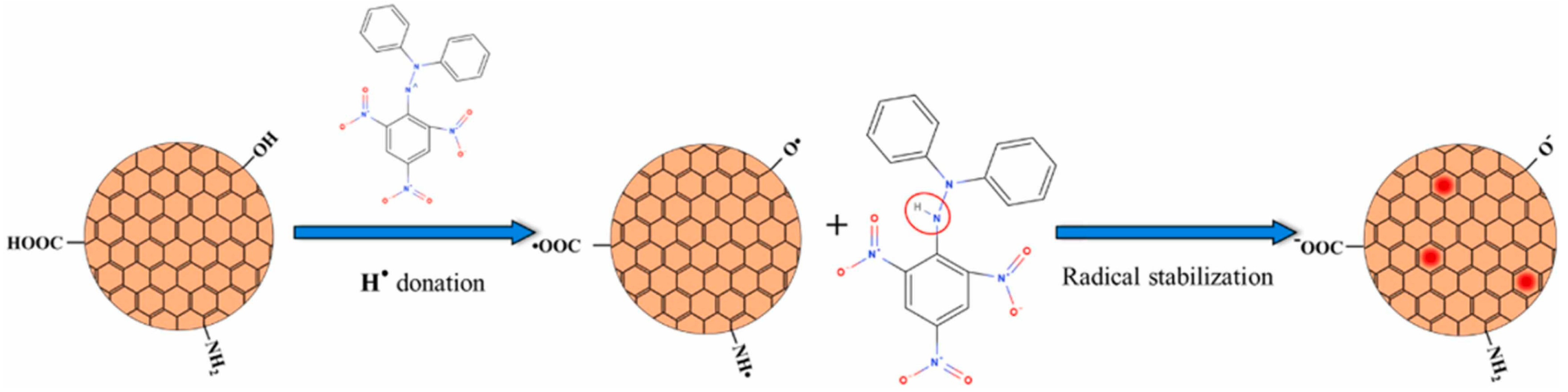
Figure 21. Mechanism of DPPH• reduction by antioxidant C-dots in aqueous media. Copyright permission from reference [27].
Mechanism of DPPH• reduction by antioxidant C-dots in aqueous media. Copyright permission from reference [2].
Recently, several studies have investigated the antioxidant properties of C-dots derived from plant extracts (Table 1). Gudimella et al. (2022) prepared fluorescent C-dots from Carica papaya leaves with significant antioxidant and anti-inflammatory properties [32][7]. The results confirmed that the synthetic C-dots were a significant alternative antioxidant agent with half the maximal effective concentration (EC50) of DPPH radical scavenging activity of the C-dots was 27.6 μg/mL [32][7]. The antioxidant capacity was quantified using a phosphomolybdate assay with an EC50 of 23 μg/mL. The oxidation–reduction reactions occurred because of the electron-donating groups on the surface of C-dots, leading to the reduction of molybdenum (VI to V). Li et al. (2021) used biomass Salvia miltiorrhiza to produce fluorescent C-dots via the hydrothermal method [33][8]. The surface of the C-dots containing S. miltiorrhiza as a polymer contributed to their high antioxidant capacity. The C-dots scavenged •OH, DPPH• and O•−2O2•−, with excellent scavenging efficiencies of 71.4, 88.9 and 95.6%, respectively [33][8].
Skin aging is a natural and gradual process influenced by both intrinsic (hormonal changes, genetics, and metabolism) and extrinsic (environmental exposures and lifestyle choices) factors [11,34,35][1][9][10]. The signs of aging include wrinkles, fine lines, uneven pigmentation, and loss of skin elasticity, which are caused by key factors such as genetics, the degradation of collagen and elastin, the loss of hyaluronic acid, oxidative stress, glycation, and inflammation (Figure 32) [34][9]. It is well known that antioxidants (e.g., vitamins C, E, A, flavonoids and phenolics, and hydroxycinnamates) can effectively inhibit these oxidation pathways by strongly stimulating old keratinocytes, eliminating ROS radicals, increasing collagen synthesis, and regulating gene expression to improve facial skin conditions [11,34][1][9]. Therefore, C-dots with antioxidant properties can effectively scavenge ROS radicals and prevent cell damage, helping maintain the health and function of cells, which is essential for the overall anti-aging effects. Moreover, C-dots are generally considered biocompatible, indicating that they can interact with biological systems without causing significant side effects. Biocompatibility is key factor for promising therapeutic applications, including those related to anti-aging. Recently, fluorescent carbon dots developed from tannic acid using the microwave-assisted pyrolysis method exhibited high biocompatibility (cell viability of 99.7 ± 0.8%) and excellent free ROS radical scavenging (82.8 ± 4.3%) [36][11]. The tannic acid-derived C-dots (10 μg/mL) significantly inhibited skin aging-related tyrosinase, elastase, and collagenase by 44.2 ± 1.3%, 52.6 ± 1.0%, and 77.6 ± 4.8%, respectively. The results suggested that tannic acid with low toxicity and superior anti-aging and antioxidant properties could be an excellent anti-aging material for cosmetic applications [36][11]. Moon et al. (2020) produced C-dots from Opuntia humifusa using a microwave-assisted method (800 W for 10 min) [10][12]. Synthetic C-dots exhibited outstanding antioxidant and anti-pollutant activities by suppressing AhR degradation, ROS production, and MMP-9 and COX-2 expressions in human keratinocytes (HaCaT cells), suggesting potential cosmeceutical properties [10][12]. Plant extract-derived C-dots with potential antioxidant properties are excellent candidates for anti-aging applications.
2. Anti-Inflammatory Activity
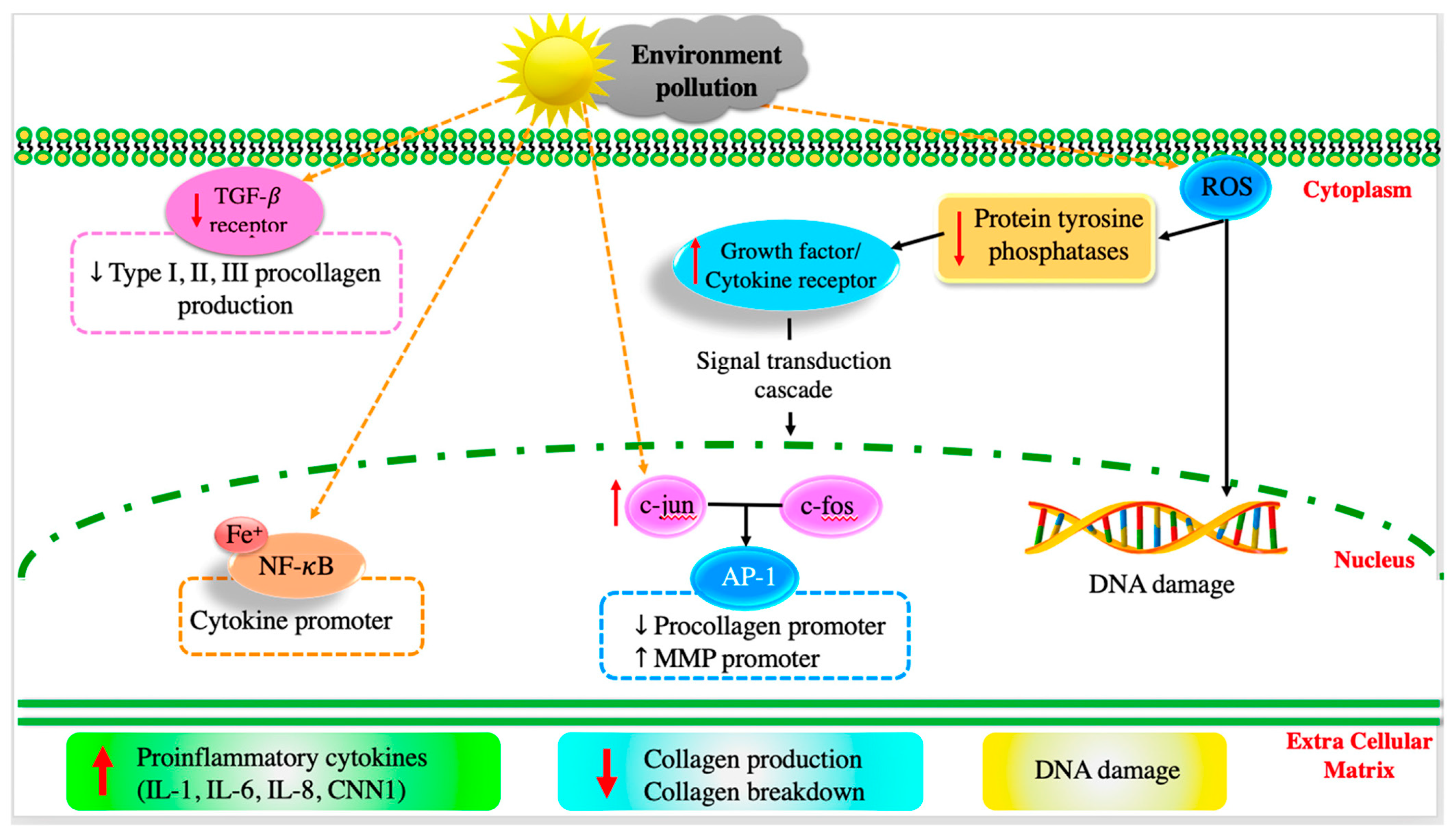
After long-term exposure to external harmful factors (UV irradiation, pollutants, and smoke), ROS radicals are generated on the skin, mediating inflammatory responses that can kill a significant number of skin cells [35][10]. Generally, ROS radicals downregulate receptor protein tyrosine phosphatases’ (RPTPs) activity, thereby enhancing the expression of phosphorylated receptor tyrosine kinases (RTKs), activating downstream signaling pathways (e.g., transforming growth factor (TGF-β), transcription factor activator protein-1 (AP-1), subsequent nuclear factor-𝜅B (NF-κB), and the activation of mitogen-activated protein kinase (MAPK)) [11,37][1][13]. Additionally, the external factors upregulate the expression of tumor necrosis factor-𝛼 (TNF-α), which is important in the pro-inflammatory process, resulting in the inhibition of collagen synthesis and stimulation of matrix metalloproteinase (MMP-9) production [11][1]. It was also found that long-term exposure to UV radiation can upregulate the expression of other cytokines, such as cysteine-rich protein (CCN1) and interleukins (IL-1, IL-6, and IL-8), which promote aging-related and skin-inflammatory processes [11,38][1][14].
The potential anti-inflammatory effects of plant extract-derived C-dots can be attributed to their ability to scavenge ROS radicals, modulate immune responses, and interact with inflammatory signaling pathways. In particular, the C-dots can effectively scavenge ROS radicals and downregulate pro-inflammatory mediators (e.g., TNF-α, IL-1α, IL-1β, IL-2, IL-6, IL-8, IL-12, and IFN-γ receptors), which are useful for improving the immune system [10,39,40][12][15][16]. Deng et al. (2023) reported the powerful anti-inflammatory activities of C-dots produced from Broccoli water extract using a hydrothermal method (180 °C and 8 h) [41][17]. The synthetic C-dots remarkably reduced the expression of IL-6 and TNF-α as the concentration of C-dots increased, compared to the control group [41][17]. C-dots showed a good ability to regulate NO production by significantly reducing NO−3NO3− in zebrafish after treatment with C-dots for 2 h [41][17]. In addition, the synthetic C-dots upregulated the expressions of superoxide dismutase (SOD) and glutathione peroxidase (GPX-4), which, in turn, scavenge excessed ROS and reduce inflammation [41][17]. Therefore, plant extract-derived C-dots can effectively control inflammation-induced skin conditions (e.g., redness, irritation, and premature aging) and even certain skin conditions (such as acne, rosacea, and eczema). Studies on the anti-inflammatory activity of C-dots are presented in Table 1.
Acne is often caused by inflammation due to an immune response triggered by excess oil and bacteria [42][18]. C-dots with anti-inflammatory properties can help alleviate the inflammation associated with acne lesions, promoting faster healing and reducing the risk of scarring [40,43][16][19]. Secondly, controlling inflammation can prevent aging signals, because chronic inflammation can contribute to premature aging by breaking down collagen and elastin fibers [11][1]. As anti-inflammatory agents, C-dots can potentially slow this process and contribute to maintaining skin elasticity and firmness [36,44][11][20]. In addition, inflammation can compromise the natural barrier function of the skin, inducing moisture loss and vulnerability to external irritants [45][21]. C-dots enhance skin barrier function, helping the skin stay hydrated and protected [40,45][16][21].
3. UV Absorption Properties
One of the critical properties of C-dots is their UV absorption behavior, which exhibits interesting optical properties that are useful for various applications, including sensing, bioimaging, optoelectronics, and cosmetics [9,46][22][23]. The UV absorption of C-dots involves electronic transitions within the nanoscale carbon-based structure, which varies depending on their size of C-dots, surface functionalization, and synthesis method [9,47][22][24]. The absorption bands of the carbon core are attributed the π–π* transition of aromatic C==C bonds or the n–π* transition of C==O/C==N bonds [47][24]. The absorption properties of C-dots are affected by the types and content of surface groups, the size of π-conjugated domains, and the oxygen/nitrogen content in carbon cores [47][24]. The exited-state intramolecular proton transfer through O–H–O and O–H–N tunnels and ample conjugated structures are responsible for the barrier property of C-dots [47][24]. In addition, due to quantum confinement, smaller-sized C-dots have a larger bandgap and energy separation between electronic energy levels, resulting in higher-energy electronic transitions that lead to UV absorption. Ezati et al. (2022) demonstrated the ultraviolet-blocking property of pectin/carbon dots fluorescent film [47][24]. The C-dot-added composite films exhibited a light UV absorption at wavelength of 230–320 nm and prevented more than 90% of UV transmission at 280 nm [47][24]. Min et al. (2023) successfully applied gelatin/poly(vinyl alcohol)-based functional films integrated with spent coffee ground-derived carbon dots and grapefruit seed extract for active packaging application based on the UV-absorption properties of C-dots [48][25]. The C-dot-added film completely blocked UV rays but sacrificed a little transparency of the film [48][25]. The results suggested that the intramolecular protein transport through O–H–N, O–H–O tunnels and appropriate junction structures resulted in the UV protection properties of film-containing C-dots [48][25]. Overall, the UV absorption properties can be harnessed for UV-blocking materials, sensors, and photovoltaic devices. For instance, C-dots with strong UV absorption can be incorporated into sunscreens or other materials to provide UV protection (Table 1).
Table 1.
Plant extract-derived C-dots with high antioxidants, anti-inflammatory properties, and UV protection for further cosmetic application.
| Plant Sources | Production Techniques | Dimensions (nm) | Cosmetic Properties | Ref. | |
|---|---|---|---|---|---|
| Beta vulgaris | Hydrothermal (160 °C for 8 h) |
3–5 | Antioxidant | DPPH radical scavenging 94.5% | [49][26] |
| Broccoli water extract | Hydrothermal (160 °C for 8 h) |
5–15 | Antioxidants | DPPH radicals scavenging 47.5% ABTS radicals scavenging 52.6% Low cytotoxicity (100% cell viability at 100 μg/mL C-dots) |
[41][17] |
| UV protection | UV absorption at 265 nm | ||||
| Carica Papaya leave | Sand bath method (160 °C for 24 h) |
2–20 | Antioxidant | DPPH free radical scavenging 85% | [32][7] |
| Anti-inflammatory | Inhibition the lysis of overloaded red blood cells (RBC) 91% | ||||
| Red Korean ginseng | Microwave-assisted method (700 W and 14 min) |
1–4 | Antioxidant | DPPH radical scavenging 85.4% H2O2 free radical scavenging 87% Low cytotoxicity (80% cell survival at 100 μLow cytotoxicity (80% cell survival at 100 �g/mL C-dots) |
[50][27] |
| Salvia miltiorrhiza | Hydrothermal (160 °C for 6 h) |
1.53–16.94 | Antioxidant | SOD scavenging 95.6% Hydroxyl radical scavenging 71.4% DPPH radical scavenging 88.9% Low cytotoxicity (90% cell survival at 2 mg/mL C-dots) |
[33][8] |
| UV protection | UV absorption peaks at 280 and 325 nm | ||||
| Gynostemma | Calcination | ~~2.49 | Antioxidant | H2O2 radical scavenging 80% | [51][28] |
| UV protection | UV absorption peak at 268 nm Long-term stability (30 d) at room temperature |
||||
| Ocimum tenuiflorum | Hydrothermal (200 °C and 20 h) |
1–3 | Antioxidant | DPPH radical scavenging 87% Low cytotoxicity (95% cell survival at 200 μLow cytotoxicity (95% cell survival at 200 �g/mL C-dots) |
[52][29] |
| UV protection | UV absorption peaks at 254 and 315 nm | ||||
| Centella asiatica | Microwave-assisted method (800 W and 10 min) |
2.97–5.26 | Antioxidant | DPPH radical scavenging 78.97% H2O2 scavenging 89.47% |
[53][30] |
| UV protection | UV absorption peak at 213 nm | ||||
| Anti-inflammatory | Inhibition of protein denaturation of 85.84% | ||||
| Annona squamosa leaves | Hydrothermal (140 °C and 10 h) | 2–3 | UV protection | UV absorption peaks at 223 and 281 nm | [54][31] |
| Anti-inflammatory | Inhibition they lysis of overloaded RBC 20–50% | ||||
| Rose-heart radish | Hydrothermal (180 °C and 3 h) |
1.2–6 | UV protection | UV absorption peak at 282 nm | [55][32] |
| Red cabbage | Hydrothermal (200 °C and 36 h) |
2–7 | Antioxidant | DPPH radical scavenging 61% •OH radical scavenging 56% |
[56][33] |
| UV protection | UV absorption peak at 365 nm | ||||
| Trapa bispinosa peel | Pyrolysis | 5–10 | UV protection | UV absorption peak at 538 nm | [57][34] |
| Solanum lycopersic | Hydrothermal (160 °C for 3 h) |
~~9 | Antioxidant | DPPH radical scavenging 63.8% | [1][35] |
| UV protection | UV absorption peak at 265 nm | ||||
| Ananas comosus | Hydrothermal (200 °C for 6 h) |
1.0–4.0 | Antioxidant | DPPH radical scavenging 50.2% H2O2 scavenging 93.4% |
[58][36] |
| Echinops persicus | Hydrothermal (200 °C for 10 h) |
4–6 | Antioxidant | DPPH radical scavenging 95% | [59][37] |
| Opuntia humifusa | Microwave-assisted method (800 W and 10 min) |
5–10 | Antioxidant | DPPH radical scavenging 94% •OH radical scavenging 81% |
[10][12] |
| Leathesia difformis | Hydrothermal (180 °C for 4 h) |
4–15 | UV protection | UV absorption peak at 365 nm Long-term photostability UV blocking 99.96% |
[60][38] |
| Dunaliella salina | Hydrothermal (200 °C for 3 h) |
3.0–3.5 | UV protection | UV absorption peak at 270 nm SPF values of 35 |
[61][39] |
References
- Ngoc, L.T.N.; Moon, J.; Lee, Y. Antioxidants for improved skin appearance: Intracellular mechanism, challenges and future strategies. Int. J. Cosmet. Sci. 2023, 45, 299–314.
- Innocenzi, P.; Stagi, L. Carbon dots as oxidant-antioxidant nanomaterials, understanding the structure-properties relationship. A critical review. Nano Today 2023, 50, 101837.
- Chen, M.; Zhou, S.; Guo, L.; Wang, L.; Yao, F.; Hu, Y.; Li, H.; Hao, J. Aggregation behavior and antioxidant properties of amphiphilic fullerene C60 derivatives cofunctionalized with cationic and nonionic hydrophilic groups. Langmuir 2019, 35, 6939–6949.
- Zu, F.; Yan, F.; Bai, Z.; Xu, J.; Wang, Y.; Huang, Y.; Zhou, X. The quenching of the fluorescence of carbon dots: A review on mechanisms and applications. Microchim. Acta 2017, 184, 1899–1914.
- Song, Y.; Zhu, S.; Xiang, S.; Zhao, X.; Zhang, J.; Zhang, H.; Fu, Y.; Yang, B. Investigation into the fluorescence quenching behaviors and applications of carbon dots. Nanoscale 2014, 6, 4676–4682.
- Chen, M.; Yin, K.; Zhang, G.; Liu, H.; Ning, B.; Dai, Y.; Wang, X.; Li, H.; Hao, J. Magnetic and Biocompatible Fullerenol/Fe(III) Microcapsules with Antioxidant Activities. ACS Appl. Bio Mater. 2020, 3, 358–368.
- kanthi Gudimella, K.; Gedda, G.; Kumar, P.S.; Babu, B.K.; Yamajala, B.; Rao, B.V.; Singh, P.P.; Kumar, D.; Sharma, A. Novel synthesis of fluorescent carbon dots from bio-based Carica Papaya Leaves: Optical and structural properties with antioxidant and anti-inflammatory activities. Environ. Res. 2022, 204, 111854.
- Li, Y.; Li, W.; Yang, X.; Kang, Y.; Zhang, H.; Liu, Y.; Lei, B. Salvia Miltiorrhiza-Derived Carbon Dots as Scavengers of Reactive Oxygen Species for Reducing Oxidative Damage of Plants. ACS Appl. Nano Mater. 2021, 4, 113–120.
- Baumann, L. How to use oral and topical cosmeceuticals to prevent and treat skin aging. Facial Plast. Surg. Clin. 2018, 26, 407–413.
- Gu, Y.; Han, J.; Jiang, C.; Zhang, Y. Biomarkers, oxidative stress and autophagy in skin aging. Ageing Res. Rev. 2020, 59, 101036.
- Son, M.H.; Park, S.W.; Jung, Y.K. Antioxidant and anti-aging carbon quantum dots using tannic acid. Nanotechnology 2021, 32, 415102.
- Moon, J.Y.; Ngoc, L.T.N.; Chae, M.; Tran, V.V.; Lee, Y.C. Effects of microwave-assisted Opuntia humifusa extract in inhibiting the impacts of particulate matter on human keratinocyte skin cell. Antioxidants 2020, 9, 271.
- Panich, U.; Sittithumcharee, G.; Rathviboon, N.; Jirawatnotai, S. Ultraviolet radiation-induced skin aging: The role of DNA damage and oxidative stress in epidermal stem cell damage mediated skin aging. Stem Cells Int. 2016, 2016, 7370642.
- Kim, M.; Park, H.J. Molecular mechanisms of skin aging and rejuvenation. In Molecular Mechanisms of the Aging Process and Rejuvenation; Intechopen Limited: London, UK, 2016; p. 450.
- Kong, B.; Yang, T.; Cheng, F.; Qian, Y.; Li, C.; Zhan, L.; Li, Y.; Zou, H.; Huang, C. Carbon dots as nanocatalytic medicine for anti-inflammation therapy. J. Colloid Interface Sci. 2022, 611, 545–553.
- Sadique, M.A.; Yadav, S.; Ranjan, P.; Khan, R.; Singh, J.; Singh, R.P. Carbon dots as a potent anti-inflammatory agent. In Carbon Dots: Next-Generation Materials for Biomedical Applications; IOP Publishing: Bristol, UK, 2022; pp. 1–2.
- Deng, W.; Zang, C.; Li, Q.; Sun, B.; Mei, X.; Bai, L.; Shang, X.; Deng, Y.; Xiao, Y.; Ghiladi, R.A. Hydrothermally derived green carbon dots from broccoli water extracts: Decreased toxicity, enhanced free-radical scavenging, and anti-inflammatory performance. ACS Biomater. Sci. Eng. 2023, 9, 1307–1319.
- Zaenglein, A.L. Acne vulgaris. N. Engl. J. Med. 2018, 379, 1343–1352.
- Chuna, Z.; Changmin, K.; Peiping, W. Clinical observation of superpulsed carbon dioxide dot matrix laser in the treatment of superficial acne scars. Front. Med. Sci. Res. 2020, 2, 10–15.
- Ye, W.; Qiu, J.; Xu, Z.; Chen, C.; Hu, G.; Hu, C.; Zhuang, J.; Li, W.; Zhang, X.; Lei, B. Oil-Soluble Ultraviolet-Absorbing Carbon Dots Dispersed in Polyethylene Films as Antiaging Materials. ACS Appl. Nano Mater. 2023, 6, 12933–12945.
- Dong, C.; Xu, M.; Wang, S.; Ma, M.; Akakuru, O.U.; Ding, H.; Wu, A.; Zha, Z.; Wang, X.; Bi, H. Fluorescent carbon dots with excellent moisture retention capability for moisturizing lipstick. J. Nanobiotechnol. 2021, 19, 299.
- Marseille, M. The Prospect Use of Carbon Dots in Skin Care. Ph.D. Thesis, University of Miami, Coral Gables, FL, USA, 2023.
- Geng, Y.; Xiang, Z.; Lv, C.; Wang, Y.; Xin, X.; Yang, Y. High efficiency gold extraction through photo-luminenscent vesicles self-aggregated by sodium dodecyl sulfate and carbon quantum dots with a visual fluorescent method for Au (III) detection. Sep. Purif. Technol. 2019, 222, 60–67.
- Ezati, P.; Rhim, J.-W. Pectin/carbon quantum dots fluorescent film with ultraviolet blocking property through light conversion. Colloids Surfaces B Biointerfaces 2022, 219, 112804.
- Min, S.; Ezati, P.; Yoon, K.S.; Rhim, J.-W. Gelatin/poly(vinyl alcohol)-based functional films integrated with spent coffee ground-derived carbon dots and grapefruit seed extract for active packaging application. Int. J. Biol. Macromol. 2023, 231, 123493.
- Smrithi, S.P.; Kottam, N.; Muktha, H.; Mahule, A.M.; Chamarti, K.; Vismaya, V.; Sharath, R. Carbon dots derived from Beta vulgaris: Evaluation of its potential as antioxidant and anticancer agent. Nanotechnology 2021, 33, 45403.
- Tejwan, N.; Kundu, M.; Ghosh, N.; Chatterjee, S.; Sharma, A.; Abhishek Singh, T.; Das, J.; Sil, P.C. Synthesis of green carbon dots as bioimaging agent and drug delivery system for enhanced antioxidant and antibacterial efficacy. Inorg. Chem. Commun. 2022, 139, 109317.
- Wei, X.; Li, L.; Liu, J.; Yu, L.; Li, H.; Cheng, F.; Yi, X.; He, J.; Li, B. Green Synthesis of Fluorescent Carbon Dots from Gynostemma for Bioimaging and Antioxidant in Zebrafish. ACS Appl. Mater. Interfaces 2019, 11, 9832–9840.
- Shukla, D.; Pandey, F.P.; Kumari, P.; Basu, N.; Tiwari, M.K.; Lahiri, J.; Kharwar, R.N.; Parmar, A.S. Label-Free Fluorometric Detection of Adulterant Malachite Green Using Carbon Dots Derived from the Medicinal Plant Source Ocimum tenuiflorum. ChemistrySelect 2019, 4, 4839–4847.
- Thokchom, B.; Bhavi, S.M.; Abbigeri, M.B.; Shettar, A.K.; Yarajarla, R.B. Green synthesis, characterization and biomedical applications of Centella asiatica-derived carbon dots. Carbon Lett. 2023, 33, 1057–1071.
- Vibhute, A.; Patil, T.; Malavekar, D.; Patil, S.; Lee, S.; Tiwari, A.P. Green synthesis of fluorescent carbon dots from annona squamosa leaves: Optical and structural properties with bactericidal, anti-inflammatory, anti-angiogenesis applications. J. Fluoresc. 2023, 33, 1619–1629.
- Liu, W.; Diao, H.; Chang, H.; Wang, H.; Li, T.; Wei, W. Green synthesis of carbon dots from rose-heart radish and application for Fe3+ detection and cell imaging. Sens. Actuators B Chem. 2017, 241, 190–198.
- Sharma, N.; Das, G.S.; Yun, K. Green synthesis of multipurpose carbon quantum dots from red cabbage and estimation of their antioxidant potential and bio-labeling activity. Appl. Microbiol. Biotechnol. 2020, 104, 7187–7200.
- Mewada, A.; Pandey, S.; Shinde, S.; Mishra, N.; Oza, G.; Thakur, M.; Sharon, M.; Sharon, M. Green synthesis of biocompatible carbon dots using aqueous extract of Trapa bispinosa peel. Mater. Sci. Eng. C 2013, 33, 2914–2917.
- Rodríguez-Varillas, S.; Fontanil, T.; Obaya, Á.J.; Fernández-González, A.; Murru, C.; Badía-Laíño, R. Biocompatibility and antioxidant capabilities of carbon dots obtained from tomato (Solanum lycopersicum). Appl. Sci. 2022, 12, 773.
- Rajamanikandan, S.; Biruntha, M.; Ramalingam, G. Blue emissive carbon quantum dots (CQDs) from bio-waste peels and its antioxidant activity. J. Clust. Sci. 2022, 33, 1045–1053.
- Nasseri, M.A.; Keshtkar, H.; Kazemnejadi, M.; Allahresani, A. Phytochemical properties and antioxidant activity of Echinops persicus plant extract: Green synthesis of carbon quantum dots from the plant extract. SN Appl. Sci. 2020, 2, 670.
- Kim, K.W.; Kwon, Y.M.; Kim, S.Y.; Kim, J.Y.H. One-pot synthesis of UV-protective carbon nanodots from sea cauliflower (Leathesia difformis). Electron. J. Biotechnol. 2022, 56, 22–30.
- Chatzimitakos, T.G.; Kasouni, A.; Troganis, A.; Leonardos, I.; Tzovenis, I.; Ntzouvaras, A.; Stalikas, C. Carbon nanodots synthesized from Dunaliella Salina as sun protection filters. C 2020, 6, 69.
More