PD-1/PD-L1 Inhibitors in Ovarian Cancer: Comparison
Please note this is a comparison between Version 1 by Sanda Maria Cretoiu and Version 2 by Catherine Yang.

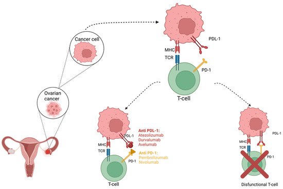
Ovarian cancer is considered one of the most aggressive and deadliest gynecological malignancies worldwide. Unfortunately, the therapeutic methods that are considered the gold standard at this moment are associated with frequent recurrences. Survival in ovarian cancer is associated with the presence of a high number of intra tumor infiltrating lymphocytes (TILs). Therefore, immunomodulation is considered to have an important role in cancer treatment, and immune checkpoint inhibitors may be useful for restoring T cell-mediated antitumor immunity. PD-1 is a 50–55 kDa, 288 aa transmembrane glycoprotein that lacks the membrane-proximal cysteine residue required for homodimerization that is present in the structure of the other members of the CD28 family, thus rendering PD-1 monomeric, both in solution and on cell surface, as structural and biochemical analyses show [26]. The cytoplasmic domain presents two tyrosine residues, with the membrane-proximal one constituting an immunoreceptor tyrosine-based inhibitory motif (ITIM) and the second one constituting an immunoreceptor tyrosine-based switch motif (ITSM).

  • PD-1/PD-L1
  • ovarian cancer
  • gynecological malignancy
  • miRNAs
  • lncRNAs
1. PD-L1 Targeted Immunotherapy in Cancers
Currently, there is an increased number of studies regarding the use of PD-1/PD-L1 pathway inhibitors. Malignant epithelial tumors are the most common neoplasm of the ovary, accounting for 90% of all cases of ovarian cancers [1]. Several molecular studies have been carried out, in order to determine the pathogenesis of these malignant lesions, thus revealing several aspects that subdivided the tumors into two subtypes. Type I tumors are derived from benign, extra-ovarian precursors [2]. The group includes clear-cell and endometrioid carcinomas, both of which are associated with underlining endometriosis. Mucinous carcinoma, low-grade serous ovarian carcinoma, and Brenner tumors are also included in these groups, although they are less common [3]. They tend to present as a unilateral cystic mass. Type II malignant neoplasm develops from intraepithelial lesions in the fallopian tube. High-grade ovarian carcinoma, carcinosarcoma, and undifferentiated carcinoma are considered type II lesions and characterized by a highly aggressive clinical behavior and poor prognosis. The patients are frequently discovered with advanced disease at the moment of diagnosis [4].
The expression of PD-1 on the surface of many cell types, and especially on tumors, makes it possible, through the interaction between PD-1 and PD-L1, though difficult, to get rid of cancer cells by the mechanisms of the immune system, which leads to the emergence of therapeutic resistance [5][6][7][8]. Moreover, there was an increased therapeutic resistance in certain histological types that were initially associated with a poor prognosis, but in which case immunotherapy brought an optimistic perspective [9]. Immune checkpoint inhibitors were successfully used as a battleline treatment in melanomas, non-small cell lung cancer, renal or urinary tract cancers, and breast, head, and neck cancers; they were recently introduced as an option in malignant hematological proliferations [10]. The pioneer therapy in checkpoint immune inhibitors is represented by ipilimumab, a monoclonal antibody that blocks the CTLA-4 molecule [11][12]. Ever since, novel molecules have been developed that obstruct the perpetuation of other inflammatory cascades, such as the ones provoked by the PD-1 and PDL-1 complexes. For example, nivolumab and pembrolizumab are anti-PD-1 antibodies that presented a successful rate in treating patients who were suffering from metastatic melanoma, as well as cemiplimab, from the same drug class, and they were utilized in cutaneous squamous cell carcinoma (Figure 13) [11][12][13][14]. Other examples of checkpoint inhibitors that interfere with PDL-1 incorporation are durvalumab, atezolizumab, and avelumab, which support the usual treatment protocol, especially in urothelial or non-small lung cancers [7][10][14].
Figure 13. Antibody blockade of PD-1 and PD-L1. Anti-PD-1 and PD-L1 antibodies block the PD-1/PD-L1 signalling and enhance antitumor immune activity. Created with BioRender.com (Last accessed on 26 September 2022).

2. Efficacy of PD-1/PD-L1 Inhibitors in Ovarian Cancer

Despite numerous therapeutic protocols, ovarian cancer remains a major cause of neoplastic mortality [15][16]. Therefore, there are now several trials conducted that introduce the latest class of checkpoint immune inhibitors that target PD-1/PDL-1 complexes into the therapeutic protocol of FIGO stage III or IV or relapsed ovarian cancer. Understanding the programmed cell death pathway associated with the PD-1 receptor allowed for the design of clinical trials for treating ovarian cancer using antibodies against the PD-1 receptors (nivolumab and pembrolizumab) and PD-L1 ligands (avelumab, BMS-936559, durvalumab, and atezolizumab).
The efficacy of PD-1/PD-L1 inhibitors, as promising immunotherapeutic agents, was analyzed by Chen et al. in a meta-analysis based on 91 published clinical trials, phases I to III, in different cancer types, including gynecological cancers. This meta-analysis revealed that PD-1/PD-L1 inhibitors, combined with chemotherapy, had a statistically significantly higher objective response rate, compared with immunotherapy alone, and the duration of response was significantly reduced in the case of the same combination [17].
There are many factors that determine how a tumor will respond to this type of treatment. For example, pembrolizumab, not approved by FDA for ovarian cancer, showed a modest result for immunotherapy in stage II clinical trials [15][18]. This can be explained by the fact that efficiency is dependent on the presence of microsatellite instability [19][20]. In certain circumstances, pembrolizumab could be used for patients with MSI-H (microsatellite instability-high), dMMR (mismatch repair-deficient), solid tumors, and no satisfactory alternative treatment options. This was indicated in a study by Le et al., which was performed to evaluate the efficacy of the PD-1 blockade across 12 different tumor types, with MSI-H and dMMR, which were found to be sensitive to immune checkpoint blockade [21]. The incidence of dMMR in ovarian cancer is reported to be between 2–29%, with the higher frequencies being associated with younger ages of diagnosis and the presence of an increased tumoral lymphocytic infiltrate [22].
Another predictive biomarker of the efficiency of anti-PDL-1 antibodies is the overexpression of PDL-1 on the cell membranes of the malignant cells, as suggested in a study published by Chin et al. [7][8][10]. They showed that FIGO stage III and IV ovarian tumors, with a raised immune reactivity molecular subtype, expressed a high quantity of PDL-1 on the surface of tumoral cells. Understanding the fact that not all patients who undergo anti-PDL-1 checkpoint inhibitor and present an upregulation of PDL-1 on tumor cells and infiltrated inflammatory cells have a good response to therapy, they raised the dilemma of the necessity of other molecular biomarkers for an improved description of the molecular pattern of the high-grade ovarian tumors [15]. Moreover, it has been displayed in several preclinical trials that the favorable outcome from checkpoint inhibitors therapies is strongly connected to the perfect timing of initiating the treatment. It is considered that the ideal moment to administer the therapy is when the tumoral contingent reaches the maximum level of intraepithelial tumor-infiltrating lymphocytes (TILs) [19]. It has been demonstrated that PDL-1 overexpression itself downregulates the T cytotoxic CD8+ lymphocytes action and, therefore, decreases the protection of the host immune system against tumor overgrowth [23].
However, as remarkable as the response after checkpoint immune inhibitors treatment in melanoma may be, the trials results for urothelial and non-small lung cancers in ovarian tumors show a modest improvement of the median survival rate of 10–15%, until nowadays, with disease control observed in approximately 50% of patients. [23][24][25][26][27][28]. One possibility for enhancing the response to anti-PDL-1 antibodies in these types of cancers is to associate them with radiotherapy, which is known to boost the effect of anti-PDL-1 checkpoint inhibitors in other malignancies [5]. The JAVELINE 200 trial, a phase III clinical study, included 566 patients who were platinum resistant or had refractory ovarian cancer. The study compared avelumab monotherapy with the avelumab-pegylated liposomal doxorubicin combination. A therapeutic advantage was observed in patients who were treated with the combination and had PDL-1 expression on their tumoral samples [23][29]. The study presented, as main objectives, the evaluation of progression-free and overall survival; as secondary objectives, the study presents the objective response and its duration, as well as aspects related to the tolerance of the therapy and pharmacokinetics of the drug. At the moment, several clinical trials have been conducted on small-scale populations of patients for ovarian cancer, and they used the following molecules in their research: ipilimumab, tremelimumab, nivolumab, pembrolizumab, and other anti-PDL-1 antibodies, such as BMS-936559, MEDI4736, MPDL33280A, and MSB0010718C (avelumab) [30].
The first study aimed at the efficacy of PD-1 inhibitors was published by Hamanishi et al. and aimed at evaluating the role of nivolumab (a fully humanized monoclonal antibody against the PD-1 receptor) in ovarian cancer that is resistant to treatment with platinum agents, as other previously suggested [31][32][33]. The same team demonstrated the association between the high level of PD-L1 and reduced survival rate, as well as the low number of CD(+) cytotoxic lymphocytes [34].
The findings published by Webb et al., which found PD-L1 expression mainly in CD68+ macrophages, approached the topic in a different light [35]. They quantified the expression of PDL-1 in different subtypes of ovarian cancers, as well as the relationship between the expression of PDL-1 in tumor cells and the marker in immune cells associated with tumor proliferation. The authors also verified the prognostic significance of this expression, placing a special emphasis on PDL-1 expression in high-grade ovarian serous carcinomas. Their study revealed a positive relationship between PD-L1 expression and the presence of tumor-infiltrating regulatory T cells and/or lymphocytes with the CD8+ CD103+ PD-1+ phenotype. The level of PD-L1 expression in the different histopathological subtypes of ovarian cancer varied greatly with highest expression of PD-L1 in serous ovarian carcinoma (57.4%), followed by mucinous ovarian carcinoma (26.7%) and endometrioid ovarian carcinoma (24%) [35]. Clear-cell ovarian carcinoma was associated with the lowest level of PD-L1 (16.2%) [35]. The authors also demonstrated a positive correlation between PD-L1 expression and the overall survival of patients with high-grade ovarian serous carcinoma (HGSC), thus endorsing the concept of adaptive resistance of tumor cells [35]. At first glance, the studies performed by Hamanishi and Webb seem to be contradictory, and the major differences in PDL-1 expression reported by the authors seem to be related to the clones used in the immunohistochemical studies, as suggested by Webb et al. For this reason, the evaluation of PDL-1 expression in tumor cells from ovarian carcinomas (regardless of type), as well as from immune cells associated with tumors, seems to require an appropriate protocol and reporting system, similar to that used for lung carcinomas, for better traceability of results. In this sense, in addition to the classic detection of PDL-1 expression through immunohistochemical tests, alternative, innovative approaches are probably needed that could detect the presence of PDL-1 in much smaller amounts of tissue or biological fluids. Such a method was validated by Grel et al. [36]. Some studies suggest a positive correlation between the TILs and survival rate [37]. Others have underlined a relationship between high PD-L1 expression in the peritoneal fluid of these patients, as well as the formation of metastases within the peritoneal cavity, knowing that malignant serous ovarian tumors have a special predisposition for the invasion of the peritoneum [38].
Pembrolizumab is a humanized anti-PD-1 monoclonal antibody that blocks the PD-1 on the cell surface, thus preventing the PD-1/PD-L1 interaction. A phase Ib study developed by Varga et al. evaluated the role of pembrolizumab in 26 patients with advanced ovarian cancer, fallopian tube cancer, and peritoneal invasion who did not respond to first-line oncological treatment. [39]. The results of the treatment seem to be modest, since the overall response rate was only 11.5% and disease control rate was 34.6%. Complete response to treatment was obtained in only one patient, partial response occurred in two patients, and disease stabilization was confirmed in six patients [39][40][41]. A similar study using pembrolizumab as a single therapeutic agent for patients with recurrent ovarian carcinoma demonstrated a similar, modest therapeutic response [18].
In addition to pembrolizumab, other therapeutic agents have been studied, in order to diversify the immunotherapy-type therapeutic options for ovarian cancer patients. Avelumab is a fully humanized anti-PD-L1 IgG1 monoclonal antibody that inhibits the PD-L1 interaction with the PD-1 receptor [41]. The study conducted with avelumab is one of the largest studies to date involving the programmed death pathway [42]. The overall objective responses, in a group of 124 women with recurrent or refractory ovarian cancer, was 9.7%, and the disease control rate was noted in about half of the patients. PD-1 expression was assessed in 74 cases, and 57 women (77%) showed increased PD-L1 expression. However, it seems that the results of therapy with avelumab are quite modest and comparable to those with pembrolizumab; the objective response rate was 12.3%, while, in the group of women without excessive PD-L1 expression, it was 5.9% [42]. Contrary to expectations that anti-PD-L1/PD-1 therapies could be effective in ovarian cancers, clinical trials showed that their performance remains restricted—first of all, because the expression of PDL-1 is modest in ovarian carcinomas [43].
Another monoclonal anti-PD-L1 antibody, BMS-936559, was studied. This study included 17 patients with ovarian cancer, among which, 5.9% responded partially to treatment, and 17.6% achieved disease stabilization. All patients received doses of 10 mg/kg. There are also studies on the effectiveness of durvalumab—a monoclonal antibody directed against the PD-L1 protein [41].
Despite the promising results in the treatment of solid tumors, the anti PD-1/anti-PD-L1 therapies are characterized by an increased number of adverse effects, especially immune-mediated side effects. The increased activation of the immune system leads to systemic effects in a high number of cases, with the most frequent being represented by colitis, myocarditis, encephalitis, pneumonitis, hepatitis, etc. Fatal events have also been reported, but with a decreased incidence (0.3–1.3%). The frequency and the number of associated symptoms increase in the case of an association with two immune checkpoint inhibitors [44].

References

  1. Breast Cancer—Statistics. Available online: https://www.cancer.net/cancer-types/breast-cancer/statistics (accessed on 14 May 2021).
  2. Lim, D.; Oliva, E. Precursors and pathogenesis of ovarian carcinoma. Pathology 2013, 45, 229–242.
  3. Kurman, R.J.; Shih Ie, M. The Dualistic Model of Ovarian Carcinogenesis: Revisited, Revised, and Expanded. Am. J. Pathol. 2016, 186, 733–747.
  4. Kurman, R.J.; Shih Ie, M. Pathogenesis of ovarian cancer: Lessons from morphology and molecular biology and their clinical implications. Int. J. Gynecol. Pathol. 2008, 27, 151–160.
  5. Sharma, P.; Allison, J.P. The future of immune checkpoint therapy. Science 2015, 348, 56–61.
  6. Okazaki, T.; Honjo, T. PD-1 and PD-1 ligands: From discovery to clinical application. Int. Immunol. 2007, 19, 813–824.
  7. Nhokaew, W.; Kleebkaow, P.; Chaisuriya, N.; Kietpeerakool, C. Programmed Death Ligand 1 (PD-L1) Expression in Epithelial Ovarian Cancer: A Comparison of Type I and Type II Tumors. Asian Pac. J. Cancer Prev. 2019, 20, 1161–1169.
  8. Wilczynski, N.L.; Morgan, D.; Haynes, R.B.; Hedges, T. An overview of the design and methods for retrieving high-quality studies for clinical care. BMC Med. Inform. Decis. Mak. 2005, 5, 20.
  9. Pedoeem, A.; Azoulay-Alfaguter, I.; Strazza, M.; Silverman, G.J.; Mor, A. Programmed death-1 pathway in cancer and autoimmunity. Clin. Immunol. 2014, 153, 145–152.
  10. Alsaab, H.O.; Sau, S.; Alzhrani, R.; Tatiparti, K.; Bhise, K.; Kashaw, S.K.; Iyer, A.K. PD-1 and PD-L1 Checkpoint Signaling Inhibition for Cancer Immunotherapy: Mechanism, Combinations, and Clinical Outcome. Front. Pharmacol. 2017, 8, 561.
  11. Michot, J.M.; Bigenwald, C.; Champiat, S.; Collins, M.; Carbonnel, F.; Postel-Vinay, S.; Berdelou, A.; Varga, A.; Bahleda, R.; Hollebecque, A.; et al. Immune-related adverse events with immune checkpoint blockade: A comprehensive review. Eur. J. Cancer 2016, 54, 139–148.
  12. Hodi, F.S.; O’Day, S.J.; McDermott, D.F.; Weber, R.W.; Sosman, J.A.; Haanen, J.B.; Gonzalez, R.; Robert, C.; Schadendorf, D.; Hassel, J.C.; et al. Improved survival with ipilimumab in patients with metastatic melanoma. N. Engl. J. Med. 2010, 363, 711–723.
  13. Pardoll, D.M. The blockade of immune checkpoints in cancer immunotherapy. Nat. Rev. Cancer 2012, 12, 252–264.
  14. Lee, H.T.; Lee, S.H.; Heo, Y.S. Molecular Interactions of Antibody Drugs Targeting PD-1, PD-L1, and CTLA-4 in Immuno-Oncology. Molecules 2019, 24, 1190.
  15. Chin, C.D.; Fares, C.M.; Campos, M.; Chen, H.W.; Shintaku, I.P.; Konecny, G.E.; Rao, J. Association of PD-L1 expression by immunohistochemistry and gene microarray with molecular subtypes of ovarian tumors. Mod. Pathol. 2020, 33, 2001–2010.
  16. Cancer Statistics Review, 1975-2015—Previous Version—SEER Cancer Statistics Review. Available online: https://seer.cancer.gov/archive/csr/1975_2015/index.html (accessed on 16 May 2021).
  17. Chen, S.; Zhang, Z.; Zheng, X.; Tao, H.; Zhang, S.; Ma, J.; Liu, Z.; Wang, J.; Qian, Y.; Cui, P.; et al. Response Efficacy of PD-1 and PD-L1 Inhibitors in Clinical Trials: A Systematic Review and Meta-Analysis. Front. Oncol. 2021, 11, 562315.
  18. Matulonis, U.A.; Shapira-Frommer, R.; Santin, A.D.; Lisyanskaya, A.S.; Pignata, S.; Vergote, I.; Raspagliesi, F.; Sonke, G.S.; Birrer, M.; Provencher, D.M.; et al. Antitumor activity and safety of pembrolizumab in patients with advanced recurrent ovarian cancer: Results from the phase II KEYNOTE-100 study. Ann. Oncol. 2019, 30, 1080–1087.
  19. Doo, D.W.; Norian, L.A.; Arend, R.C. Checkpoint inhibitors in ovarian cancer: A review of preclinical data. Gynecol. Oncol. Rep. 2019, 29, 48–54.
  20. Hinchcliff, E.; Hong, D.; Le, H.; Chisholm, G.; Iyer, R.; Naing, A.; Hwu, P.; Jazaeri, A. Characteristics and outcomes of patients with recurrent ovarian cancer undergoing early phase immune checkpoint inhibitor clinical trials. Gynecol. Oncol. 2018, 151, 407–413.
  21. Le, D.T.; Durham, J.N.; Smith, K.N.; Wang, H.; Bartlett, B.R.; Aulakh, L.K.; Lu, S.; Kemberling, H.; Wilt, C.; Luber, B.S.; et al. Mismatch repair deficiency predicts response of solid tumors to PD-1 blockade. Science 2017, 357, 409–413.
  22. Leskela, S.; Romero, I.; Cristobal, E.; Perez-Mies, B.; Rosa-Rosa, J.M.; Gutierrez-Pecharroman, A.; Caniego-Casas, T.; Santon, A.; Ojeda, B.; Lopez-Reig, R.; et al. Mismatch Repair Deficiency in Ovarian Carcinoma: Frequency, Causes, and Consequences. Am. J. Surg. Pathol. 2020, 44, 649–656.
  23. Gonzalez-Martin, A.; Sanchez-Lorenzo, L. Immunotherapy with checkpoint inhibitors in patients with ovarian cancer: Still promising? Cancer 2019, 125 (Suppl. 24), 4616–4622.
  24. Borella, F.; Ghisoni, E.; Giannone, G.; Cosma, S.; Benedetto, C.; Valabrega, G.; Katsaros, D. Immune Checkpoint Inhibitors in Epithelial Ovarian Cancer: An Overview on Efficacy and Future Perspectives. Diagnostics 2020, 10, 146.
  25. Sato, E.; Olson, S.H.; Ahn, J.; Bundy, B.; Nishikawa, H.; Qian, F.; Jungbluth, A.A.; Frosina, D.; Gnjatic, S.; Ambrosone, C.; et al. Intraepithelial CD8+ tumor-infiltrating lymphocytes and a high CD8+/regulatory T cell ratio are associated with favorable prognosis in ovarian cancer. Proc. Natl. Acad. Sci. USA 2005, 102, 18538–18543.
  26. Zhang, L.; Conejo-Garcia, J.R.; Katsaros, D.; Gimotty, P.A.; Massobrio, M.; Regnani, G.; Makrigiannakis, A.; Gray, H.; Schlienger, K.; Liebman, M.N.; et al. Intratumoral T cells, recurrence, and survival in epithelial ovarian cancer. N. Engl. J. Med. 2003, 348, 203–213.
  27. Hwang, W.T.; Adams, S.F.; Tahirovic, E.; Hagemann, I.S.; Coukos, G. Prognostic significance of tumor-infiltrating T cells in ovarian cancer: A meta-analysis. Gynecol. Oncol. 2012, 124, 192–198.
  28. Ghisoni, E.; Imbimbo, M.; Zimmermann, S.; Valabrega, G. Ovarian Cancer Immunotherapy: Turning up the Heat. Int. J. Mol. Sci. 2019, 20, 2927.
  29. A Study Of Avelumab Alone Or In Combination With Pegylated Liposomal Doxorubicin Versus Pegylated Liposomal Doxorubicin Alone In Patients with Platinum Resistant/Refractory Ovarian Cancer (JAVELIN Ovarian 200). Available online: https://clinicaltrials.gov/ct2/show/NCT02580058 (accessed on 26 September 2021).
  30. De Felice, F.; Marchetti, C.; Palaia, I.; Musio, D.; Muzii, L.; Tombolini, V.; Panici, P.B. Immunotherapy of Ovarian Cancer: The Role of Checkpoint Inhibitors. J. Immunol. Res. 2015, 2015, 191832.
  31. Hamanishi, J.; Mandai, M.; Ikeda, T.; Minami, M.; Kawaguchi, A.; Murayama, T.; Kanai, M.; Mori, Y.; Matsumoto, S.; Chikuma, S.; et al. Safety and Antitumor Activity of Anti-PD-1 Antibody, Nivolumab, in Patients With Platinum-Resistant Ovarian Cancer. J. Clin. Oncol. 2015, 33, 4015–4022.
  32. Topalian, S.L.; Hodi, F.S.; Brahmer, J.R.; Gettinger, S.N.; Smith, D.C.; McDermott, D.F.; Powderly, J.D.; Carvajal, R.D.; Sosman, J.A.; Atkins, M.B.; et al. Safety, activity, and immune correlates of anti-PD-1 antibody in cancer. N. Engl. J. Med. 2012, 366, 2443–2454.
  33. Crotzer, D.R.; Sun, C.C.; Coleman, R.L.; Wolf, J.K.; Levenback, C.F.; Gershenson, D.M. Lack of effective systemic therapy for recurrent clear cell carcinoma of the ovary. Gynecol. Oncol. 2007, 105, 404–408.
  34. Pawlowska, A.; Suszczyk, D.; Okla, K.; Barczynski, B.; Kotarski, J.; Wertel, I. Immunotherapies based on PD-1/PD-L1 pathway inhibitors in ovarian cancer treatment. Clin. Exp. Immunol. 2019, 195, 334–344.
  35. Webb, J.R.; Milne, K.; Kroeger, D.R.; Nelson, B.H. PD-L1 expression is associated with tumor-infiltrating T cells and favorable prognosis in high-grade serous ovarian cancer. Gynecol. Oncol. 2016, 141, 293–302.
  36. Grel, H.; Ratajczak, K.; Jakiela, S.; Stobiecka, M. Gated Resonance Energy Transfer (gRET) Controlled by Programmed Death Protein Ligand 1. Nanomaterials 2020, 10, 1592.
  37. Santoiemma, P.P.; Powell, D.J., Jr. Tumor infiltrating lymphocytes in ovarian cancer. Cancer Biol. Ther. 2015, 16, 807–820.
  38. Abiko, K.; Mandai, M.; Hamanishi, J.; Yoshioka, Y.; Matsumura, N.; Baba, T.; Yamaguchi, K.; Murakami, R.; Yamamoto, A.; Kharma, B.; et al. PD-L1 on tumor cells is induced in ascites and promotes peritoneal dissemination of ovarian cancer through CTL dysfunction. Clin. Cancer Res. 2013, 19, 1363–1374.
  39. Varga, A.P.-P.; Piha-Paul, S.A.; Ott, P.A.; Mehnert, J.M.; Berton-Rigaud, D.; Johnson, E.A.; Cheng, J.D.; Yuan, S.; Rubin, E.H.; Matei, D.E. Antitumor Activity and Safety of Pembrolizumab in Patients (Pts) with PD-L1 Positive Advanced Ovarian Cancer: Interim Results from a Phase Ib Study. J. Clin. Oncol. 2015, 33, 5510.
  40. Gadducci, A.; Guerrieri, M.E. Immune Checkpoint Inhibitors in Gynecological Cancers: Update of Literature and Perspectives of Clinical Research. Anticancer Res. 2017, 37, 5955–5965.
  41. Bose, C.K. Immune Checkpoint Blockers and Ovarian Cancer. Indian J. Med. Paediatr. Oncol. 2017, 38, 182–189.
  42. Disis, M.L.P.; Patel, M.R.; Pant, S.; Infante, J.R.; Lockhart, A.C.; Kelly, K.; Beck, J.T.; Gordon, M.S.; Weiss, G.J.; Ejadi, S.; et al. Avelumab (MSB0010718C), an Anti-PD-L1 Antibody, in Patients with Previously Treated, Recurrent or Refractory Ovarian Cancer: A Phase Ib, Open-Label Expansion Trial. J. Clin. Oncol. 2015, 33, 5509.
  43. Monk, B.J.; Colombo, N.; Oza, A.M.; Fujiwara, K.; Birrer, M.J.; Randall, L.; Poddubskaya, E.V.; Scambia, G.; Shparyk, Y.V.; Lim, M.C.; et al. Chemotherapy with or without avelumab followed by avelumab maintenance versus chemotherapy alone in patients with previously untreated epithelial ovarian cancer (JAVELIN Ovarian 100): An open-label, randomised, phase 3 trial. Lancet Oncol. 2021, 22, 1275–1289.
  44. Martins, F.; Sofiya, L.; Sykiotis, G.P.; Lamine, F.; Maillard, M.; Fraga, M.; Shabafrouz, K.; Ribi, C.; Cairoli, A.; Guex-Crosier, Y.; et al. Adverse effects of immune-checkpoint inhibitors: Epidemiology, management and surveillance. Nat. Rev. Clin. Oncol. 2019, 16, 563–580.
More
Video Production Service