Biosynthetic Pathways of Hormones in Plants: Comparison
Please note this is a comparison between Version 1 by Andrzej Bajguz and Version 2 by Sirius Huang.

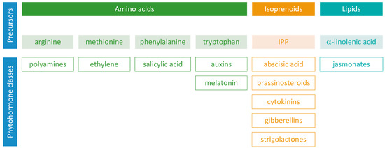
Phytohormones exhibit a wide range of chemical structures, though they primarily originate from three key metabolic precursors: amino acids, isoprenoids, and lipids. Specific amino acids, such as tryptophan, methionine, phenylalanine, and arginine, contribute to the production of various phytohormones, including auxins, melatonin, ethylene, salicylic acid, and polyamines. Isoprenoids are the foundation of five phytohormone categories: cytokinins, brassinosteroids, gibberellins, abscisic acid, and strigolactones. Furthermore, lipids, i.e., α-linolenic acid, function as a precursor for jasmonic acid. The biosynthesis routes of these different plant hormones are intricately complex. Understanding of these processes can greatly enhance our knowledge of how these hormones regulate plant growth, development, and physiology. 

  • biosynthesis
  • phytohomones

1. Introduction

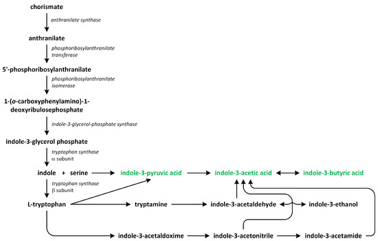
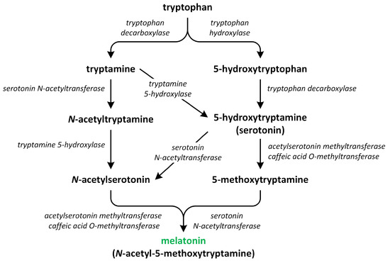
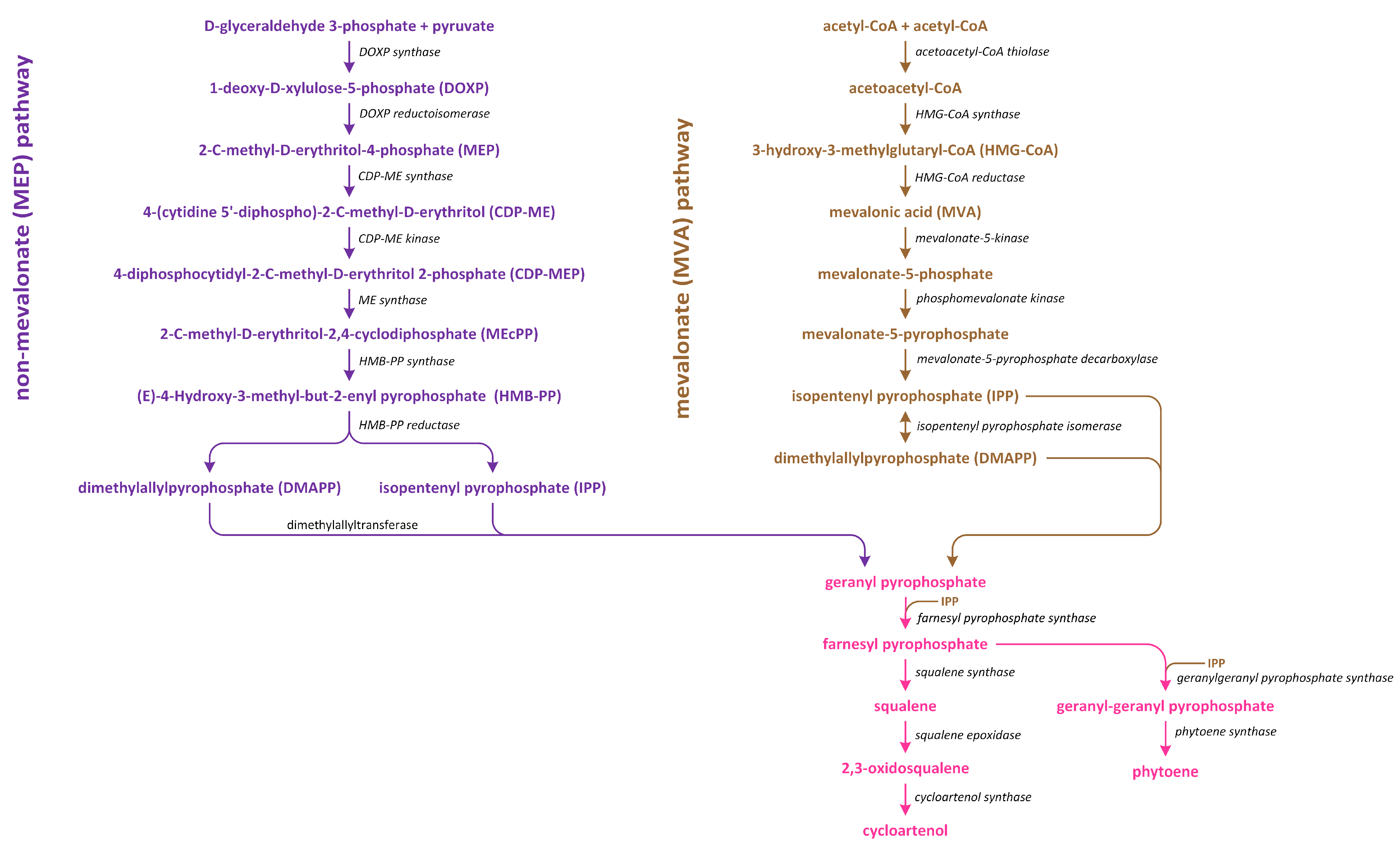
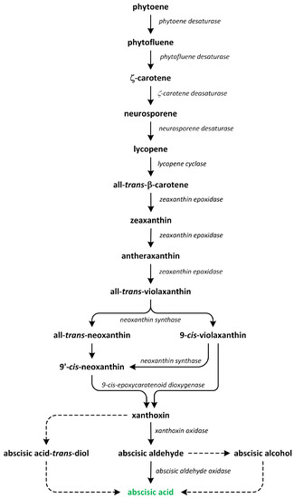
Phytohormones, which are also known as plant hormones, are small, naturally occurring organic compounds that significantly influence the growth, development, defense, productivity, and physiological mechanisms of plants. They also orchestrate various cellular activities within the plant. Even at minimal concentrations, they are operative in plant cells, tissues, and organs. They are found in all vascular plants and a substantial number of non-vascular species (Table 1). From the initial discovery of auxin to the most recent unearthing of strigolactones (SLs), 12 groups of phytohormones—auxins, cytokinins (CKs), gibberellins (GAs), abscisic acid (ABA), ethylene, brassinosteroids (BRs), salicylic acid (SA), jasmonates, polyamines (PAs), melatonin, SLs, and peptide hormones—have been identified in numerous plant species. The various chemical structures of phytohormones are pivotal for their diverse biological functions and biosynthesis (Figure 1):
Figure 1.
Precursors of phytohormone biosynthesis.
  • auxins and melatonin are indole derivatives;
  • ABA is a sesquiterpene;
  • ethylene is the simplest alkene;
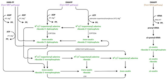
  • CKs are adenine analogues;
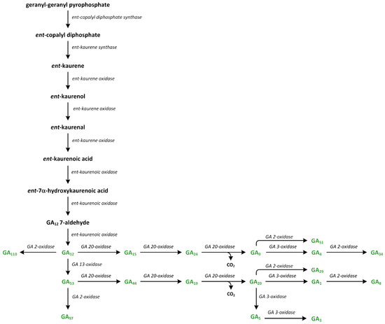
  • GAs are tetracyclic diterpenoid acids;
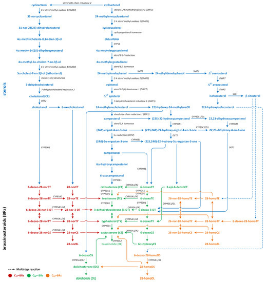
  • BRs are polyhydroxysteroids;
  • jasmonates are derived from fatty acids;
Table 1. The localization of hormones in plants [7][8].
The localization of hormones in plants [7,8].
  • PAs are aliphatic nitrogenous bases;
  • SA is a phenolic organic acid;
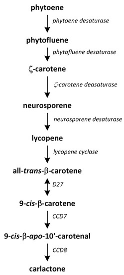
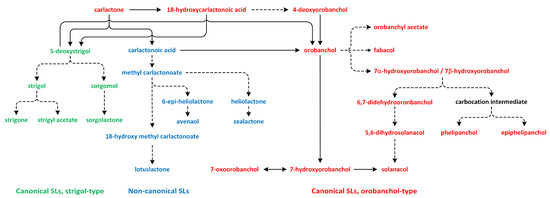
  • SLs are terpenoid lactones
  • [
  • 1
  • ]
  • [
  • 2
  • ]
  • [1,2].
In addition, peptide hormones regulate many developmental and defense processes, such as meristem maintenance, xylem and phloem differentiation, stomata patterning, pollination, embryo and endosperm development, cell division, nodulation, and systematic response [3]. They are divided into secreted and non-secreted types. Secreted peptide hormones are further divided into post-translationally modified peptides and cysteine-rich peptides. Peptide hormones are synthesized as larger precursor molecules, which are then cleaved to produce active peptides, e.g., CLAVATA3 (CLV3)/Embryo Surrounding Region-Related (CLE), Phytosulfokine (PSK), Plant Peptide-Containing Sulfated Tyrosine (PSY) peptides belong to post-translationally modified peptides; Rapid Alkalinization Factor (RALF) peptides belong to cysteine-rich peptides; Plant Elicitor Peptides (PEP)—to non-secreted peptides. Peptide hormones are also generated by plant pathogens, symbionts, and microbes that interact with plants. They are crucial in establishing a molecular interface that allows them to co-exist with the host plant. These organisms produce effectors that mimic peptide phytohormones and other effectors of pathogens and symbionts. Plant receptors recognize these effectors, which primarily regulate growth rather than defense responses. However, the origin of non-plant peptide phytohormones is still a subject of controversy [3][4][5][6][3,4,5,6]
As such, plants host diverse phytohormone pathways. Significant advancements have been made in the study of phytohormone biology and synthesis over the past decade. An assortment of new tools and methods has been developed, resulting in the discovery of phytohormone substrates, intermediates, and final products. The biosynthetic pathways of plant hormones have been elucidated, and extensive research has been carried out into the genes found in the plant genome, encoding the enzymes that catalyze the various stages of phytohormone synthesis [1][2][1,2].

2. Polyamines

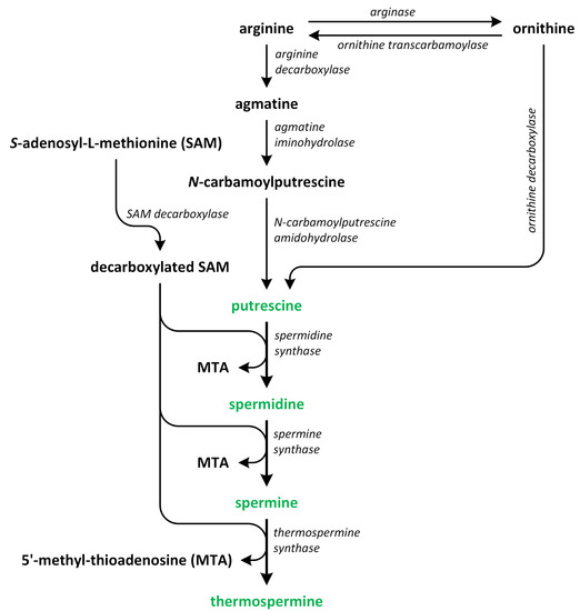
Polyamines (PAs) play a crucial role in plant growth, metabolism, and development. Structurally, these compounds are organic polycationic alkylamines that contain two or more amino groups. PAs such as putrescine (Put), spermidine (Spd), and spermine (Spm) are the most well-studied and recognized PAs found in plants. These three PAs regulate various physiological activities, such as photosynthesis, flower generation, embryogenesis, and organogenesis. They are also accountable for maintaining the stability of nucleic acids, various protein molecules, and the membrane structure. Additionally, they play a substantial role in enhancing the tolerance of many plants to the presence of a variety of abiotic and biotic stress factors, thereby impacting crop yield. Putrescine is a key compound involved in the biosynthesis of PAs. It serves as a common intermediate in the creation of Spd, Spm, and thermospermine. The biosynthesis pathways of Put have been identified in many plants (Figure 2) [9][10][11][12][9,10,11,12].
Figure 2.
The biosynthetic pathways of polyamines (phytohormones are green).

3. Ethylene

The volatile phytohormone ethylene plays a key role in controlling various developmental and physiological activities, including seed dormancy and germination, growth of vegetation, flowering, maturation of climacteric fruit, and aging. Moreover, ethylene is seen as a critical component that protects plants from both biological and non-biological stressors. Being a gaseous plant hormone, ethylene has the ability to easily spread from its creation sites without needing any biochemical alterations or metabolic processes, allowing it to be sensed promptly [13][29].
Ethylene production is intricately controlled by internal cues throughout its development. Additionally, ethylene production can be swayed by environmental triggers, including biological triggers, like an onslaught of pathogens, and non-biological triggers, like injury, low oxygen levels, exposure to ozone, cold temperatures, or freezing conditions [14][30].
The biosynthesis of ethylene is a relatively straightforward process that involves two key enzymatic reactions (Figure 3).
Figure 3.
The biosynthetic pathways of ethylene (phytohormone is green).

4. Salicylic Acid

Salicylic acid (SA) participates in plant growth and development. In heat-generating plants, it initiates the process of heat production by triggering an alternative respiration pathway, leading to the release of foul-smelling compounds that lure pollinators. In terms of plant defense, SA acts as a messenger, managing the expression of plant pathogenesis-related genes and fostering disease resistance. Additionally, it helps to orchestrate plant reactions to diverse types of non-biological stress factors, like extreme temperatures, saline conditions, and oxidative stress. SA also has a role in controlling a range of other activities in plants, including photosynthesis, respiration, growth of vegetation, seed germination, flowering, and aging [15][16][17][42,43,44].
In plants, SA occurs via two pathways: (i) the isochorismate synthase (ICS) (major fraction) and (ii) the phenylalanine ammonia-lyase (PAL) pathways (Figure 4). Both biosynthetic pathways start in chorismate plastids and vary between plant species [15][42]. Although plants employ both pathways simultaneously, the ICS pathway is the primary contributor, accounting for approximately 95% of SA synthesis. Chorismate is a primary metabolic precursor of both pathways [18][45]. This compound is the end product of the shikimate pathway, which is initiated by erythrose-4-phosphate and phosphoenolpyruvate [19][20][46,47].
Figure 4.
The biosynthetic pathways of salicylic acid (phytohormone is green).

5. Auxins

Auxins play a crucial role in the regulation of various aspects of plant growth and development. They are involved in controlling processes such as cell division, elongation, differentiation, tropisms (response to external stimuli), flowering, apical dominance (suppression of lateral bud growth), lateral root formation, senescence (aging), abscission (shedding of plant parts), and responses to environmental stresses [21][56]. The most well-studied auxin in plants is indole-3-acetic acid (IAA). The process of producing auxins in plants is highly intricate, and understanding this process can significantly enhance our comprehension of auxins’ biological role and contribution to the regulation of plant growth and physiology [22][57]. Although different plant species employ distinct strategies to optimize their metabolic pathways, it seems that there could be shared mechanisms for auxin biosynthesis, given that IAA is an phytohormone essential to plant life. Auxins are primarily generated in the plant’s apical meristems, young leaves, and flower buds, and they are then quickly transported throughout the plant via the phloem. Nonetheless, some research suggests that auxins may also be locally synthesized within the roots [23][24][25][58,59,60].
Two major routes have been assumed to contribute to de novo IAA biosynthesis in plants: the tryptophan (Trp)-dependent and Trp-independent pathways (Figure 5). Several pathways for Trp-dependent IAA biosynthesis have been proposed, including:
Figure 5.
The biosynthetic pathways of auxins (phytohormones are green).
  • the indole-3-acetamide (IAM) pathway;
  • the indole-3-pyruvic acid (IPA) pathway;
  • the tryptamine (TAM) pathway;
  • the indole-3-acetaldoxime (IAOX) pathway [26][27][28][29][30][61,62,63,64,65].
Even though IAA was the first natural auxin to be identified, our understanding of the genes that encode all of the enzymes involved in auxin biosynthesis remains limited. It is also uncertain whether all of these pathways exist in all plant species [31][66].

6. Melatonin

Melatonin, which is formally known as N-acetyl-5-methoxytriptamine, is an indolamine plant hormone that plays a key role in controlling growth and various physiological responses. Affected functions include root structure and development, flowering, leaf aging, fruit maturation, and determining the levels of chlorophyll, proline, and carbohydrates. It also functions as a signaling molecule during non-biological stress situations, like cold, drought, heavy metal exposure, UV radiation, and salinity, and it helps to direct plant defense responses against pathogen invasions [32][33][80,81]. The highest levels of melatonin in plants are found in the mitochondria, endoplasmic reticulum, and chloroplasts, suggesting that these organelles are the primary sites involved in its biosynthesis. The varied locations of melatonin within the plant could potentially influence its mode of action in different ways [34][82]. The biosynthesis of melatonin begins with an essential aromatic amino acid tryptophan, which is synthesized de novo via the shikimate pathway (Figure 6).
Figure 6.
The biosynthetic pathways of melatonin (phytohormone is green).
The pathway involved in the synthesis of this phytohormone occurs through various four-step reactions catalyzed by six enzymes: tryptophan decarboxylase (TDC), tryptophan hydroxylase (TPH), tryptamine 5-hydroxylase (T5H), serotonin N-acetyltransferase (SNAT), N-acetylserotonin O-methyltransferase (ASMT), and caffeic acid O-methyltransferase (COMT) [35][83]. These enzymes have different cellular localization. Chloroplasts and mitochondria contain SNAT. TPH, ASMT/COMT, and TDC are present in the cytoplasm. T5H can be only found in the endoplasmic reticulum [36][84]. Recent research suggests that the synthesis of melatonin in plants primarily takes place in chloroplasts and mitochondria. These organelles have inherited the ability to produce melatonin from their bacterial forebears. Under normal circumstances, melatonin production predominantly occurs in the chloroplasts. However, if the pathway in the chloroplasts is obstructed, the production of melatonin shifts to the mitochondria. Therefore, under stress conditions, melatonin is chiefly synthesized in the mitochondria [37][38][85,86].

7. Abscisic Acid

Abscisic acid (ABA) also plays an important role in many cellular processes, including seed development, dormancy, germination, vegetative growth, and responses to environmental stresses [39][92]. ABA is ubiquitous in lower and higher plants. It was reported that young tissues contain high levels of ABA [40][93]. The biosynthesis of ABA predominantly takes place in vascular tissues, and it is subsequently transported into target tissues. This transportation process occurs through both the xylem and phloem, enabling bidirectional transport between the roots and shoots. The naturally occurring form of ABA is S-(+)-ABA, which has a side chain defined as 2-cis, 4-trans. The trans-ABA is biologically inactive, whereas R-(−)-ABA, which can potentially result from racemization through the catabolite ABA-trans-diol, exhibits biological activity [41][42][94,95]. The initial stages of ABA biosynthesis occur within plastids. In plants, two distinct ABA biosynthesis pathways have been identified. The first pathway is a direct route, while the second pathway is indirect and involves the conversion of a C15 compound (i.e., farnesyl pyrophosphate) and a C40 carotenoid [43][96]. Through the characterization of ABA-deficient mutants and the isolation of relevant mutated genes, it has been determined that the indirect pathway is the primary route involved in ABA biosynthesis in plants [43][44][96,97]. Investigations into the biosynthesis of carotenoids, as well as their pathways and mechanisms, have indicated that mevalonate (MVA) plays a minor role as a precursor of carotenoid biosynthesis in chloroplasts [45][46][98,99]. In plants, there are two distinct pathways involved in isoprenoid production (Figure 7). The first pathway, which is known as the MVA pathway, is shared by animals, fungi, and a few bacteria. It operates in both the cytosol and mitochondria, and it is responsible for generating precursor molecules used in the synthesis of sterols, certain sesquiterpenes, and the side chain of ubiquinone. On the other hand, the second pathway, which is called the methylerythritol phosphate (MEP) pathway, is localized within plastids [47][100].
Figure 7. The methylerythritol phosphate (MEP) and mevalonate (MVA) pathways lead to the conversion of isopentenyl diphosphate to cycloartenol and phytoene.
The conversion of geranylgeranyl pyrophosphate (GGPP) into phytoene, which is a C40 carotenoid, is the initial and rate-limiting step involved in carotenoid synthesis. This reaction is catalyzed by the enzyme phytoene synthase (Figure 8).
Figure 8.
The biosynthetic pathways of abscisic acid (phytohormone is green).

8. Brassinosteroids

Brassinosteroids (BRs) are steroid phytohormones that elicit a wide spectrum of morphological and physiological responses, as well as a tolerance of abiotic and biotic stress [48][49][50][51][52][53][54][108,109,110,111,112,113,114]. BRs are categorized into three primary groups based on the number of carbon atoms present in each steroid molecule, and BRs are divided into three main groups on the basis of each steroid molecule, i.e., C27, C28, and C29 [55][56][115,116]. The basic structure of C27-BRs is the 5α-cholestane skeleton; 5α-ergostane serves as the foundational structure of C28-BRs, while 5α-stigmastane forms the basis of C29-BRs. The structures of these hormones vary due to the type and orientation of oxygenated functions in the A- and B-rings, as well as the number and position of functional groups in the side chain. These variations arise from oxidation and reduction reactions during their synthesis in plants [57][58][59][117,118,119].
Three pathways of BR biosynthesis that lead to the production of BRs of type C27, C28, or C29 were identified in plants. Early steps involved in their synthesis, which are common for all BRs, occur via the MVA or non-MVA (MEP) pathways (Figure 7). Later steps differentiate between the biosynthesis pathways (cycloartenol- and cycloartanol-dependent) of this group of plant hormones (Figure 9) [56][60][61][116,120,121].
Figure 9.
The biosynthetic pathways of different types of brassinosteroids (phytohormones are red, green, and orange).

9. Cytokinins

Cytokinins (CKs) regulate plant development, nutrient uptake, cell division, cell differentiation, chlorophyll senescence, apical dominance, embryonic development, and general aspects of plant growth [62][136]. Cytokinins are N6-substituted adenine derivative compounds that can be divided into two groups: (i) isoprenoid and (ii) aromatic CKs. Isoprenoid CKs more abundant than aromatic compounds [63][64][137,138]. Isoprenoid CKs include compounds such as N6-(∆2-isopentenyl adenine (iP), dihydrozeatin, and trans- and cis-zeatin (tZ and cZ, respectively), while N6-benzyladenine and its hydroxyl derivatives, such as ortho- and meta-topolin, are representatives of aromatic CKs [65][139].
Cytokinin levels are spatially and temporally regulated. Cytokinins are abundant in the apical meristems, root tips, and immature seeds. It is generally assumed that the root tip is the main site of the biosynthesis of CKs. On the other hand, literature data indicate that the shoot apex, cambium, and immature seeds are also capable of synthesizing CKs [66][140]. Changes in CK levels are related to the phase of cell cycle, consequences of influence of environmental factors, presence or lack of mineral nutrients, and effect of abiotic or biotic stress factors. The levels of active CKs in plants are highly regulated by the rates of production, interconversion, transport, and degradation [67][141].
Cytokinins can be synthesized de novo as nucleotide mono-, di-, or tri-phosphates, which are characterized by low biological activity. Moreover, tRNA is a source of CKs. The release of cytokinins from this nucleic acid leads to the creation of nucleotide monophosphates. The conversions of nucleotides, nucleosides, and free bases are catalyzed by enzymes involved in the metabolism of adenine [68][142]. In the next step, an isoprenoid moiety is added to the adenine present in the ATP and/or ADP molecule [69][143]. An alternative pathway, in which a hydroxylated side chain is added to the adenine moiety, has also been proposed [70][144].
DMAPP and HMBDP are the common isoprenoid side chain donors in biosynthetic pathway of CKs [71][72][102,145]. In the case of formation of isopentenyladenine-type CKs via the precipitation of DMAPP, the side chain is hydroxylated by cytochrome P450 mono-oxygenase (Figure 7) [73][146]. The CK nucleotides that include nucleotides released from tRNA are then hydrolyzed to free bases [71][102].
Cytokinin biosynthesis is catalyzed by the enzyme isopentenyl transferase (IPT) (Figure 10). There are two types of the adenylate IPT, which are responsible for attachment of an isopentenyl group to the N6 atom present in AMP, ADP, or ATP and tRNA. IPT does not add isopentenyl group to adenosine or adenine. IPT acts in the same way on adenine present in the structure of tRNA [69][143]. Adenylate IPT can synthesize iP and tZ nucleotides [72][74][145,147].
Figure 10.
The biosynthetic pathways of cytokinins (phytohormones are green).

10. Gibberellins

Gibberellins (GAs) are tetracyclic and diterpenoid plant hormones that form a large group of carboxylic acids [75][153]. GAs are present in all vascular plants and lower plants, such as lycophytes and ferns. GAs are involved in the promotion of organ growth, enhance cell elongation and/or division, and activate most developmental processes (i.e., seed germination, induction of flowering, and maturation) in many plant species [76][77][154,155]. GAs were first isolated from the fungus Gibberella fujikuroi (reclassified as Fusarium fujikuroi), which stimulated growth in infected vascular plants. Next, their presence in plants as phytohormones was confirmed in the late 1950s. The system that numbers GAs, starting with GA1, assigns GAs in order of their discovery and structural characterization [78][156]. The most well-known and biologically active GAs in plants are GA1, GA3, GA4, and GA7. There are three structural properties that alter these GAs: the presence of hydroxyl group in C-3β, carboxyl group in C-6, and lactone between C-4 and C-10. The 3β-hydroxyl group can be replaced by other functional groups at positions C-2 and/or C-3. However, GA5 and GA6 are representatives of bioactive GAs that lack a hydroxyl group at C-3β [75][153].
The knowledge of GA biosynthesis has developed rapidly in recent years. Quantita-tive analysis shows that biosynthesis of GAs occurs in actively growing tissues, i.e., young leaves, shoot apices, and flowers. In contrast, there are some reports that indicate that xylem and phloem exudates contain Gas, suggesting the presence of long-distance transportation of these phytohormones in plants [76][79][80][154,157,158].
In plants, GAs are formed from GGPP through IPP, which represents the C-5 building moiety involved in the synthesis of terpenoid (isoprenoid) compounds (Figure 7) [77][81][155,159]. In the green tissues of plants, IPP is produced via two pathways: (i) the MVA pathway in the cytoplasm and (ii) the methylerythritol phosphate (MEP) pathway in plastids in two-step reactions. Such a metabolism is ancient and leads to the synthesis of 12,000 natural diterpenoid products in plant tissues. Recent data suggest that this compound is cyclized to the tetracyclic hydrocarbon precursor ent-kaurene in plastids by ent-copalyl diphosphate, which predominantly occurs through the MEP pathway [79][157]. However, the MVA pathway may also be involved in the synthesis of isoprenoid intermediates of GGPP and their transport from the cytosol into the plastids [82][160]. The formation of ent-kaurene from GGPP takes place in the stroma of proplastids or in the developing chloroplasts, except those of mature chloroplasts [83][161]. Ten functional GGPP synthase (GGPPS) genes were discovered in A. thaliana. Among these genes, seven encode enzymes were localized in the plastid. However, GGPPS11 is expressed most strongly and constitutively in plants that produce most of the substrate utilized in the biosynthesis of various terpenoids in plastids [84][162]. In contrast to A. thaliana, rice is reported to contain one functional GGPPS that is present in the plastid, which is involved in the biosynthesis of all diterpenoid compounds, including ent-kaurene, which is a key intermediate involved in the formation of GAs [85][163].
Firstly, the synthesis of ent-kaurene from GGPP starts via cyclization, which initiated by the proton of the dicyclic ent-copalyl diphosphate (CPP), which is catalyzed by a type II diterpene cyclase: ent-copalyl diphosphate synthase (CPS) (Figure 11). CPS contains a conserved DXDD motif, in which aspartate gives a proton to initiate this reaction via cyclization. Additionally, a water molecule attached to amino acids (histidine and asparagine) plays role as the catalytic base that accepts a proton and terminates the reaction [86][164]. CPS activity is inhibited by high concentrations of both Mg2+ and GGPP, which are promoted by light, leading to reduced flux into the GA pathway during de-etiolation. This process is part of the mechanism used to decrease GA biosynthesis and its cellular concentration [87][165].
Figure 11.
The biosynthetic pathways of gibberellins (phytohormones are green).
The second step of the transformation of ent-copalyl diphosphate to ent-kaurene is catalyzed by a type I cyclase, which is known as ent-kaurene synthase (KS) (Figure 11). Cyclization reaction is initiated via metal-dependent heterolytic cleavage of the O-C bond. A pimeren-8-yl carbocation takes place, which then undergoes rearrangement. The loss of H+ is important step that leads to tetracyclic ent-kaurene [88][166]. Enzyme KS contains RLX(N,D)DXX(S,T,G)XXX(E,D) and DDXXD motifs, which can bind to ion Mg2+, which is associated with the diphosphate residue and participates in its ionization [89][167]. The lycophyte Selaginella moellendorfii, which is one of the first plants to have evolved the ability to synthesize GAs, possesses monofunctional CPS and KS enzymes, which are characteristic of vascular plants. In S. moellendorffii, KS is characterized by low substrate specificity, as it converts different stereoisomeric forms of CPP to various organic compounds, while KS enzymes in angiosperms are specific only for ent-CPP [90][168]. In angiosperms, CPS and KS possess the functional diversification required to produce the diterpenoids involved in plants’ defense responses [79][91][157,169].

11. Strigolactones

Strigolactones (SLs), which are carotenoid-derived terpenoids, were initially characterized as germination stimulants for root parasitic plant witchweed (Striga spp.). SLs are a class of carotenoid-derived terpenoids that are involved in diverse developmental processes, such as seed germination, shoot branching, leaf senescence, and root development, as well as responses to various environmental conditions (e.g., light stress and high-temperature stress) [92][179]. They are also known as rhizosphere chemical signals that can regulate interactions between arbuscular mycorrhizal fungi and root parasitic plants. These groups of phytohormones also affect photosynthesis, synthesis, and action of other phytohormones, as well as regulating the levels of different metabolic compounds [93][94][180,181].
According to their chemical structures, SLs can be classified into two groups: canon-ical and non-canonical SLs. Canonical SLs contain a tricyclic lactone structure composed of three rings (ABC-rings) connected to a butenolide group (D-rings) through an enol-ether bridge, which is critical for the performance of their biological activities in plants. In contrast, non-canonical SLs do not have typical ABC-rings, though they contain both an enol-ether bridge and D-ring moieties [95][182].
Strigolactone biosynthesis mainly occurs in roots. The intermediates in the SL synthetic pathway have been also found in the stem. The biosynthesis of this group of phytohormones is tightly regulated by environmental conditions, such as starvation of phosphate or drought stress [96][183]. The early steps of SL biosynthesis are common for each type and may occur via the MVA or MEP pathways that lead to IPP synthesis, which is sequentially condensed to create DMAPP, geranyl pyrophosphate (GPP), farnesyl pyrophosphate (FPP), and GGPP (Figure 7) [56][116]. Thus, the first part of the SL biosynthetic pathway starts in chloroplasts. The condensation of two GGPP molecules by phytoene synthase generates phytoene (Figure 12), which is the first uncolored carotenoid [97][184]. Next, phytoene is transformed via a sequence of reactions of desaturation and cis/trans-isomerization into all-trans-lycopene. Cyclization reactions include conversion of all-trans-lycopene into all-trans-β-carotene and all-trans-α-carotene [98][99][185,186]. Dwarf27 (D27) isomerizes all-trans-β-carotene to 9-cis-β-carotene, followed by cleavage induced by carotenoid removal of enzyme dioxygenase 7 (CCD7) from 9-cis-β-apo-10′-carotenal, which undergoes cleavage and rearrangement reactions to create carlactone (CL), which is the central intermediate in SL biosynthesis (Figure 12). This reaction is catalyzed by the CCD8 enzyme [100][187]. CCD7 is encoded by gene MAX3 and its orthologs genes: RMS5 and D17/HTD1. Gene MAX4 and its orthologs RMS1, D10, and Defective in Anther Dehiscence 1 (DAD1) encode CCD8. Both enzymes act in a progressive manner [101][188].
Figure 12.
The biosynthetic pathways of carlactone.
Carlactone (CL) contains only A- and D-rings that have an enol-ether bridge and exhibits SL-like properties. For example, CL inhibits shoot branching in SL biosynthetic mutants (i.e., rice, and A. thaliana) and promotes seed germination of Striga hermonthica. CL has been identified as an endogenous precursor in synthetic routes of both canonical and non-canonical SLs [99][102][103][186,189,190]. Furthermore, a new compound, which is identified as 3-hydroxyCL, is formed in vitro using 9-cis-3-hydroxy-β-apo-10′-carotenal in a reaction catalyzed by enzymes D27, CCD7, and CCD8. This compound was identified in plants such as rice, A. thaliana, and N. benthamiana [104][191].
The SL biosynthesis activates the biochemical conversions of CL through the participation of cytochrome P450 mono-oxygenases and other enzymes, which are responsible for the biological and structural diversity of these phytohormones. Subsequently, CL is transported from the chloroplast to the cytoplasm, where the reactions related to SL biosynthesis occur (Figure 13) [103][190].
Figure 13.
The biosynthetic pathways of different types of strigolactones (phytohormones are green, blue, and red).

12. Jasmonates

Jasmonates are plant hormones that originate from oxylipins involved in plant devel-opment and stress responses [105][198]. Their major representatives are the isomers of jasmonic acid (JA): (+)-7-iso-JA and (−)-JA. JA can be metabolized into methyl jasmonate (MeJA), which is a volatile phytohormone. Jasmonates have previously been defined as JA and its diverse metabolites derived from various reactions created via esterification, methylation, sulfation, glycosylation, conjugation, de-carboxylation, hydroxylation, and carboxylation [106][199]. Recently, an alternative and probably more ancient pathway was proposed. In this route, JA originates from dinor-OPDA (2,3-dinor-12-oxo-10,15(Z)-phytodienoic acid [dn-OPDA]) [107][200]. Thus, 12-oxo-10,15(Z)-phytodienoic acid (OPDA), dn-OPDA, and their derivatives are members of JAs family. JA and MeJA are the best characterized group of jasmonates in plants, and they are regarded as one of the major phytohormones that regulate both defense responses and the development process [108][201].
Jasmonate biosynthesis pathways have been extensively investigated in dicotyledonous plants, such as A. thaliana, tobacco, and tomato (Figure 14).
Figure 14.
The biosynthetic pathways of jasmonic acid (phytohormones are green).
ScholarVision Creations