Your browser does not fully support modern features. Please upgrade for a smoother experience.
Please note this is a comparison between Version 2 by Rita Xu and Version 1 by Liming Hu.
Peroxynitrite (ONOO−) is a crucial reactive oxygen species that plays a vital role in cellular signal transduction and homeostatic regulation. Determining and visualizing peroxynitrite accurately in biological systems is important for understanding its roles in physiological and pathological activity.
- peroxynitrite
- fluorescent probes
- intracellular imaging
1. Introduction
Peroxynitrite (ONOO−) is a kind of reactive oxygen species (ROS) generated by the rapid reaction of nitric oxide (NO) and a superoxide anion free radical (O2·−) in the absence of enzyme catalysis, which has strong oxidation, nucleophilic, and nitration properties [1]. It occupies crucial roles in the transformations of other major reactive species. (Scheme 1) Its pKa value is 6.8 [2], and the half-life is approximately 1 s [3,4][3][4] at pH 7.4. ONOO− can react with a variety of bioactive substances (such as protein, nucleic acid, lipid, etc.) with very high reactivity. In addition to its oxidation, nucleophilic, and nitration properties, ONOO− can also be converted into higher activity secondary free radicals, including hydroxyl radicals (·OH), nitro radicals (·NO2), and carbonate radicals (CO3·−), which further react with biomolecules and ultimately lead to cell death.
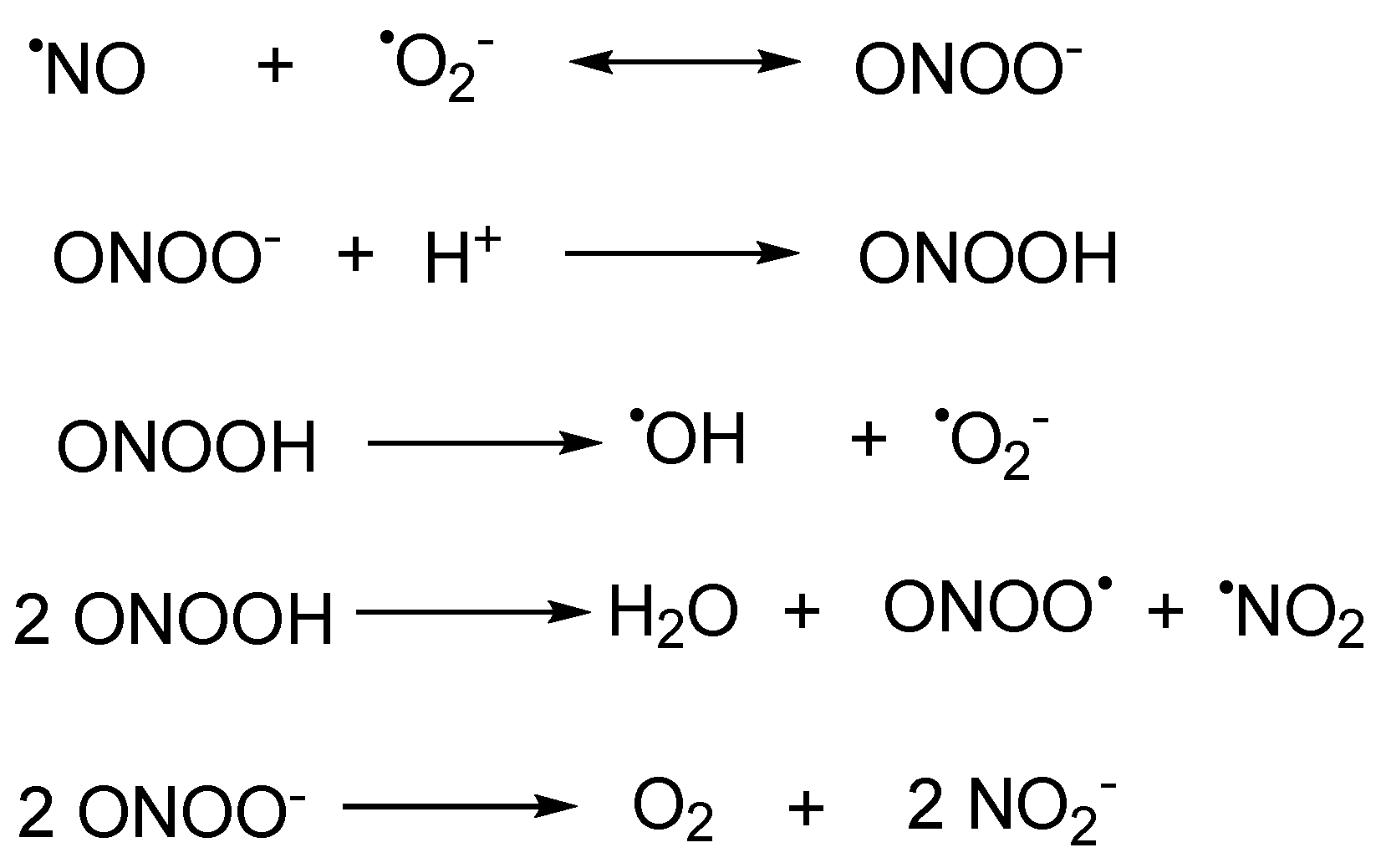
Scheme 1. The biogenesis of peroxynitrite and its transformations with other major reactive species.
Based on these properties, peroxynitrite exhibits two effects with different directions. In the living system, when the ONOO− remains at a level which is under normal physiological conditions, it serves as an indispensable physiological activator and signaling molecule. However, when the concentration of ONOO− elevates, the excess ONOO− will turn the redox state of the cell to a pro-oxidant state [5,6][5][6]. Eventually, serious inflammation and disease will be induced, for example, rheumatism, hepatic disease, neurodegenerative disease, cancer, and so on [7,8,9,10][7][8][9][10]. Therefore, it would be of great significance to develop a method which could accurately detect ONOO− and explore the physiological role of ONOO− in living systems.
In comparison to other ONOO− detection methods (positron emission computed tomography (PET), computed tomography (CT), magnetic resonance imaging (MRI), and genetically encoded indicators) [11], spectroscopic detections, especially fluorescent probes, possess advantages such as excellent temporal and spatial resolution, simple operation, high sensitivity and selectivity, and non-destructive and in situ real-time visualization of biological samples [12,13,14,15][12][13][14][15].
2. Fluorescent Probes
2.1. Xanthene as Fluorophore Core
Xanthene dyes can be categorized as fluorescein, rhodol, and rhodamine based on the type of the substituents on the 3- and 6-position [16]. They are well known because of their switchable fluorescent off–on flexibility. Xanthene dyes can produce fluorescence wavelengths above 510 nm, reaching far-red areas depending on the conjugative substituents. Thus, they are of widespread use in optical diagnostic research [17]. The triggers of ONOO−-responsive probes with xanthene as a fluorophore core were generally built on (1) oxidation of the hydrazide (Xan1–Xan15) [18[18][19][20][21][22][23][24][25][26][27][28][29][30][31][32],19,20,21,22,23,24,25,26,27,28,29,30,31,32], (2) oxidative cleavage of the substituents at the hydroxyl or amino group (Xan 16–Xan 28) [33[33][34][35][36][37][38][39][40][41][42][43][44][45],34,35,36,37,38,39,40,41,42,43,44,45], (3) oxidation of pyrylium (Xan 29–Xan 33) [46,47[46][47][48][49][50],48,49,50], and (4) oxidation of the hydrogenated xanthene (Xan 34–Xan 37) [51,52,53,54][51][52][53][54] and others (Xan 38–Xan 41) [55,56,57,58][55][56][57][58].2.1.1. Hydrazide Oxidative Xanthene Probes
In 2002, Guo et al. reported a spiro form hydrazide rhodamine (Xan 1) [18] as the ONOO− fluorescent probe. The hydrazide probe was colorless and non-fluorescent. Upon treating with ONOO−, the spiro hydrazide group was oxidized, releasing a highly fluorescent rhodamine B. The response finished in as fast as 30 s. Meanwhile, the detection limit was only 24 nM. The response avoids interference from the 10−5 M Cu(II) ion. Thus, it represents the rapid, sensitive, and specific fluorescent detection of ONOO−. Based on the recognized pattern and the easy structurally modification character of rhodamine, a series of related probes were developed, aiming to improve the performance of different aspects of the response (Figure 1). Longer emissive wavelengths (up to the NIR range) were obtained with more conjugate groups installed in Xan 2–5 [19[19][20][21][22],20,21,22], Xan 8 [25], and Xan 10 [27]. Dual-channel fluorescence was afforded when coumarins were introduced to the rhodamine ring (515/700 nm for Xan 5 and 631/669 nm for Xan 10), making the response produce more information. Ratiometric fluorescence was realized in Xan 6 [23] and Xan 7 [24] with the introduction of a 2-(2′-hydroxyphenyl)benzothiazole group and a 4-hydroxycarbazole group, respectively, in which the intensity of the original band disappeared with the generation of a new band with a longer wavelength. Large Stokes shift and excellent lysosome-targeting ability were achieved with the engineering of a fused tetrahydroquinoxaline ring, making Xan 9 [26] capable of detecting both peroxynitrite and lysosomal pH. Sodium-dependent multivitamin transporter (SMVT)-targeted ability was acquired by introducing the biotin group for Xan 9, making it possible to detect the peroxynitrite in head and neck cancer cells.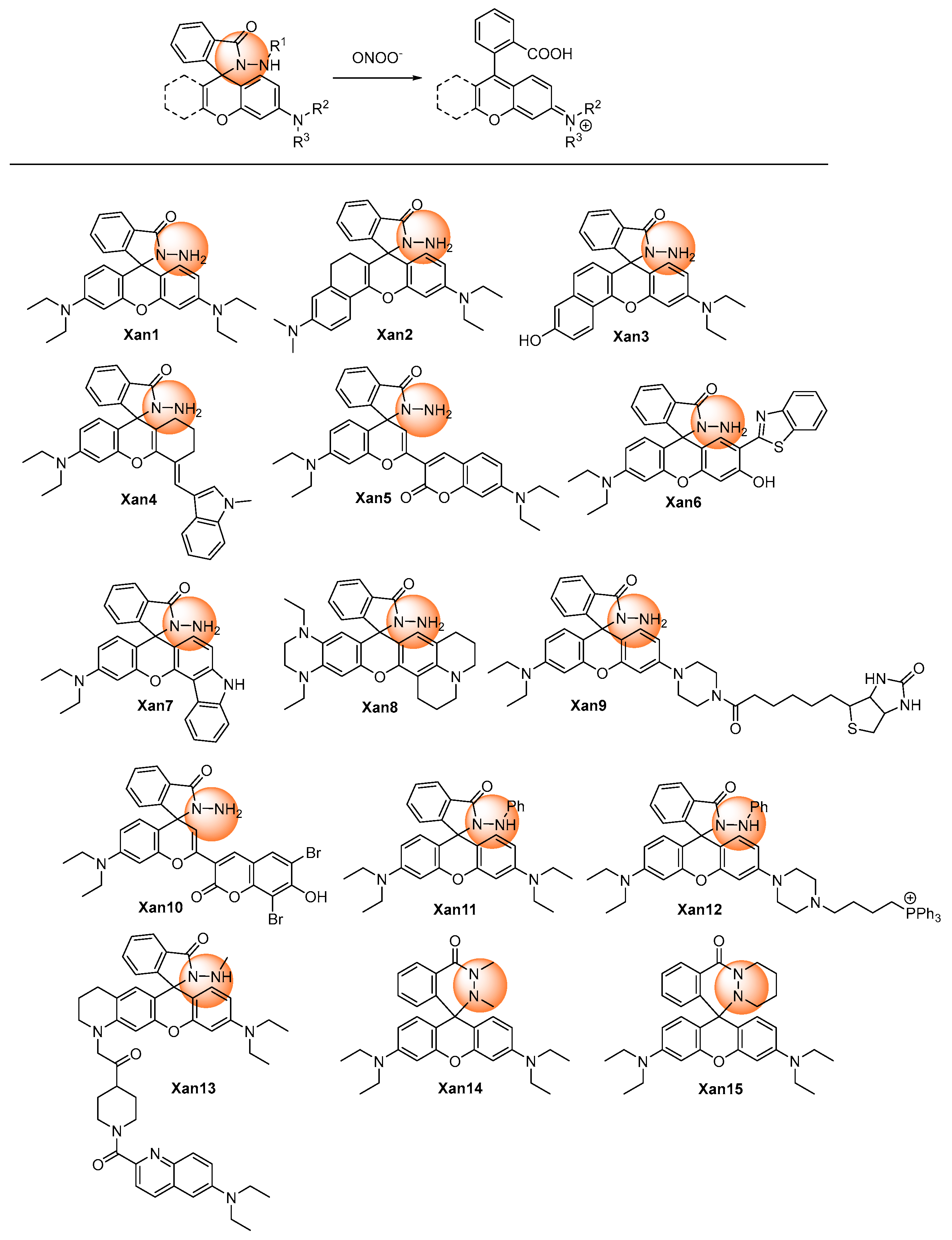
Figure 1. Chemical structures of the hydrazide oxidative xanthene probes.
2.1.2. Oxidative Cleavage of the Recognition Groups to Release Xanthene Probes
Utilizing the oxidative ability of peroxynitrite, the recognition groups at the 2- or 6-hydroxyl or amino group of xanthenes derivative could be cleaved to release xanthenes with high fluorescence (Figure 2).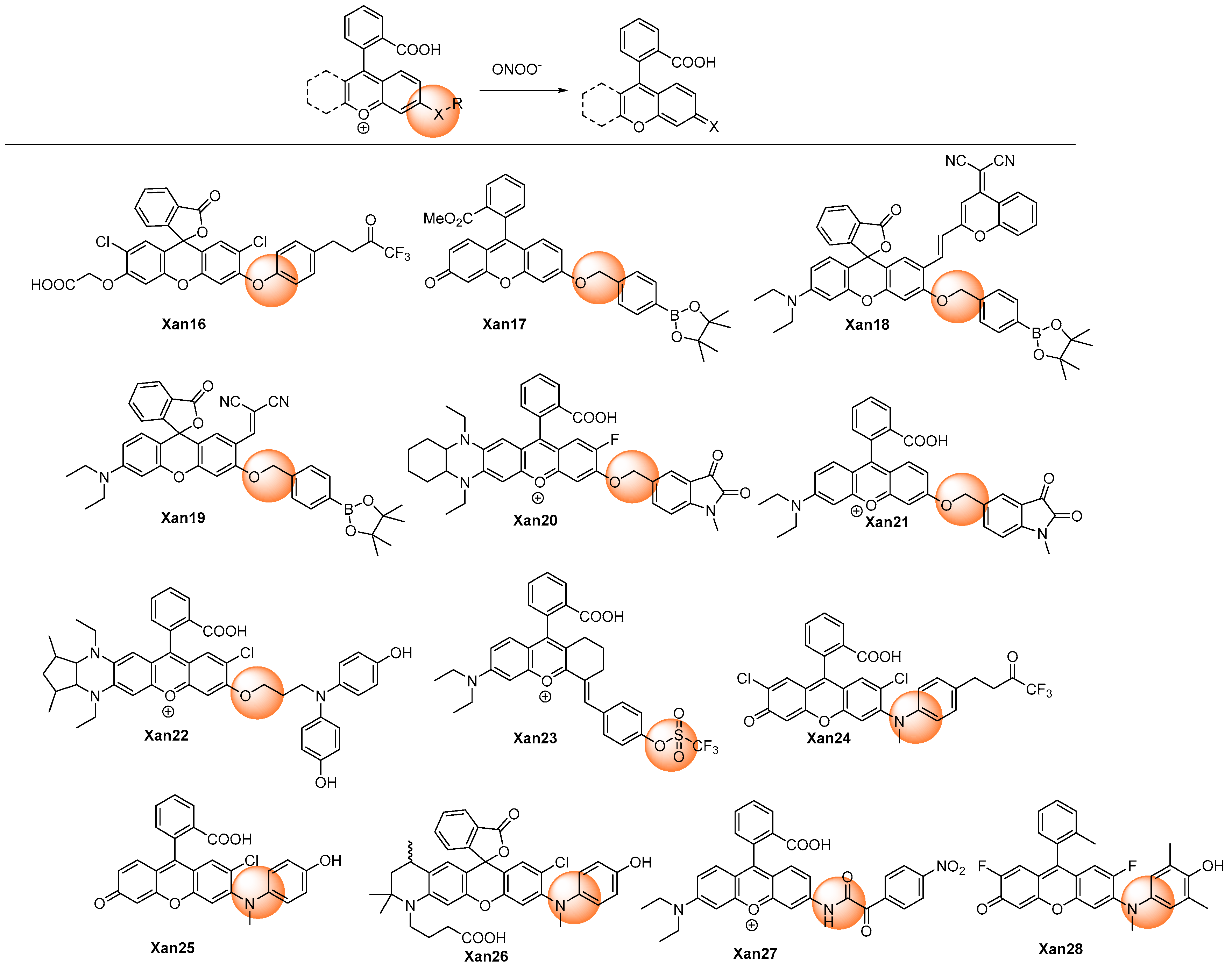
Figure 2. Chemical structures of the oxidative cleavage xanthene probes.
2.1.3. Oxidation of Pyrylium
Yuan et al. discovered an aminophenyl-substituted pyrylium as a highly sensitive and selective scaffold towards peroxynitrite after the screening of nineteen dyes and then further modified it to a FRET probe (Xan 29) [46] with TP absorption. After the response, the pyrylium emission band at 651 nm disappeared, and a coumarin characteristic emission band at 473 nm was enhanced. Detailed response mechanisms involving nucleophilic addition, oxidation, elimination, and hydrolysis reactions on chromenylium fluorophore were proposed and verified by MS spectra. Although the destroyed-type response led to the decrease of the emission wavelength, the combination technique of the ratiometric measure and TP imaging made it possible to specifically and rapidly visualize the peroxynitrite in an inflamed mouse model. Furthermore, the detection limit was as low as 11.3 nM, which was at a super level among the peroxynitrite probes. Subsequently, similar structures were synthesized for different applications. Gong et al. reported esterified Xan 30 [47] with better membrane penetrability and mitochondria targeting ability, which could image the peroxynitrite in the acute liver injury model in living cells. Li et al. introduced a piperazine ring to respond to the pH and finally realized the fluorescent imaging of the cellular peroxynitrite level as well as the mitophagy behavior [48]. Yuan et al. performed an original structure–activity relationship study of the substituents at the recognition site. (Figure 3) They discovered that pyrylium involving aryl substituents with strong electron-withdrawing groups could improve the sensitivity; meanwhile, pyrylium involving aryl substituents with strong electron-donating groups could improve the selectivity. Hence, they designed a coumarin, which was a not strong electron-withdrawing and -donating group, substituted pyrylium (Xan 32) [49] to satisfy the high requirements of both selectivity and sensitivity, and the results showed that an outstanding detection sensitivity of 4.1 nM of the detection limit as well as a high 130-fold ratiometric emission signal were realized. Employing the probe, the changing content of peroxynitrite in the diseases model involving nonalcoholic fatty liver and drug-induced liver injury was successfully visualized to unfold the functionality of a related enzyme. Zhou et al. introduced a naphthimide fluorophore in the xanthene carboxylic position. After the response, both coumarin and naphthimide fluorescence were produced to output a multicolor signal. The probe Xan 33 was applied for the early detection and evaluation of arthritis [50].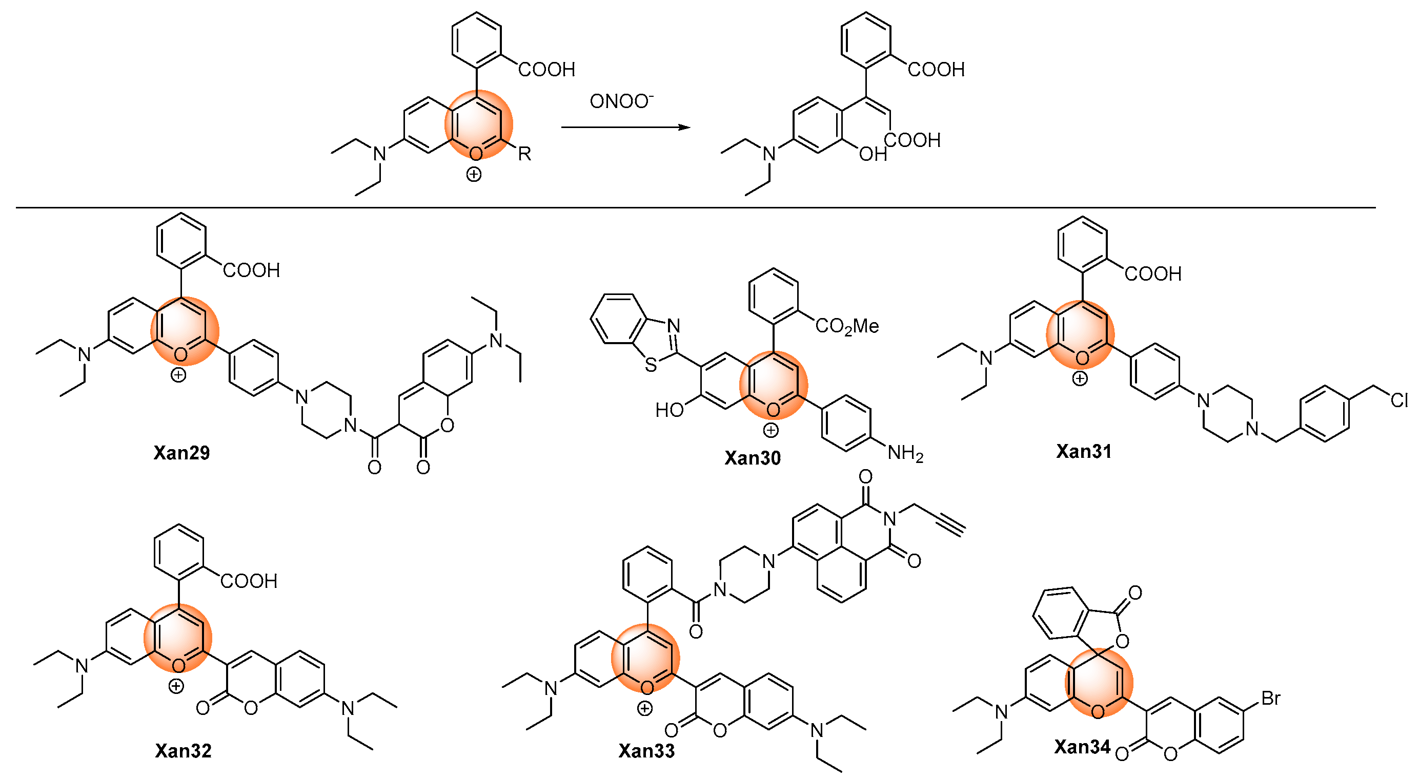
Figure 3. Chemical structures of the pyrylium oxidative xanthene probes.
2.1.4. Oxidation of Hydrogenated Xanthene
Peroxynitrite can oxidize t non-fluorescent hydrogenated xanthene to produce highly fluorescent aromatic products. (Figure 4) Gong et al. developed 9,10-dihydroacridine Xan 35 as the peroxynitrite detection probe. An over 100-fold fluorescence enhancement could be achieved after reacting with peroxynitrite. The probe was utilized to detect intracellular peroxynitrite [52]. Similar O-, Si-, and P- hydrogenated rhodamine systems were also reported. Xan 36–37 [53,54][53][54] displayed a very fast response speed (<20 s); for Xan 38 [55], the relatively low response speed was probably due to the low reactivity caused by the presence of the electron-withdrawing phosphonic group. Nevertheless, Xan 35–38 [52,53,54,55][52][53][54][55] all exhibited very low detection limits at the nanomolar level, and they were all applied to fluorescent imaging of cell endogenous peroxynitrite.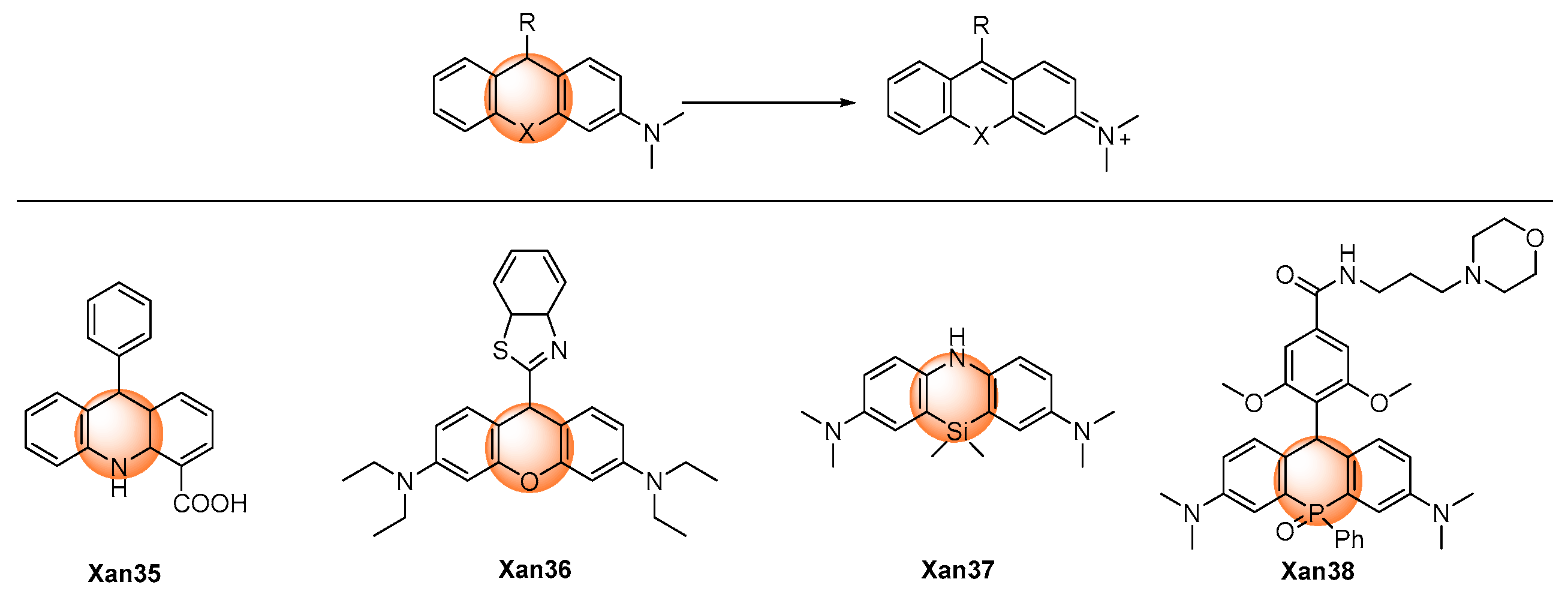
Figure 4. Chemical structures of the oxidative hydrogenated xanthene probes.
2.1.5. Others
Wu et al. described a Rhodol-based probe, Xan 39 [56], which introduced 1,1-dimethylhydrazone as a peroxynitrite recognition group. (Figure 5) The probe was non-fluorescent as a result of the rotational vibration of the C=N bond. Using the oxidative ability of peroxynitrite, the hydrazine was oxidatively cleaved into the corresponding aldehyde with significant fluorescence. The response exhibited a low detection limit (57 nM) with a short response time (<60 s). The probe was applied in the fluorescent imaging of exogenous and endogenous peroxynitrite in living cells.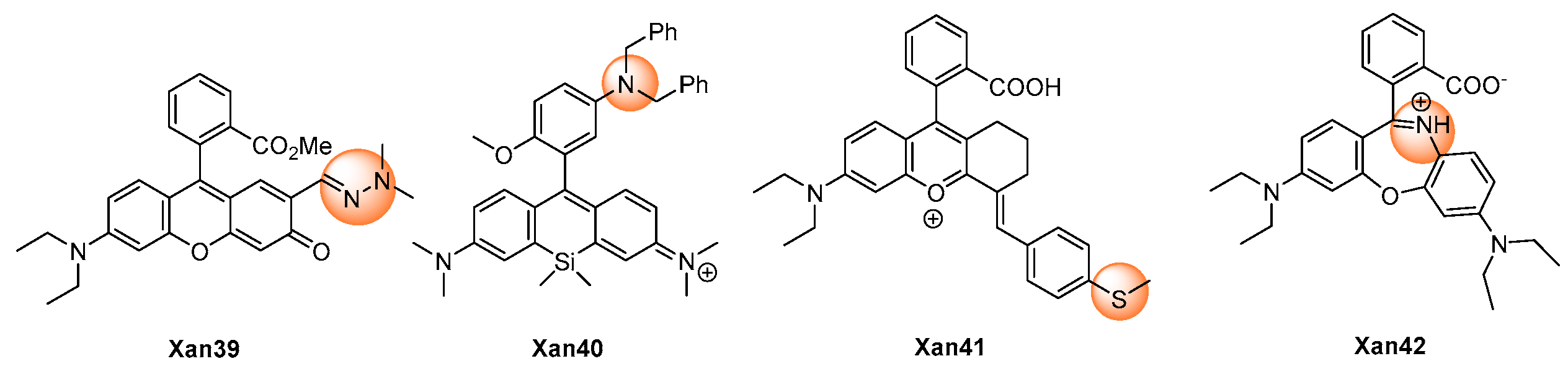
Figure 5. Chemical structures of the other xanthene probes.
2.2. Dicyano-Based Compounds as Fluorophore Core
Dicyano-based compounds are characteristic of their donor–π–acceptor structure, which endows them with large Stokes shifts and excellent photostability as a result of the ICT process. In addition, this sort of chromophore was generally easily synthesized and structurally modified. Thus, great attention has been attracted towards dicyano-based compounds to build probes with different functionalities [60]. The designing rule for dicyano-based peroxynitrite probes was a consensus, which was described in Figure 6. In general, the responsive groups, such as diphenylphosphonyl, benzyl boronates, and 4-hydroxyphenyl, were modified on the donor moiety to stop the ICT process. The response with peroxynitrite would break the links between the donor moiety and the response groups and release the dicyano-based chromophores with strong fluorescence.
Figure 6. Chemical structures of the dicyano-based probes.
2.3. Coumarin as Fluorophore Core
The research history of coumarin (also known as 1-benzopyran-2-one or 2H-chromen-2-one) was more than 200 years. Plenty of extensive investigations have been performed to modify the weak fluorescent parent coumarin to its derivatives with different desired photophysical properties, with a considerable amount of them now very active in the commercial market [75]. Inserting the electron-donors in the 7-position leads to a bathochromic shift to the emission wavelength; in addition, a donor–π–donor structure was formed, which facilitates the use of itself to design the ICT type probes by further introducing an electron-acceptor recognition group. (Figure 7) Xie et al. adopted this strategy and synthesized Cou 1 [76]. The 4-nitrophenyl oxoacetyl recognition group reacted with peroxynitrite rapidly and produced the deprotected product Cou 2 [77]. They used Cou 1, together with the two-photon fluorescent imaging technology, to visualize the peroxynitrite produced in the mitochondria in an anthracycline-induced cardiotoxicity mouse model. However, Li et al. reported that the deprotected product, Cou 2, also further reacted with peroxynitrite in 5 s in the concentration range of 0.064–0.64 μM, and the resulting nitration products were confirmed by ESI-MS analysis. The 3-position of coumarin could also be introduced with electron-donors to generate the donor–π–donor structure. Wei et al. developed Cou 3 as the peroxynitrite probe using the 4-nitrophenyl oxoacetyl group as a recognition moiety [78]. The fluorescence of Cou 3 was quenched but could be quickly recovered with eight-fold enhancement after the response with peroxynitrite. The probe was used to image exogenous peroxynitrite formation in living cells in a biosystem.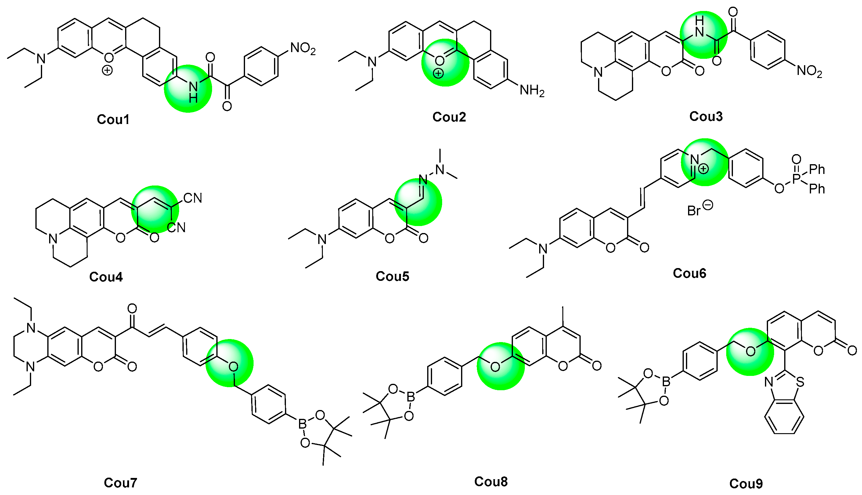
Figure 7. Chemical structures of the coumarin probes.
2.4. N-Substituted Coumarin as Fluorophore Core
As analogues for coumarin dyes, 2-(benzo[d]thiazol-2-yl)phenylacrylonitrile derivatives exhibited longer emission wavelengths than the related coumarins. (Figure 8) In particular, 2-(benzo[d]thiazol-2-yl)-3-(2-hydroxyphenyl)acrylonitrile derivatives (NCou 1–3) [85,86,87][85][86][87] served as the precursors for iminocoumarin, and they exhibited aggregation-induced emission luminogens (AIEgens) in aqueous conditions. Upon the response with peroxynitrite, 2-(benzo[d]thiazol-2-yl)-3-(2-hydroxyphenyl)acrylonitrile would be generated, which would further transform into iminocoumarin in situ. The probes were applied for the fluorescent imaging of cell exogenous and endogenous peroxynitrite, though the response rate was usually relatively slow.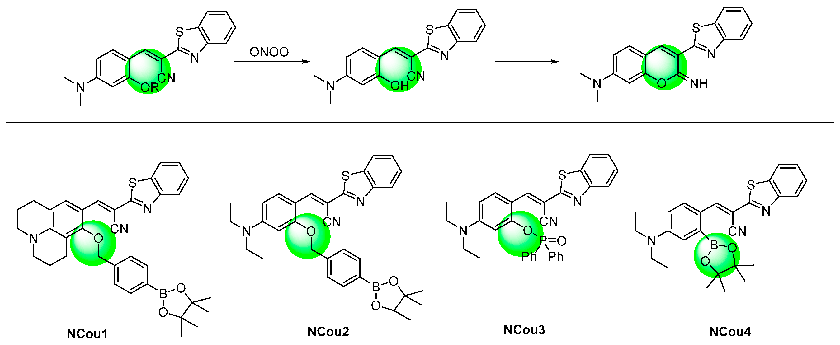
Figure 8. Chemical structures of the N-substituted coumarin probes and their responsive mechanism.
References
- Radi, R. Peroxynitrite, a Stealthy Biological Oxidant. J. Biol. Chem. 2013, 288, 26464–26472.
- Radi, R.; Beckman, J.S.; Bush, K.M.; Freeman, B.A. Peroxynitrite oxidation of sulfhydryls. The cytotoxic potential of superoxide and nitric oxide. J. Biol. Chem. 1991, 266, 4244–4250.
- Ducrocq, C.; Blanchard, B.; Pignatelli, B.; Ohshima, H. Peroxynitrite: An endogenous oxidizing and nitrating agent. Cell. Mol. Life Sci. 1999, 55, 1068–1077.
- Masumoto, H.; Kissner, R.; Koppenol, W.H.; Sies, H. Kinetic study of the reaction of ebselen with peroxynitrite. FEBS Lett. 1996, 398, 179–182.
- Butler, A.R.; Feelisch, M. Therapeutic uses of inorganic nitrite and nitrate: From the past to the future. Circulation 2008, 117, 2151–2159.
- Singh, D.K.; Winocour, P.; Farrington, K. Oxidative stress in early diabetic nerphropathy: Fueling the fire. Nat. Rev. Endocrinol. 2011, 7, 176–184.
- Migita, K.; Yamasaki, S.; Ida, H.; Kita, M.; Hida, A.; Shibatomi, K.; Kawakami, A.; Aoyagi, T.; Eguchi, K. The role of peroxynitrite in cyclooxygenase-2 expression of rheumatoid synovium. Clin. Exp. Rheumatol. 2002, 20, 59–62.
- Mahdi, A.; Tengbom, J.; Alvarsson, M.; Wernly, B.; Zhou, Z.; Pernow, J. Red Blood Cell Peroxynitrite Causes Endothelial Dysfunction in Type 2 Diabetes Mellitus via Arginase. Cells 2020, 9, 1712.
- Pandey, V.K.; Amin, P.J.; Shankar, B.S. G1-4A, a polysaccharide from Tinospora cordifolia induces peroxynitrite dependent killer dendritic cell (KDC) activity against tumor cells. Int. Immunopharmacol. 2014, 23, 480–488.
- Vana, L.; Kanaan, N.M.; Hakala, K.; Weintraub, S.T.; Binder, L.I. Peroxynitrite-induced nitrative and oxidative modifications alter tau filament formation. Biochemistry 2011, 50, 1203–1212.
- Tarpey, M.M.; Fridovich, I. Methods of detection of vascular reactive species: Nitric oxide, superoxide, hydrogen peroxide, and peroxynitrite. Circ. Res. 2001, 89, 224–236.
- Ma, Q.; Xu, S.; Zhai, Z.; Wang, K.; Liu, X.; Xiao, H.; Zhuo, S.; Liu, Y. Recent Progress of Small-Molecule Ratiometric Fluorescent Probes for Peroxynitrite in Biological Systems. Chem. Eur. J. 2022, 28, e202200828.
- Wang, S.; Chen, L.; Jangili, P.; Sharma, A.; Li, W.; Hou, J.T.; Qin, C.; Yong, J.; Kim, J.S. Design and applications of fluorescent detectors for peroxynitrite. Coord. Chem. Rev. 2018, 374, 36–54.
- Mao, Z.; Xiong, J.; Wang, P.; An, J.; Zhang, F.; Liu, Z.; Kim, J.S. Activity-based fluorescence probes for pathophysiological peroxynitrite fluxes. Coord. Chem. Rev. 2022, 454, 214356.
- Cui, W.-L.; Wang, M.-H.; Yang, Y.-H.; Wang, J.-Y.; Zhu, X.; Zhang, H.; Ji, X. Recent advances and perspectives in reaction-based fluorescent probes for imaging peroxynitrite in Biological Systems. Coord. Chem. Rev. 2023, 474, 214848.
- Khan, Z.; Sekar, N. Far-red to NIR emitting xanthene-based fluorophores. Dye. Pigment. 2022, 208, 110735.
- Poronik, Y.M.; Vygranenko, K.V.; Gryko, D.; Gryko, D.T. Rhodols—Synthesis, photophysical properties and applications as fluorescent probes. Chem. Soc. Rev. 2019, 48, 5242–5265.
- Yang, X.F.; Guo, X.Q.; Zhao, Y.B. Development of a novel rhodamine-type fluorescent probe to determine peroxynitrite. Talanta 2002, 57, 883–890.
- Wu, D.; Ryu, J.C.; Chung, Y.W.; Lee, D.; Ryu, J.H.; Yoon, J.H.; Yoon, J. A Far-Red-Emitting Fluorescence Probe for Sensitive and Selective Detection of Peroxynitrite in Live Cells and Tissues. Anal. Chem. 2017, 89, 10924–10931.
- Zhu, B.C.; Zhang, M.; Wu, L.; Zhao, Z.Y.; Liu, C.Y.; Wang, Z.K.; Duan, Q.X.; Wang, Y.W.; Jia, P. A highly specific far-red fluorescent probe for imaging endogenous peroxynitrite in the mitochondria of living cells. Sens. Actuators B Chem. 2018, 257, 436–441.
- Liu, D.; Feng, S.; Feng, G. A rapid responsive colorimetric and near-infrared fluorescent turn-on probe for imaging exogenous and endogenous peroxynitrite in living cells. Sens. Actuators B Chem. 2018, 269, 15–21.
- Feng, S.M.; Liu, D.D.; Feng, G.Q. A dual-channel probe with green and near-infrared fluorescence changes for invitro and invivo detection of peroxynitrite. Anal. Chim. Acta 2019, 1054, 137–144.
- Jia, P.; Zhuang, Z.H.; Liu, C.Y.; Wang, Z.K.; Duan, Q.X.; Li, Z.L.; Zhu, H.C.; Zhang, F.F.; Sheng, W.L.; Zhu, B.C. Development of a ratiometric fluorescent probe with a large emission shift for imaging ONOO− in live cells and zebrafish. Dye. Pigment. 2020, 173, 107942.
- Ding, H.Y.; Peng, L.P.; Yuan, G.Q.; Zhou, L.Y. Design, synthesis and bioimaging application of a novel two-photon xanthene fluorescence probe for ratiometric visualization of endogenous peroxynitrite in living cells and zebrafish. Dye. Pigment. 2020, 176, 108232.
- Xia, Q.F.; Feng, S.M.; Hong, J.X.; Feng, G.Q. One probe for multiple targets: A NIR fluorescent rhodamine-based probe for ONOO− and lysosomal pH detection in live cells. Sens. Actuators B Chem. 2021, 337, 129732.
- Wu, Y.; Zhang, X.; Lu, X.Y.; Chen, Y.; Ju, J.D.; Wu, H.W.; Zhu, B.C.; Huang, S.Y. An SMVT-targeting and peroxynitrite-activating fluorescent probe for head and neck cancer imaging and peroxynitrite detection. Sens. Actuators B Chem. 2021, 348, 130677.
- Huang, W.M.; Du, X.M.; Zhang, C.J.; Zhang, S.R.; Zhang, J.J.; Yang, X.F. Rational Design of a Dual-Channel Fluorescent Probe for the Simultaneous Imaging of Hypochlorous Acid and Peroxynitrite in Living Organisms. Anal. Chem. 2022, 94, 17485–17493.
- Ambikapathi, G.; Kempahanumakkagari, S.K.; Lamani, B.R.; Shivanna, D.K.; Maregowda, H.B.; Gupta, A.; Malingappa, P. Bioimaging of Peroxynitrite in MCF-7 Cells by a New Fluorescent Probe Rhodamine B Phenyl Hydrazide. J. Fluoresc. 2013, 23, 705–712.
- Li, H.Y.; Li, X.H.; Wu, X.F.; Shi, W.; Ma, H.M. Observation of the Generation of ONOO– in Mitochondria under Various Stimuli with a Sensitive Fluorescence Probe. Anal. Chem. 2017, 89, 5519–5525.
- Chen, S.Y.; Vurusaner, B.; Pena, S.; Thu, C.T.; Mahal, L.K.; Fisher, E.A.; Canary, J.W. Two-Photon, Ratiometric, Quantitative Fluorescent Probe Reveals Fluctuation of Peroxynitrite Regulated by Arginase 1. Anal. Chem. 2021, 93, 10090–10098.
- Liu, C.Y.; Zhang, X.; Li, Z.L.; Chen, Y.N.; Zhuang, Z.H.; Jia, P.; Zhu, H.C.; Yu, Y.M.; Zhu, B.C.; Sheng, W.L. Novel Dimethylhydrazine-Derived Spirolactam Fluorescent Chemodosimeter for Tracing Basal Peroxynitrite in Live Cells and Zebrafish. J. Agric. Food Chem. 2019, 67, 6407–6413.
- Zhang, X.; Chen, Y.N.; Liu, C.Y.; Zhuang, Z.H.; Li, Z.; Jia, P.; Zhu, H.C.; Yu, Y.M.; Zhu, B.C.; Sheng, W. A novel hexahydropyridazin-modified rhodamine fluorescent probe for tracing endogenous/exogenous peroxynitrite in live cells and zebrafish. Dye. Pigment. 2019, 170, 107573.
- Yang, D.; Wang, H.L.; Sun, Z.N.; Chung, N.W.; Shen, J.G. A Highly Selective Fluorescent Probe for the Detection and Imaging of Peroxynitrite in Living Cells. J. Am. Chem. Soc. 2006, 128, 6004–6005.
- Dębowska, K.; Dębski, D.; Michałowski, B.; Dybala-Defratyka, A.; Wójcik, T.; Michalski, R.; Jakubowska, M.; Selmi, A.; Smulik, R.; Piotrowski, Ł.; et al. Characterization of Fluorescein-Based Monoboronate Probe and Its Application to the Detection of Peroxynitrite in Endothelial Cells Treated with Doxorubicin. Chem. Res. Toxicol. 2016, 29, 735–746.
- Wen, L.; Ma, X.Y.; Yang, J.; Jiang, M.M.; Peng, C.; Ma, Z.Y.; Yu, H.; Li, Y.H. A New Ratiometric Design Strategy Based on Modulation of π-Conjugation Unit for Developing Fluorescent Probe and Imaging of Cellular Peroxynitrite. Anal. Chem. 2022, 94, 4763–4769.
- Peng, C.; Yang, J.F.; Li, W.; Lin, D.; Fei, Y.X.; Chen, X.L.; Yuan, L.; Li, Y.H. Development of Probes with High Signal-to-Noise Ratios Based on the Facile Modification of Xanthene Dyes for Imaging Peroxynitrite during the Liver Ischemia/Reperfusion Process. Anal. Chem. 2022, 94, 10773–10780.
- Wang, W.W.; Xiong, J.H.; Song, X.J.; Wang, Z.; Zhang, F.; Mao, Z.Q. Activatable Two-Photon Near-Infrared Fluorescent Probe Tailored toward Peroxynitrite In Vivo Imaging in Tumors. Anal. Chem. 2020, 92, 13305–13312.
- Xiong, J.H.; Wang, W.W.; Wang, C.X.; Zhong, C.; Ruan, R.Q.; Mao, Z.Q.; Liu, Z.H. Visualizing Peroxynitrite in Microvessels of the Brain with Stroke Using an Engineered Highly Specific Fluorescent Probe. ACS Sens. 2020, 5, 3237–3245.
- Wang, P.Z.; Yu, L.; Gong, J.K.; Xiong, J.H.; Zi, S.Y.; Xie, H.; Zhang, F.; Mao, Z.Q.; Liu, Z.H.; Kim, J.S. An Activity-Based Fluorescent Probe for Imaging Fluctuations of Peroxynitrite (ONOO−) in the Alzheimer’s Disease Brain. Angew. Chem. 2022, 61, e202206894.
- Shi, A.; Zeng, Y.L.; Xin, D.X.; Zhou, Y.Y.; Zhao, L.Z.; Peng, J.J. Real-Time Visualization of the Antioxidative Potency of Drugs for the Prevention of Myocardium Ischemia-Reperfusion Injury by a NIR Fluorescent Nanoprobe. ACS Sens. 2022, 7, 3867–3875.
- Peng, T.; Yang, D. HKGreen-3: A Rhodol-Based Fluorescent Probe for Peroxynitrite. Org. Lett. 2010, 12, 4932–4935.
- Peng, T.; Wong, N.K.; Chen, X.; Chan, Y.K.; Ho, D.H.H.; Sun, Z.N.; Hu, J.J.; Shen, J.Q.; El-Nezami, H.; Yang, D. Molecular Imaging of Peroxynitrite with HKGreen-4 in Live Cells and Tissues. J. Am. Chem. Soc. 2014, 136, 11728–11734.
- Peng, T.; Chen, X.M.; Gao, L.; Zhang, T.; Wang, W.; Shen, J.G.; Yang, D. A rationally designed rhodamine-based fluorescent probe for molecular imaging of peroxynitrite in live cells and tissues. Chem. Sci. 2016, 7, 5407–5413.
- Cheng, D.; Xu, W.; Yuan, L.; Zhang, X.B. Investigation of Drug-Induced Hepatotoxicity and Its Remediation Pathway with Reaction-Based Fluorescent Probes. Anal. Chem. 2017, 89, 7693–7700.
- Knewtson, K.E.; Rane, D.; Peterson, B.R. Targeting Fluorescent Sensors to Endoplasmic Reticulum Membranes Enables Detection of Peroxynitrite During Cellular Phagocytosis. ACS Chem. Biol. 2018, 13, 2595–2602.
- Cheng, D.; Pan, Y.; Wang, L.; Zeng, Z.B.; Yuan, L.; Zhang, X.B.; Chang, Y.T. Selective Visualization of the Endogenous Peroxynitrite in an Inflamed Mouse Model by a Mitochondria-Targetable Two-Photon Ratiometric Fluorescent Probe. J. Am. Chem. Soc. 2017, 139, 285–292.
- Gong, X.Y.; Cheng, D.; Li, W.; Shen, Y.; Peng, R.; Shi, L.W.; He, L.; Yuan, L. A highly selective ratiometric molecular probe for imaging peroxynitrite during drug-induced acute liver injury. J. Mater. Chem. B 2021, 9, 8246–8252.
- Li, M.L.; Huang, Y.; Song, S.M.; Shuang, S.M.; Dong, C. Piperazine-Based Mitochondria-Immobilized pH Fluorescent Probe for Imaging Endogenous ONOO– and Real-Time Tracking of Mitophagy. ACS Appl. Bio Mater. 2022, 5, 2777–2785.
- Cheng, D.; Gong, X.Y.; Wu, Q.; Yuan, J.; Lv, Y.; Yuan, L.; Zhang, X.B. High-Selectivity Fluorescent Reporter toward Peroxynitrite in a Coexisting Nonalcoholic Fatty Liver and Drug-Induced Liver Diseases Model. Anal. Chem. 2020, 92, 11396–11404.
- Xu, W.Z.; Yang, Q.M.; Zeng, J.Q.; Tan, L.B.; Zhou, L.Y.; Peng, L.P.; Zhou, Y.Z.; Xie, C.; Luo, K.; Zhang, Z. A biomarker (ONOO−)-activated multicolor fluorescent probe for early detection and assessment of arthritis. Sens. Actuators B Chem. 2022, 359, 131565.
- Li, Y.; Zhao, Z.W.; Xiao, Y.S.; Wang, X.; Jiao, X.Y.; Xie, X.L.; Zhang, J.; Tang, B. Reactivity Modulation of Benzopyran-Coumarin Platform by Introducing Electron-Withdrawing Groups: Specific Detection of Biothiols and Peroxynitrite. Anal. Chem. 2019, 91, 6097–6102.
- Li, Z.H.; Liu, R.; Tan, Z.L.; He, L.; Lu, Z.L.; Gong, B. Aromatization of 9,10-Dihydroacridine Derivatives: Discovering a Highly Selective and Rapid-Responding Fluorescent Probe for Peroxynitrite. ACS Sens. 2017, 28, 501–505.
- Ren, M.H.; Wang, L.F.; Lv, X.; Liu, J.; Chen, H.; Wang, J.J.; Guo, W. Development of a benzothiazole-functionalized red-emission pyronin dye and its dihydro derivative for imaging lysosomal viscosity and tracking endogenous peroxynitrite. J. Mater. Chem. B 2019, 7, 6181–6186.
- Wang, L.F.; Liu, J.; Zhao, S.W.; Zhang, H.X.; Sun, Y.Q.; Wei, A.; Guo, W. Fluorescence imaging of hypochlorous acid and peroxynitrite in vitro and in vivo with emission wavelength beyond 750 nm. Chem. Commun. 2020, 56, 7718–7721.
- Lin, X.F.; Fan, M.T.; Li, N.; Yang, J.J.; Zhu, H.D.; Chen, B.; Zhu, J.R.; Zhang, D.Z.; Wang, T.; Cui, X.Y. Phosphorus-substituted rhodamines for bioimaging of the lysosomal peroxynitrite in vivo. Dye. Pigment. 2022, 201, 110201.
- Wu, J.C.; Lin, Y.F.; Yu, Y.T.; Li, Y.Q.; Ye, T.Q.; Zhou, H.W.; Li, L.; Wang, J.B. A highly selective and sensitive fluorescence probe based on Rhodol for imaging of endogenous peroxynitrite in living cells. Dye. Pigment. 2022, 206, 110597.
- Miao, J.F.; Huo, Y.Y.; Shi, H.; Fang, J.R.; Wang, J.J.; Guo, W. A Si-rhodamine-based near-infrared fluorescent probe for visualizing endogenous peroxynitrite in living cells, tissues, and animals. J. Mater. Chem. B 2018, 6, 4466–4473.
- Li, Z.; Lu, J.; Pang, Q.; You, J. Construction of a Near-Infrared Fluorescent Probe for Ratiometric Imaging of Peroxynitrite during Tumor Progression. Analyst 2021, 146, 5204–5211.
- Zhang, H.; Xu, Y.; Li, H.; Shi, W.; Li, X.; Ma, H. New Rhodamines with Changeable π-Conjugation for Lengthening Fluorescence Wavelengths and Imaging Peroxynitrite. Chem 2022, 8, 287–295.
- Zhang, W.; Huo, F.; Yin, C. Recent advances of dicyano-based materials in biology and medicine. J. Mater. Chem. B 2018, 6, 6919–6929.
- Mulay, S.V.; Kim, Y.; Lee, K.J.; Yudhistira, T.; Park, H.S.; Churchill, D.G. A fluorogenic and red-shifted diphenyl phosphinate-based probe for selective peroxynitrite detection as demonstrated in fixed cells. New J. Chem. 2017, 41, 11934–11940.
- Gu, B.; Liu, C.F.; Wu, Y.; Zhang, C.X.; Shen, Y.M.; Liu, M.Q. Application of a Colorimetric and Near-Infrared Fluorescent Probe in Peroxynitrite Detection and Imaging in Living Cells. ACS Omega 2020, 5, 27530–27535.
- Zhang, Y.B.; Ma, D.G. Selective detection of peroxynitrite in living cells by a near-infrared diphenyl phosphinate-based dicyanoisophorone probe. Spectrochim. Acta A Mol. Biomol. Spectrosc. 2021, 244, 118890.
- Luo, X.Z.; Cheng, Z.Y.; Wang, R.; Yu, F.B. Indication of Dynamic Peroxynitrite Fluctuations in the Rat Epilepsy Model with a Near-Infrared Two-Photon Fluorescent Probe. Anal. Chem. 2021, 93, 2490–2499.
- Yin, X.Y.; Feng, W.Y.; Gong, S.Y.; Feng, G.Q. Near-infrared fluorescent probe with rapid response and large Stokes shift for imaging peroxynitrite in living cells, zebrafish and mice. Dye. Pigment. 2020, 172, 107820.
- Han, X.J.; Yang, X.P.; Zhang, Y.R.; Li, Z.P.; Cao, W.B.; Zhang, D.; Ye, Y. A novel activatable AIEgen fluorescent probe for peroxynitrite detection and its application in EC1 cells. Sens. Actuators B Chem. 2020, 321, 128510.
- Kang, H.; Shu, W.; Yu, J.; Gao, M.X.; Han, R.B.; Jing, J.; Zhang, R.B.; Zhang, X.L. A near-infrared fluorescent probe for ratiometric imaging peroxynitrite in Parkinson’s disease model. Sens. Actuators B Chem. 2022, 359, 131393.
- Chen, C.Y.; Yang, Y.S.; Chen, H.; Fan, X.J.; Zhu, H.L.; Li, Z. Imaging pulmonary fibrosis with a practical probe for the detection of peroxynitrite in living cells and mice. Dye. Pigment. 2022, 204, 110443.
- Xu, J.Q.; Gao, M.J.; Guo, J.S.; Wang, Y.H.; Wei, R.; Meng, Y.L.; Kang, Y.F. A highly selective probe for ratiometric imaging peroxynitrite in living cells and in vivo. Bioorg. Chem. 2022, 128, 106055.
- Wu, L.L.; Tian, X.; Han, H.H.; Wang, J.; Groleau, R.R.; Tosuwan, P.; Wannalerse, B.; Sedgwick, A.C.; Bull, S.D.; He, X.P.; et al. A Simple Near-Infrared Fluorescent Probe for the Detection of Peroxynitrite. ChemistryOpen 2019, 8, 1407–1409.
- Jia, P.; Liu, D.M.; Zhuang, Z.H.; Liu, C.Y.; Li, Z.L.; Yu, C.; Chen, Y.N.; Zhu, H.C.; Zhang, X.; Yu, Y.M.; et al. Dicyanoisophorone-Derived Near-Infrared Fluorescent Probe for Ultrasensitive Detection of Peroxynitrite in Living Cells and Zebrafish. Ind. Eng. Chem. Res. 2019, 58, 19778–19784.
- Zhu, M.Y.; Zhou, H.; Ji, D.D.; Li, G.; Wang, F.; Song, D.Y.; Deng, B.; Li, C.; Qiao, R.Z. A near-infrared fluorescence probe for ultrafast and selective detection of peroxynitrite with large Stokes shift in inflamed mouse models. Dye. Pigment. 2019, 168, 77–83.
- Jin, C.; Wu, P.F.; Yang, Y.S.; He, Z.X.; Zhu, H.L.; Li, Z. A novel fluorescent probe for the detection of peroxynitrite and its application in acute liver injury model. Redox Biol. 2021, 46, 102068.
- Sun, Q.; Xu, J.J.; Ji, C.L.; Shaibani, M.S.S.; Li, Z.; Lim, K.; Zhang, C.W.; Li, L.; Liu, Z.P. Ultrafast Detection of Peroxynitrite in Parkinson’s Disease Models Using a Near-Infrared Fluorescent Probe. Anal. Chem. 2020, 92, 4038–4045.
- Cao, D.X.; Liu, Z.Q.; Verwilst, P.; Koo, S.; Jangjili, P.; Kim, J.S.; Lin, W.Y. Coumarin-Based Small-Molecule Fluorescent Chemosensors. Chem. Rev. 2019, 119, 10403–10519.
- Xie, X.L.; Tang, F.Y.; Liu, G.Z.; Li, Y.; Su, X.X.; Jiao, X.Y.; Wang, X.; Tang, B. Mitochondrial Peroxynitrite Mediation of Anthracycline-Induced Cardiotoxicity as Visualized by a Two-Photon Near-Infrared Fluorescent Probe. Anal. Chem. 2018, 90, 11629–11635.
- Li, M.L.; Huang, Y.; Song, S.M.; Shuang, S.M.; Wang, R.B.; Dong, C. Sensitive monitoring mitochondrial peroxynitrite based on a new reaction site and cell imaging by anthracycline-based red emitting fluorescence probe. Dye. Pigment. 2021, 195, 109727.
- Wei, W.P.; Li, R.; Zhu, M.; Zhao, L.L.; Ran, H.Y.; Pang, M.L.; Zhu, G.H. Coumarin-based fluorescence turn-on probes for high selectivity peroxynitrite detection and imaging in living cells and γ-carrageenan-induced inflammatory tissue and mice. Microchem. J. 2022, 183, 108003.
- Fang, Y.; Chen, R.X.; Qin, H.F.; Wang, J.J.; Zhang, Q.; Chen, S.J.; Wen, Y.H.; Wang, K.P.; Hu, Z.Q. A chromene based fluorescence probe: Accurate detection of peroxynitrite in mitochondria, not elsewhere. Sens. Actuators B Chem. 2021, 334, 129603.
- Kim, S.; Ko, C.W.; Lim, T.; Yoo, S.; Ham, H.J.; Kang, S.Y.; Kang, S.; Cho, S.K.; Han, M.S. A hydrazone-based turn-on fluorescent probe for peroxynitrite detection and live-cell imaging. Dye. Pigment. 2019, 171, 107762.
- Shen, Y.M.; Dai, L.C.; Zhang, Y.Y.; Li, H.T.; Chen, Y.D.; Zhang, C.X. A novel pyridinium-based fluorescent probe for ratiometric detection of peroxynitrite in mitochondria. Spectrochim. Acta A Mol. Biomol. Spectrosc. 2020, 228, 117762.
- Parthiban, C.; Manivannan, R.; Son, Y.A. A novel near-infrared fluorescent probe for rapid detection of peroxynitrite with large stokes shift and imaging in living cells. J. Photochem. Photobiol. A 2022, 423, 113579.
- Palanisamy, S.; Wu, P.Y.; Wu, S.C.; Chen, Y.J.; Tzou, S.C.; Wang, C.H.; Chen, C.Y.; Wang, Y.M. In vitro and in vivo imaging of peroxynitrite by a ratiometric boronate-based fluorescent probe. Biosens. Bioelectron. 2017, 91, 849–856.
- Wang, M.M.; Wang, C.; Song, W.W.; Zhong, W.T.; Sun, T.M.; Zhu, J.L.; Wang, J. A novel borate fluorescent probe for rapid selective intracellular peroxynitrite imaging. Spectrochim. Acta A Mol. Biomol. Spectrosc. 2021, 251, 119398.
- Kim, J.; Park, J.; Lee, H.; Choi, Y.; Kim, Y. A boronate-based fluorescent probe for the selective detection of cellular peroxynitrite. Chem. Commun. 2014, 50, 9353–9356.
- Xia, L.L.; Tong, Y.; Li, L.S.; Cui, M.Y.; Gu, Y.Q.; Wang, P. A selective fluorescent turn-on probe for imaging peroxynitrite in living cells and drug-damaged liver tissues. Talanta 2019, 204, 431–437.
- Jiang, G.Y.; Li, C.B.; Lai, Q.F.; Liu, X.; Chen, Q.Q.; Zhang, P.F.; Wang, J.G.; Tang, B.Z. An easily available ratiometric AIE probe for peroxynitrite in vitro and in vivo imaging. Sens. Actuators B Chem. 2021, 329, 129223.
- Zhang, J.; Li, Y.P.; Zhao, J.J.; Guo, W. An arylboronate-based fluorescent probe for selective and sensitive detection of peroxynitrite and its applications for fluorescence imaging in living cells. Sens. Actuators B Chem. 2016, 237, 67–74.
More