Exploitation of Neutrophil Functions to Combat Disease: Comparison
Please note this is a comparison between Version 1 by Patricia A Olofsen and Version 2 by Jessie Wu.

Neutrophils are crucial innate immune cells and comprise 50–70% of the white blood cell population under homeostatic conditions. Upon infection and in cancer, blood neutrophil numbers significantly increase because of the secretion of various chemo- and cytokines by, e.g., leukocytes, pericytes, fibroblasts and endothelial cells present in the inflamed tissue or in the tumor microenvironment (TME). The function of neutrophils in cancer has recently gained considerable attention, as they can exert both pro- and anti-tumorigenic functions, dependent on the cytokine milieu present in the TME.  Here, swe address several promising therapeutic options are addressed, such as cytokine therapy, immunocytokines and immunotherapy, which aim to exploit the anti-tumorigenic potential of neutrophils in cancer treatment  or block excessive neutrophil-mediated inflammation in autoimmune diseases.

  • neutrophils
  • cytokines
  • tissue-resident neutrophils
  • autoimmune diseases
  • cancer
  • tumor microenvironment
  • TME
  • NETs
  • cytokine

1. Introduction

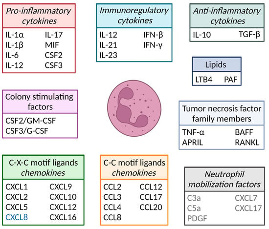
Neutrophils are the body’s first line of defense against pathogens, e.g., bacteria and fungi, and comprise 50–70% of the white blood cell population in human circulation. They are essential immune cells, and patients that lack (mature) neutrophils often succumb to severe opportunistic bacterial infections [1]. Neutrophils contain at least four types of granules: azurophilic/primary-, specific/secondary-, gelatinase/tertiary-, and secretory-granules and are, therefore, together with eosinophils and basophils, also known as granulocytes [2]. The different classes of granules are formed sequentially during neutrophil differentiation and contain different proteins important to pathogen killing (Figure 1) [2]. In addition to granule proteins, neutrophils also produce various cytokines and chemokines important for, e.g., pathogen killing and the attraction of leukocytes, respectively (Figure 2). Cytokines comprise a large group of secreted pro- and anti-inflammatory factors, grouped based on their structural homology, the similarity of their receptors and/or function (Figure 2). Chemokines are a subgroup of cytokines whose generic function is to induce cell migration.
Figure 1. Neutrophil development in the bone marrow. Long-term hematopoietic stem cells give rise to mature neutrophils via several stem and progenitor cell stages, promyelocytes, myelocytes, metamyelocytes and band cells. Granule content differs between various stages of differentiation and comprises proteins like neutrophil elastase, collagenase and gelatinase. LT-HSC: long-term hematopoietic stem cell; GMP: granulocyte–monocyte progenitor; MMP: matrix metalloproteinase.
Figure 2. Cytokines and chemokines produced by neutrophils and/or other cells that affect neutrophil function, which are discussed in the full version of the review. IL = interleukin; MIF = macrophage migration inhibitory factor; CSF = colony-stimulating factor; IFN = interferon; TGF = transforming growth factor; LTB4 = leukotriene B4; PAF = platelet-activating factor; CXCL = C-X-C motif ligand; CCL = C-C motif ligand; TNF = tumor necrosis factor; APRIL = a proliferation-inducing ligand; BAFF = B cell-activating factor; RANKL = receptor activator of NF-κB ligand; C3a/C5a = complement factor 3a/5a; PDGF = platelet-derived growth factor. Of note, the CXCL8 gene (indicated in blue) is lacking in mice, and the neutrophil mobilization factors (shown in gray) are not produced by neutrophils but do affect neutrophil mobilization.
Neutrophils have a short half-life in blood, ranging from 13 to 19 hours under homeostatic conditions [3]. Given their rapid turnover, approximately 1 billion neutrophils per kilogram of body weight are produced daily, which can be extended to 10 billion under disease conditions, e.g., inflammation and cancer [4][5][6]. For a long time, it was believed that neutrophils were specialized cells that existed to prevent infections and could not be more versatile because of their short half-life. However, since several reports showed the prominent pro- and anti-tumorigenic roles of neutrophils in cancer, they have gained increased attention [7][8][9][10].
With increasing knowledge about neutrophil plasticity and function, as reviewed in the full version of this entry [11], there is growing interest in exploring new therapeutic interventions to harness neutrophils’ innate capabilities to target and eliminate pathogens and cancer cells. Such potential strategies could target neutrophil recruitment and polarity; modulate neutrophil activation; or reduce excessive inflammation. As the field of immunotherapy is continuously evolving, several innovative therapeutic approaches have been developed or are being developed and could be used to leverage the anti-tumorigenic potential of neutrophils and block excessive neutrophil-mediated inflammation in autoimmune diseases.

23. Cytokine Therapeutics

In conditions of exacerbated cytokine production, e.g., inflammatory and autoimmune disease, the inhibition of cytokine functions caused by monoclonal antibodies or receptor blockers has been successfully used in the clinic. For example, patients with rheumatoid arthritis and Crohn’s disease are effectively treated with various TNF-blocking monoclonal antibodies, while a human IL-12/IL-23 monoclonal antibody is used to treat psoriasis patients, both resulting in reduced neutrophil infiltration into affected tissues [12][13]. Cytokines can also be therapeutically administered, as is the case for, e.g., CSF3 in congenital neutropenia patients and IFN-α for hepatitis B [14][15]. However, cytokines are pleiotropic, resulting in unwanted systemic effects, and have a narrow therapeutic range because of, among other things, a short blood half-life and unfavorable tissue distribution, making cytokine therapy challenging [16]. The cytokine engineering field has progressed tremendously over the last few years because of the development of novel techniques and a better understanding of cytokine biology, making it possible to alter cytokines so that they, e.g., can bind specific receptors with a higher affinity, leading to reduced dosing and fewer off-target effects caused by binding to other receptors, as is performed for the IL-2 “superkine” (MDNA11), currently being tested in clinical trials [17]. Furthermore, the half-life of cytokines can be extended by employing polyethylene glycol (PEG), a process that increases the molecular weight of the protein. This modification reduces renal clearance, protecting cytokines from degradation due to proteolytic enzymes and reducing their interaction with plasma constituents, thereby diminishing immunogenicity [18]. Another strategy often used to circumvent the limitations of cytokine drugs is the creation of synthetic cytokines (synthekines) using computational tools, overcoming things like pleiotropy, redundancy, poor pharmacokinetics and toxicity [19]. Multiple engineered cytokines are currently in clinical trials, as reviewed by Deckers et al. [20].

34. Immunocytokines

Genetically fusing a cytokine to another protein can help reshape the cytokine’s biodistribution profile, overcome poor pharmacokinetic properties and help promote tumor localization. This application is especially interesting in cancer, where a cytokine can be fused to a therapeutic antibody, specifically recognizing a tumor-associated antigen. These fusion constructs, called immunocytokines, hold promise as potential treatments and are currently undergoing evaluation in clinical trials [20]. For example, the CD38–IFNα2b immunocytokine TAK-573 is being tested in a phase I/II clinical trial for refractory multiple myeloma. Despite their potential, some immunocytokines have a lot of side effects because of the off-target binding of the cytokine to its receptor, resulting in the so-called “sink effect”, requiring high doses of the drug. Several engineering strategies are being developed to make the cytokine only active when it is near the tumor, one of which is Orionis Biosciences’ Activity-on-Target cytokines (AcTakines). These AcTakines are engineered to have a reduced receptor affinity, hampering cytokine activity until the immunocytokine accumulates near a target cell [21].

45. Immunotherapy

In addition to the cytokine part of immunocytokines, the antibody itself can also affect neutrophils by initiating neutrophil-mediated tumor cell killing via ADCC. All antibodies used for immunotherapy purposes are of the IgG isotype, which can bind various Fc gamma receptors on immune cells. Human neutrophils express the activating Fc-gamma receptors FcγRI (CD64), FcγRIIa (CD32a) and FcγRIIIa (CD16a) [22][23]. In addition, they also express the inhibitory receptor FcγRIIb (CD32b) and the GPI-linked and, therefore, signaling dead, receptor FcγRIIIb (CD16b), of which the latter is by far the highest expressed FcγR in neutrophils [22][23]. Therefore, IgG antibodies are not very efficient in engaging neutrophils in tumor cell killing by themselves. However, a recent in vivo study showed the effective, neutrophil-mediated killing of B16 melanoma cells when combining an IgG antibody targeting gp75 (a protein expressed on B16 melanoma cells), an CD40 antagonist and TNF [24]. The findings indicated that a combination of all three components was necessary for successful tumor cell killing. This suggests that a multimodal approach combining immunotherapy with cytokine therapy could hold great potential for engaging neutrophils in tumor cell killing and could contribute to the development of novel strategies for cancer treatment.
In contrast to IgG antibodies, IgA antibodies strictly bind the activating FcαR (CD89), making them very efficient in activating neutrophils and inducing ADCC [22]. However, IgA antibodies have a short half-life because of fast clearance via the asialoglycoprotein and mannose receptors, recognizing the extensive glycosylation of IgA antibodies [25][26][27]. In addition, IgA antibodies lack a binding site for the neonatal Fc receptor (FcRn), which recycles IgG antibodies, thereby contributing to the short half-life compared with IgG antibodies [28]. The antibody engineering of IgA has been described and resulted in an IgA3.0 molecule with an increased stability and half-life, overcoming some major hurdles of IgA immunotherapy [29][30]. In addition to being effective in activating neutrophils from healthy donors and mice, preliminary data suggest that suppressive neutrophils are as capable as normal neutrophils in killing tumor cells with IgA antibodies, making them ideal candidates to induce all neutrophil subsets to kill cancer.


References

  1. Skokowa, J.; Dale, D.C.; Touw, I.P.; Zeidler, C.; Welte, K. Severe congenital neutropenias. Nat. Rev. Dis. Primers 2017, 3, 17032.
  2. Faurschou, M.; Borregaard, N. Neutrophil granules and secretory vesicles in inflammation. Microbes Infect. 2003, 5, 1317–1327.
  3. Lahoz-Beneytez, J.; Elemans, M.; Zhang, Y.; Ahmed, R.; Salam, A.; Block, M.; Niederalt, C.; Asquith, B.; Macallan, D. Human neutrophil kinetics: Modeling of stable isotope labeling data supports short blood neutrophil half-lives. Blood 2016, 127, 3431–3438.
  4. Dancey, J.T.; Deubelbeiss, K.A.; Harker, L.A.; Finch, C.A. Neutrophil kinetics in man. J. Clin. Investig. 1976, 58, 705–715.
  5. Price, T.H.; Chatta, G.S.; Dale, D.C. Effect of recombinant granulocyte colony-stimulating factor on neutrophil kinetics in normal young and elderly humans. Blood 1996, 88, 335–340.
  6. Ley, K.; Hoffman, H.M.; Kubes, P.; Cassatella, M.A.; Zychlinsky, A.; Hedrick, C.C.; Catz, S.D. Neutrophils: New insights and open questions. Sci. Immunol. 2018, 3, eaat4579.
  7. Masucci, M.T.; Minopoli, M.; Carriero, M.V. Tumor Associated Neutrophils. Their Role in Tumorigenesis, Metastasis, Prognosis and Therapy. Front. Oncol. 2019, 9, 1146.
  8. Musiani, P.; Allione, A.; Modica, A.; Lollini, P.L.; Giovarelli, M.; Cavallo, F.; Belardelli, F.; Forni, G.; Modesti, A. Role of neutrophils and lymphocytes in inhibition of a mouse mammary adenocarcinoma engineered to release IL-2, IL-4, IL-7, IL-10, IFN-alpha, IFN-gamma, and TNF-alpha. Lab. Investig. 1996, 74, 146–157.
  9. Schmielau, J.; Finn, O.J. Activated granulocytes and granulocyte-derived hydrogen peroxide are the underlying mechanism of suppression of t-cell function in advanced cancer patients. Cancer Res. 2001, 61, 4756–4760.
  10. Serafini, P.; Borrello, I.; Bronte, V. Myeloid suppressor cells in cancer: Recruitment, phenotype, properties, and mechanisms of immune suppression. Semin. Cancer Biol. 2006, 16, 53–65.
  11. Tsioumpekou, M.; Krijgsman, D.; Leusen, J.H.W.; Olofsen, P.A. The Role of Cytokines in Neutrophil Development, Tissue Homing, Function and Plasticity in Health and Disease. Cells 2023, 12, 1981.
  12. Monaco, C.; Nanchahal, J.; Taylor, P.; Feldmann, M. Anti-TNF therapy: Past, present and future. Int. Immunol. 2015, 27, 55–62.
  13. Leonardi, C.L.; Kimball, A.B.; Papp, K.A.; Yeilding, N.; Guzzo, C.; Wang, Y.H.; Li, S.; Dooley, L.T.; Gordon, K.B.; Investigators, P.S. Efficacy and safety of ustekinumab, a human interleukin-12/23 monoclonal antibody, in patients with psoriasis: 76-week results from a randomised, double-blind, placebo-controlled trial (PHOENIX 1). Lancet 2008, 371, 1665–1674.
  14. Dale, D.C.; Bonilla, M.A.; Davis, M.W.; Nakanishi, A.M.; Hammond, W.P.; Kurtzberg, J.; Wang, W.; Jakubowski, A.; Winton, E.; Lalezari, P.; et al. A Randomized Controlled Phase-Iii Trial of Recombinant Human Granulocyte-Colony-Stimulating Factor (Filgrastim) for Treatment of Severe Chronic Neutropenia. Blood 1993, 81, 2496–2502.
  15. Ye, J.Y.; Chen, J.L. Interferon and Hepatitis B: Current and Future Perspectives. Front. Immunol. 2021, 12, 733364.
  16. Baldo, B.A. Side Effects of Cytokines Approved for Therapy. Drug Saf. 2014, 37, 921–943.
  17. Levin, A.M.; Bates, D.L.; Ring, A.M.; Krieg, C.; Lin, J.T.; Su, L.; Moraga, I.; Raeber, M.E.; Bowman, G.R.; Novick, P.; et al. Exploiting a natural conformational switch to engineer an interleukin-2 ‘superkine’. Nature 2012, 484, 529–533.
  18. Harris, J.M.; Chess, R.B. Effect of pegylation on pharmaceuticals. Nat. Rev. Drug Discov. 2003, 2, 214–221.
  19. Huang, P.S.; Boyken, S.E.; Baker, D. The coming of age of de novo protein design. Nature 2016, 537, 320–327.
  20. Deckers, J.; Anbergen, T.; Hokke, A.M.; de Dreu, A.; Schrijver, D.P.; de Bruin, K.; Toner, Y.C.; Beldman, T.J.; Spangler, J.B.; de Greef, T.F.A.; et al. Engineering cytokine therapeutics. Nat. Rev. Bioeng. 2023, 1, 286–303.
  21. Huyghe, L.; Van Parys, A.; Cauwels, A.; Van Lint, S.; De Munter, S.; Bultinck, J.; Zabeau, L.; Hostens, J.; Goethals, A.; Vanderroost, N.; et al. Safe eradication of large established tumors using neovasculature-targeted tumor necrosis factor-based therapies. EMBO Mol. Med. 2020, 12, e11223.
  22. Brandsma, A.M.; Bondza, S.; Evers, M.; Koutstaal, R.; Nederend, M.; Jansen, J.H.M.; Rosner, T.; Valerius, T.; Leusen, J.H.W.; Ten Broeke, T. Potent Fc Receptor Signaling by IgA Leads to Superior Killing of Cancer Cells by Neutrophils Compared to IgG. Front. Immunol. 2019, 10, 704.
  23. Kerntke, C.; Nimmerjahn, F.; Biburger, M. There Is (Scientific) Strength in Numbers: A Comprehensive Quantitation of Fc Gamma Receptor Numbers on Human and Murine Peripheral Blood Leukocytes. Front. Immunol. 2020, 11, 118.
  24. Linde, I.L.; Prestwood, T.R.; Qiu, J.; Pilarowski, G.; Linde, M.H.; Zhang, X.; Shen, L.; Reticker-Flynn, N.E.; Chiu, D.K.; Sheu, L.Y.; et al. Neutrophil-activating therapy for the treatment of cancer. Cancer Cell 2023, 41, 356–372 e310.
  25. Boross, P.; Lohse, S.; Nederend, M.; Jansen, J.H.; van Tetering, G.; Dechant, M.; Peipp, M.; Royle, L.; Liew, L.P.; Boon, L.; et al. IgA EGFR antibodies mediate tumour killing in vivo. EMBO Mol. Med. 2013, 5, 1213–1226.
  26. Stockert, R.J.; Kressner, M.S.; Collins, J.C.; Sternlieb, I.; Morell, A.G. IgA interaction with the asialoglycoprotein receptor. Proc. Natl. Acad. Sci. USA 1982, 79, 6229–6231.
  27. Lee, S.J.; Evers, S.; Roeder, D.; Parlow, A.F.; Risteli, J.; Risteli, L.; Lee, Y.C.; Feizi, T.; Langen, H.; Nussenzweig, M.C. Mannose receptor-mediated regulation of serum glycoprotein homeostasis. Science 2002, 295, 1898–1901.
  28. Junghans, R.P.; Anderson, C.L. The protection receptor for IgG catabolism is the beta2-microglobulin-containing neonatal intestinal transport receptor. Proc. Natl. Acad. Sci. USA 1996, 93, 5512–5516.
  29. van Tetering, G.; Evers, M.; Chan, C.; Stip, M.; Leusen, J. Fc Engineering Strategies to Advance IgA Antibodies as Therapeutic Agents. Antibodies 2020, 9, 70.
  30. Stip, M.C.; Evers, M.; Nederend, M.; Chan, C.; Reiding, K.R.; Damen, M.J.; Heck, A.J.R.; Koustoulidou, S.; Ramakers, R.; Krijger, G.C.; et al. IgA antibody immunotherapy targeting GD2 is effective in preclinical neuroblastoma models. J. Immunother. Cancer 2023, 11, e006948.
More
ScholarVision Creations