Your browser does not fully support modern features. Please upgrade for a smoother experience.
Please note this is a comparison between Version 1 by Francesco Riganello and Version 2 by Catherine Yang.
Pain assessment and management in patients with disorders of consciousness (DOC) is a challenging and important aspect of care, with implications for detecting consciousness and promoting recovery.
- pain
- nociception
- disorders of consciousness
- consciousness
1. Introduction
Pain is a universal response to harmful stimuli, and is a fundamental aspect of the evolutionary process in living organisms [1][2][1,2]. Pain is a complex, multidimensional experience essential for survival and adaptation, serving as a vital communication system between the body and the brain [3]. It alerts an organism to potential threats and prompts it to take appropriate action [4]. Investigations of cellular mechanisms and behavioral responses related to nociceptor activation, tissue injury, inflammation, and the environmental context of these responses are starting to reveal the evolution of mechanisms and behaviors important for pain [2]. Consequently, pain has emerged as a universal response across diverse living beings, facilitating their ability to thrive and propagate their genetic material across generations [5][6][5,6].
2. Pain in DOC
Pain is a subjective experience, and its definition may vary from person to person. The International Association for the Study of Pain (IASP) approved a definition of pain in 1979, encompassing both the sensory and emotional dimensions of the experience and the association between tissue injury and pain [7][76]. The IASP modified its basic pain terminology in 2007, introducing new terms to describe the various aspects of pain [8][77]. However, the subjective nature of pain remains a fundamental aspect of the experience, and reporting on it becomes crucial, with the narrative approach being recommended to assess pain in subjects who can communicate (Figure 1, Table 2). Considering the current definition of pain, assessing it in non-communicative patients remains challenging [9][78].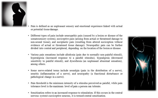
Figure 1.
Overview of various terminologies related to pain.
Table 2.
Classification and Description of Pain Types and Associated Terminology.
| Term | Definition of Pain |
|---|---|
| Pain | An unpleasant sensory and emotional experience associated with actual or potential tissue damage or described in terms of such damage. |
| Neuropathic Pain | Pain caused by a lesion or disease of the somatosensory system. |
| Central Neuropathic Pain | Neuropathic pain resulting from a lesion or disease of the central somatosensory nervous system. |
| Peripheral Neuropathic Pain | Neuropathic pain resulting from a lesion or disease of the peripheral somatosensory nervous system. |
| Nociceptive Pain | Pain that arises from actual or threatened damage to non-neural tissue and is due to the activation of nociceptors. |
| Nociplastic Pain | Pain that arises from altered nociception despite no clear evidence of actual or threatened tissue damage. |
| Types of Pain Sensations: | |
| Allodynia | Pain due to a stimulus that does not normally provoke pain. |
| Hyperalgesia | An increased response to a stimulus which is normally painful. |
| Hypoalgesia | Decreased sensitivity to painful stimuli. |
| Anesthesia dolorosa | Pain in an area or region which is anesthetic. |
| Dysesthesia | An unpleasant abnormal sensation, whether spontaneous or evoked. |
| Hyperesthesia | Increased sensitivity to stimulation, excluding the special senses. |
| Hyperpathia | A painful syndrome characterized by an abnormally painful reaction to a stimulus, especially a repetitive stimulus, as well as an increased threshold. |
| Hypoesthesia | Decreased sensitivity to stimulation, excluding the special senses. |
| Paresthesia | An abnormal sensation, whether spontaneous or evoked. |
| Nerve-Related Terms: | |
| Neuralgia | Pain in the distribution of a nerve or nerves. |
| Neuritis | Inflammation of a nerve or nerves. |
| Neuropathy | A disturbance of function or pathological change in a nerve. |
| Nociception | The neural process of encoding noxious stimuli. |
| Nociceptive Neuron | A neuron that is capable of detecting noxious stimuli. |
| Nociceptive Stimulus | A stimulus that is damaging or threatens damage to normal tissues. |
| Nociceptor | A receptor preferentially sensitive to a noxious stimulus or to a stimulus which would become noxious if prolonged. |
| Noxious Stimulus | A stimulus that is damaging to normal tissues. |
| Pain Threshold and Tolerance: | |
| Pain Threshold | The minimum intensity of a stimulus that is perceived as painful. |
| Pain Tolerance Level | The maximum level of pain which a subject is prepared to tolerate. |
| Sensitization: | |
| Sensitization | An increased response to stimulation. |
| Central Sensitization | Increased responsiveness of nociceptive neurons in the central nervous system to their normal or subthreshold afferent input. |
3. Pain Treatment in DOC
There is no consensus on the appropriate pharmacological treatment of pain in patients with DOC [21][90]. Medication should typically be given when there are clear behavioral indications of pain. Precise dosing of pharmacotherapy is crucial to prevent interference with the evaluation and therapy strategy for recovering consciousness. Moreover, if the strategy’s efficiency is still debatable, and yet to be proven through large-scale studies, the World Health Organization (WHO) proposes the WHO analgesic ladder, a pain management strategy developed in 1986 to provide adequate pain relief for cancer patients [22][91]. The ladder consists of three steps, with each step providing increasing levels of pain management options. The first step is for mild pain and involves the use of non-opioid analgesics such as NSAIDs or acetaminophen with or without adjuvants. The second step is for moderate pain and involves the use of weak opioids such as hydrocodone, codeine, or tramadol, with or without non-opioid analgesics and with or without adjuvants. The third step is for severe and persistent pain and involves the use of potent opioids such as morphine, methadone, fentanyl, oxycodone, buprenorphine, tapentadol, hydromorphone, or oxymorphone, with or without non-opioid analgesics and with or without adjuvants. It is essential to consider that inadequate pain control may impair intentional behavioral responses, whereas excessive treatment, using high doses of opioids to decrease pain, could negatively interfere with arousal [23][92] and may hinder cognitive recovery and attention [21][24][90,93]. The optimal drug dosage could preserve the patient’s arousal and consciousness, reducing the risk of misdiagnosis [25][26][94,95]. Different approaches are suggested in the presence of suspected symptomatic, mild, moderate, or neuropathic pain. In the case of managing pain with symptoms, the principles of proportionality and gradualness are considered, given their interactions with current therapies. In this case, treatment approaches typically involve the use of aspirin, paracetamol, nonsteroidal anti-inflammatory drugs, opioids, and γ-aminobutyric acid (GABA)-ergic agents [21][27][90,96]. In cases of suspected mild pain, administering aspirin, paracetamol, or nonsteroidal anti-inflammatory drugs is suggested [28][97]. For moderate or neuropathic pain, it is recommended to use high-dose aspirin or paracetamol, oral NSAIDs, and GABAergic agents [14][21][29][30][83,90,98,99]. Finally, for suspected severe pain the use of mixed agonists/antagonists, partial agonist opioids, parenteral opioids, antidepressants, anticonvulsants, and atypical agents is usually suggested [14][21][31][32][83,90,100,101]. Since around 89% of DOC patients are characterized by spasticity [33][102], which is associated with pain and other symptoms (i.e., increased hypertonia, altered sensorimotor control, and muscle spasms) [34][103], in cases of focal spasticity, or to treat severe or worsening cases, infiltration of botulinum [35][36][104,105] is suggested. For dystonia and diffuse spasticity, improvements were instead observed by administering intrathecal baclofen [37][106].4. Pain and Consciousness in DOC
Pain treatment is a relevant aspect of the management of DOC patients. However, pain characteristics related to the presence/absence of behavioral responses, and the modifications observed in biomarkers during noxious stimuli, can provide information on the covert content of consciousness (Table 23).Table 23.
Common Signs and Characteristics Evaluated in Pain in DOC Patients.
| Signs/Symptoms/Characteristics | Description | Evaluation in MCS Patients | Evaluation in VS/UWS Patients |
|---|---|---|---|
| Motor Response | Assessed in the NCS and NCS-R as part of the behavioral response to pain stimuli. | Higher-level responses, such as flexion or withdrawal. | Lower-level responses, such as abnormal posturing or none/flaccid. |
| Verbal Response | Evaluated in the NCS and NCS-R; factors such as crying, groaning, or intelligible verbalization are considered. | Higher-level responses, such as vocalization or intelligible verbalization. | Lower-level responses, such as groaning or no response. Necessary to consider lower responses due to tracheostomy conditions. |
| Facial Expression | Assessed as part of NCS and NCS-R, includes evaluation of reactions like grimacing. | More expressive, such as a cry or grimace. | Startled/oral reflexive movements or no response. |
| Visual Expression | Assessed as part of NCS, includes evaluation of reactions like fixation. | Higher-level responses, such as fixation and eye movements. | Startled or no response. |
| Pain Localization | Higher-level behavior indicative of pain as assessed by the NCS and NCS-R. | Observable. | Not observable. |
| Personalized Stimulation Reaction | Involves reactions to personalized stimuli, such as opening the hand, abducting the upper limbs, and mobilizing the head. | More demonstrated. | Less demonstrated. |
| Cardiac Frequency (Heart Rate Variability) | HRV can be used to assess autonomic responses to pain, such as increased sympathetic activity and reduced vagal modulation. | More stable HRV. | Increased sympathetic activity and reduced vagal modulation. |
| Galvanic Skin Response (GSR) | GSR measurements can indicate physiological responses to pain stimuli. | Trace conditioning was observed in healthy controls. No studies are present for MCS patients. | Can show trace conditioning to noxious stimuli. |
| Tracheostomy Condition | Pain assessment should consider lower cut-off values for tracheostomized patients due to lower verbal subscale scores. | Not applicable. | Lower cut-off values for nociception. |
| Spasticity | Severe spasticity can affect pain assessment in DOC patients. | Possible. | Possible. |