Your browser does not fully support modern features. Please upgrade for a smoother experience.
Please note this is a comparison between Version 2 by Rita Xu and Version 3 by Rita Xu.
Autism spectrum disorder (ASD) is a neurodevelopmental condition with a prevalence rate of 2.78%, and it is characterized by deficits in sociability and communication and restricted patterns of interests and activities. Remarkably, this psychiatric disorder exhibits a pronounced gender bias, with 80% of children diagnosed with ASD being boys.
- Autism Spectrum Disorder
- brain sexual differentiation
- neuroinflammation
1. Introduction
Autism spectrum disorder (ASD) is typically diagnosed in toddlers and young children who exhibit social impairment, deficits in communication, and restricted patterns of interests and activities [1]. The spectral nature of the disorder is remarkable, with some individuals being highly functional and presenting only mild, although challenging, symptoms, while others require substantial support and develop no language skills. In recent decades, the incidence of ASD has increased [2], lately showing a prevalence rate of 2.78% [3]. Epidemiological data suggest that ASD results from the interaction of different genes during development, while symptoms and severity largely depend on environmental factors that act upon these genes and alter the trajectory of brain development [4][5][6].
Remarkably, ASD exhibits a strong gender bias, being diagnosed 3.8 times more frequently in boys than in girls [7][8]. This intriguing bias persists even in the context of the increased incidence of ASD observed in later years [3]. Females may be underdiagnosed due to their tendency to camouflage autistic traits [9] and the possibility that ASD manifests differently in females than in males, as proposed by the female autism phenotype theory [10]. These diagnostic challenges may partially explain the male bias, but various lines of evidence suggest that biological factors contribute to female resilience and male vulnerability to ASD.
On one hand, the female protective effect model suggests that females require a greater etiologic load, such as a higher genetic mutation burden, to exhibit ASD symptoms than males [11]. This model of female resilience is supported by the higher risk of autistic impairments observed in family members of ASD females than in those of ASD males [12][13]. Furthermore, women with ASD have a higher burden of copy number variants and autosomal single-nucleotide variants than males with ASD [14].
On the other hand, the extreme male brain theory suggests that males may be more vulnerable to developing ASD because the characteristics of the disorder represent an extreme form of the male pattern, while females are less likely to exhibit autistic impairments due to their inherently higher social skills and empathetic behaviors [15]. This theory is linked to the hypothesis that fetal testosterone and estrogen can influence tendency to express altered social behavior [16][17], although this hypothesis has been challenged [18].
2. Mouse Models of ASD
2.1. Mouse Models of Psychiatric Disorders
Currently, no animal models have been proven to suffer from known psychiatric diseases, share their causes and symptoms, and respond similarly to treatments validated in humans. Thus, homologous models of psychiatric diseases are rare. Previously, the main utility of animal models in pre-clinical psychiatry was to validate drugs that target psychiatric symptoms. For example, rodents exposed to an open-field environment experience a conflict between their fear-induced thigmotaxis and their desire to explore the new surroundings [19]. A fearful animal would mainly explore the periphery of the field and avoid the center, particularly if it is brightly lit. For many years, this test, which is known as the open-field test, has been utilized to evaluate candidate anxiolytic drugs, demonstrating a high predictive value. Indeed, drugs that increased the exploration of the center of an open field in rodents demonstrated anxiolytic effects when administered to humans, although some exceptions were also observed [20]. Despite the lack of homologous models of psychiatric diseases, recent efforts have allowed the development of valuable animal models that serve as investigative tools [21][22]. Based on the original proposal of Willner [23], animal models of human mental disorders are required to fulfill construct, face, and predictive validity. Although no model completely accounts for all three requirements and other authors have proposed different levels of validity [24], researchers attempt to make their models as valuable as possible to ensure that they can translate their discoveries into theour understanding of human disorders. Construct validity ensures that the same etiological cause of the disease is responsible for the phenotype observed in the animal. To assess construct validity, it is necessary to understand the implicit or explicit hypothesis regarding the disease’s cause, such as the association between specific genes and their products and particular disorders. As a result, researchers have generated numerous genetically modified animals with genetic mutations similar to those found in humans or exposed animals to environmental factors associated with increased prevalence of certain pathologies. Behavioral and pharmacological analyses of these models are crucial in identifying associated phenotypic changes [25]. These analyses allow the comparison of different etiological hypotheses and the exploration of pathophysiological pathways [26]. Animal models are also expected to exhibit face validity, which means that the changes observed in the animal should be comparable to the symptoms seen in human patients. This requirement is based on the assumption that the phenotype observed in the animal arises from the same physiological processes as the human disease. However, this belief is not always accurate and can be difficult to confirm, especially since most psychiatric disorders have complex symptoms that may be caused by various genetic and environmental factors. To address this complexity, researchers have proposed the concept of endophenotype, which is a valuable tool that can advance theour understanding of psychiatric disorders [27]. The predictive validity of an animal model is demonstrated when a treatment tested on the animal has comparable effects on humans, as exemplified using benzodiazepines [20]. Occasionally, these effects can be observed without thorough comprehension of the biological processes that underlie them. As a result, some researchers regard predictive models as only partially beneficial and subject to undesirable side effects, particularly when the mechanisms of the disease and treatment are not fully understood. In summary, for an animal model of a psychiatric disorder to be valuable, it should have construct, face, and predictive validity, or at least some of these factors. The more the model imitates the etiological factors of a disease, manifests the phenotype/symptoms, and precisely forecasts the outcome of suggested treatments, the more advantageous it is to researchers who study the psychiatric disease.2.2. Tools to Evaluate Face Validity of Mouse Models of ASD and Limitations Related to Sex
Mouse models of ASD are primarily evaluated based on their face validity. Initially, reports referred to three core symptoms of autism for diagnosis [28], while the latest version of the Diagnostic and Statistical Manual of Mental Illnesses (DSM) focuses on two symptoms: persistent deficits in social communication and social interactions across multiple contexts, as well as the expression of restricted and repetitive behaviors, interests, and activities (including hyper- or hypo-reactivity to sensory stimuli) [1]. Importantly, the severity of these symptoms varies highly between individuals with ASD, which is not usually considered in pre-clinical studies. Behavioral tests have been designed based on these diagnostic criteria and divided into two categories to measure these behaviors in rodents: (1) sociability and communication deficits and (2) repetitive and stereotyped behaviors [29][30]. Initially, research into the influence of sex differences on behavior led to unintended consequences and the limiting of behavioral neuroscience research to male subjects because they were believed to be less influenced by the cyclic production of gonadal steroids that result from ovary function. Female behavior was only considered relevant in terms of understanding reproductive behavior, resulting in a poor understanding of sex differences in behavior and creating pre-clinical research biased in favor of male subjects [31]. However, theour current understanding suggests that female behavior may not be much different or difficult to study than male behavior [32], and the extended mandate of funding agencies to include subjects of both sexes has opened a new wave of data that shows sex’s influence on a wide variety of behaviors, besides reproductive behaviors. Researchers will discuss the tests most commonly used to evaluate ASD-relevant behaviors and specify whether there are differences in their applicability or results related to the sex of the animal (Figure 1).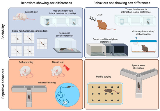
Figure 1. Sex differences in tests commonly used to assess sociability and repetitive behaviors in ASD models. The tests are categorized based on the core behavior that they measure (sociability or repetitive behaviors) and whether they yield different results in male and female animals. Created with BioRender.com.
2.2.1. Tests to Evaluate Sociability and Communication Deficits in Mice
The species Mus musculus is known to be highly social, exhibiting reciprocal social interactions, communal nesting, sexual and parenting behaviors, territorial scent marking, and aggressive behaviors [33][34][35][36]. Many social assays have been reported in the field of behavioral neuroscience. However, in researchersthis article, we will specifically discuss the tests commonly used to evaluate social impairments in mouse models of ASD. It is probable that the most popular test to assess sociability is the three-chamber social interaction test, which has been chosen by numerous scientists because it allows for automatization of measurements and can be easily standardized [37][38]. In this test, animals are initially allowed to explore a cage divided into three compartments. A stimulus animal is then placed in one of the lateral compartments, known as the “social side”, and either the time that the test mouse spends exploring the social stimulus or the time spent in the social chamber is measured. Different modifications to this test have been proposed, including changing the size of the cage and placing an object into the other lateral compartment [39][40]. Usually, a young mouse of the same sex as the test mouse is employed as a stimulus. Although strain differences have been reported, most mice will prefer the social side and, therefore, spend more time in that compartment than in that containing an inanimate object [37][39][41]. This test has the advantage of placing the stimulus mouse in a cage, which is unreachable by the test mouse, and it can be performed with aggressive animals, with many male adult mice being aggressive. In addition, this test is routinely used to measure social memory, adding a third phase in which a novel stimulus replaces the object, and the test mouse then shows a preference for such novelty. This test has been used to assess sociability in both males and females, and both sexes show similar levels of exploration of the young and same-sex social stimulus [42][43][44] (Figure 1, right panel). However, social novelty recognition is influenced by the sex and strain of the animal [44] (Figure 1, left panel). Additionally, this test can be employed to assess sex preference by presenting a female and a male adult mouse. Sex preference is also influenced by the sex and the strain of the animal [44]. The reciprocal social interaction test is certainly more ethologically relevant, as it allows the animals to freely interact with each other [30]. However, it is also more time consuming, as videos should be scored manually (although automated measurement has been achieved using machine learning approaches [45]). Besides the advantage of allowing the characterization of the mode of social engagement between the mice, a main concern is how animals should be paired, as the partner may influence the outcome of the test [46][47]. Moreover, in strains such as CF1, CBA or CD1, which have been reported to exhibit high levels of aggressive behavior [48], this test cannot be applied to adult males, as their aggression may overshadow any potential differences in sociability. A similar paradigm can be used to explore juvenile play, which is a social behavior typical of young animals. Although previous work on rats showed that females played less than males (reviewed in [49]), reports on mice are heterogenous. C57BL/6J (B6) and CD1 females solicit play activities more often than males [36][50], while outbred CF1 males performed more of these behaviors than females [43] (Figure 1, left panel). An evaluation of juvenile play in the four-core genotype (FCG) model in B6 background showed that XY males solicited play activities more often than any other group, suggesting that both gonadal hormones and sex chromosomes are relevant to the expression of this behavior [36]. In addition to performing a social novelty trial in the three-chamber social interaction test, social memory can be evaluated via a social habituation/recognition task [51]. In this test, animals are sequentially exposed to a stimulus mouse for a short period, showing habituation, and then exposed to a novel animal to evaluate their response to novelty. As for free social interaction, when aggressive animals are being assessed, stimuli can be presented in a small cage to avoid attacks. Social conditioned place preference is a useful tool for the evaluation of the rewarding nature of social interactions in young mice [52][53]. However, some strains (e.g., BALB/c) do not display social conditioning. This test shows no sex differences when performed with young animals [52]. However, female and male adult mice exhibit a differential response to social conditioning that depends on previous social conditions, such as isolation versus group housing [53]. Olfactory habituation/dishabituation to social odors is a valuable tool for the assessment of the response to a social stimulus, as it lacks the confounding effect of a second mouse with its own sociability levels. Mice tend to sniff a novel odor and then reduce their exploration as they acclimatize to the stimulus [54]. A dishabituation effect is observed when a different odor is introduced, and animals reinstate a high level of sniffing. Using social odors, it is possible to evaluate whether animals can discriminate between the same and different social odors and assess whether these odors are more salient to some animals than others [55]. No sex differences were observed in response to non-social odors [56]. Females, however, are more sensitive than male to social odors [57], possibly due to sex differences in the development of the olfactory system [58]. Although it is not yet well understood how mice communicate, ultrasonic vocalizations (USVs) appear to contribute to the communication of information and social bonding [59][60][61][62][63][64]. This observation is especially relevant to pups, and the analysis of ultrasonic vocalizations in pups separated from their dam and nest is the gold standard method for the measurement of alterations in sociability in newborns [61][64]. Pups of different strains perform different numbers of USVs, and each strain seems to have a unique repertoire of syllables [59]. In young, adolescent, and adult mice, both males and females vocalize, but the frequency and quality of the vocalizations depend on the eliciting stimulus and strain [60][65].2.2.2. Tests to Evaluate Repetitive and Stereotyped Behaviors in Mice
Mice show spontaneous motor stereotypies, including self-grooming and burying, which can develop into repetitive behaviors if they persist for prolonged periods [66]. Self-grooming is an innate behavior involved in hygiene maintenance and thermoregulation, which can be easily assessed in a home cage or novel environment [40][43][55][67][68]. Adult male mice have been reported to spend more time self-grooming than females [43]. The splash test is an alternative method used to elicit self-grooming behavior, where a sucrose solution is splashed on the back of the animal, and the sweetness of the solution sustains the grooming behavior [69]. The time spent grooming after the splash can be affected by the sex of the animal, although such an effect depends on the mouse strain being studied [70]. In the marble burying test, animals are exposed to an environment in which marble balls have been placed on top of a thick floor of bedding, which elicits the burying behavior in most mouse strains [71][72]. The test involves quantifying the marbles buried at different times, typically lasting 20 min [71]. Although most reports on marble burying behavior only involve males [72], it has been shown that the estrous cycle alters the burying response in rats [73]. Spontaneous alternation in the Y maze or T maze can also be evaluated in an attempt to measure repetitive behaviors because mice typically alternate at levels significantly above chance, indicating their willingness to explore novel environments [74][75]. These tests do not require training, though animals need to be active and explorative. However, an important confounder of these tests is that they depend on spatial working memory [76]. Male and female mice similarly alternate in the Y maze [43]. Perseverative behaviors are also relatively common in mice and can be evaluated by measuring the flexibility of a mouse in terms of switching from a learned habit to a new habit. These reversal learning tasks are usually evaluated in mazes, such as the T-maze, Morris water maze, or Clock maze [37][77]. These tests require a substantial amount of training, and they are time consuming and cannot be used in a short age period (e.g., adolescence), although some attempts have been made to develop shorter protocols [78]. Using the FCG model, it was shown that the sex–chromosome complement affects learning of a reversed task, with XY animals showing more perseverative errors [79]. The insistence on exploring a known object, subject, or area over a novel example can be interpreted as analogous to the restriction in interests or insistence on sameness observed in human subjects with ASD. To explore this observation, mice have been evaluated via the novel object recognition task [55], the social habituation/recognition task [51], or the nose poking in a hole board task [80]. Although males have mostly been evaluated in these tests, sex differences have been observed, with females exhibiting better performance when objects were similar in a novel object recognition test [81]. Hypo- and hyper-reactivity to sensory stimuli can be readily evaluated in mice. Acoustic startle, air puff startle, tail flick, and hot plate can be used to evaluate adult animals. In addition, the development of sensory capacity and its response can be evaluated during the post-natal period [43][82]. Males show a stronger startle reactivity than females [83], and some sex differences in the nociceptive response have been reported [84].2.2.3. Tests for the Evaluation of Associated Symptoms
In a subset of individuals with ASD, there are associated symptoms related to other psychiatric disorders that exhibit high comorbidity with ASD, such as anxiety and depression. Various tests can be conducted to evaluate these behaviors, many of which show sex differences (reviewed in this issue, [85]). Additionally, ASD individuals may experience seizures, intellectual disability, sleep disruption, and gastrointestinal distress. Analogous phenotypes can be assessed in mice [86]. Seizures can be observed directly or recorded via electroencephalography (EEG). Different memory tasks that measure spatial learning (e.g., Morris water maze), contextual and cued fear conditioning, shock avoidance, object recognition, and operant tasks can be used to evaluate cognitive abilities. Running wheels and home cage monitoring systems can be used to evaluate sleep and circadian activity. Many of these parameters exhibit sex differences that should be considered when evaluating mouse models of ASD. Evaluating associated behaviors can help to strengthen the phenotypes that correspond to the core symptom, though they can also identify potential confounders or artifacts. For instance, strong anxiety- or depression-related behaviors may lead to low exploration of social stimuli, rendering social interaction data meaningless. The same is true for memory deficits regarding social habituation or odor hyposensitivity and the observation of affected olfactory habituation or pup USVs. Deciding which phenotypes are relevant to associated symptoms and which phenotypes are artifacts that can confound the interpretation of tests related to diagnostic symptoms presents an internal contradiction that needs to be addressed on a case-by-case basis. Furthermore, these confounders may impact one sex more than the other, thus contributing to the sex bias observed in the model. Attention-deficit/hyperactivity disorder (ADHD) is frequently observed as a comorbid condition with ASD [87]. However, there have been prior few studies that examined the occurrence of ADHD phenotypes in rodent models of ASD. ADHD primarily affects attention, learning, hyperactivity–impulsivity, and aggressiveness [1]. Various behavioral tests can be employed to assess behaviors associated with these symptoms [88][89]. Deficits in learning and memory can be evaluated using the Barnes maze or the novel object recognition task. Hyperactivity can be measured through locomotion assessments in the open-field test. Impulsivity and attention deficits can be evaluated in tests such as the spontaneous Y maze alternation test or the continuous performance test. Aggressiveness is typically assessed using the resident–intruder test. Interestingly, animals that lack the integrin CD103 exhibit both ASD- and ADHD-related behaviors, and their phenotypes also exhibit sex specificity [90]. Investigating ADHD-related behaviors in animal models of ASD can contribute to theour understanding of the biological connections between these neurodevelopmental disorders.2.2.4. Non-Behavioral Associated Symptoms
Mouse models of ASD recapitulate other symptoms observed in individuals with ASD, which may result from etiological factors and pathophysiological pathways and are worth investigating. Human studies have shown the presence of activated glia, neuroinflammation, and expression of pro-inflammatory cytokines in the brains of individuals diagnosed with ASD [91][92], as well as elevated levels of pro-inflammatory cytokines, both basally and in response to an inflammatory stimulus, in the plasma of ASD patients [93]. Alterations in glial function, neuroinflammation, and an altered response to inflammatory stimuli have also been reported in mouse models of ASD [67][82]. In addition, immunological dysfunction, such as T cell dysfunction, autoantibody production, and augmentation of pro-inflammatory cytokines, has been proposed in the pathogenesis of ASD ([93][94][95][96], reviewed in [97]). Investigating this in topic animal models has shown that cytokines can participate in the post-natal programming of adult sociability, and they can also modulate this behavior in the adult brain [40][42][67][82]. Also, mouse models of ASD have been generated after pre- or neo-natal exposure to inflammatory stimuli (e.g., PolyI:C, LPS or virus) [77][98][99][100][101][102][103][104][105][106][107][108][109][110][111][112]. Interestingly, sex differences in inflammatory responses and glial development and function have been reported in both humans and animals [113]. The autistic brain is characterized by hyperconnectivity in local circuits and hypoconnectivity between brain regions [114]. This observation is consistent with ASD being diagnosed before the age of 5, when synaptogenesis is most active in humans, a process that is next followed by active synaptic pruning and elimination [115]. Indeed, autistic brains show increased spine density in the apical dendrites of cortical pyramidal neurons [116], and many genes identified as providing susceptibility to ASD code for synaptic proteins or affect the morphogenesis of dendritic spines (reviewed in [114][117]). Alterations in synaptic function and brain activity have been studied in some mouse models of ASD to understand the relevance of these findings [117][118][119]. Additionally, many genetic models of ASD are built on the alteration of proteins involved in synaptic function, including neuroligins [120][121][122][123][124][125][126], neurexins [127], and shank proteins [128][129][130][131][132][133][134][135][136]. As discussed below, differences in dendritic growth and synaptic formation represent a known mechanism of brain sexual differentiation.2.3. Sex Differences in Mouse Models of ASD
Animal studies on ASD have traditionally favored male animals due to its higher incidence in boys, which has led to a lack of evidence on the impact of sex on these models, and rwesearchers believe that this problem may have delayed discovery in ASD. However, recent research has identified sex differences in ASD models, shedding light on possible mechanisms involved in the etiology and pathophysiology of the disorder. ResearchersWe have included specific notations to indicate the behavioral phenotypes observed in males and females when both sexes were analyzed, and the effect of sex was considered (violet was the male symbol and was the orange female symbol). For studies in which results were obtained from both male and female subjects, but the sex effect was not reported, researcherswe used black male and female symbols. It is important to note that when only male behavioral phenotypes are reported in a model, it indicates that researcherswe did not come across any reports that specifically addressed females in those studies. Among the most extensively studied pharmacological and environmental models of ASD are those generated via pre-natal exposure to valproic acid (VPA) or maternal immune activation (MIA). Various doses of VPA can be administered to animals at different gestational ages, resulting in ASD-related behaviors (as reviewed in [137]). While most studies have used only male animals, evidence shows that VPA affects ASD-related phenotypes in males, but does not do so in females [42][138]. For example, male mice exposed to 600 mg/kg VPA at gestational day (GD) 12.5 displayed reduced sociability in the three-chamber test, while female social interaction was not affected [42]. However, VPA affects female mice, as they show signs of neuroinflammation during the post-natal period [82] and in adulthood [42]. Remarkably, repetitive behaviors were not assessed in females, and, hence, evidence of the sex-specific effect of VPA on behavior is lacking and warrants further investigation. MIA models are generated by challenging the maternal immune system using an inflammatory stimulus. Commonly used inflammatory stimuli are the polyinosinic–polycytidylic acid (PolyI:C) that mimics viral infections and bacterial lipolysaccharides (LPS) that elicit an inflammatory response similar to the one triggered by a bacterial infection. When PolyI:C is administered at GD12, there is a consistent effect on sociability (reviewed in [139]). Although pre-natal PolyI:C exposure can affect social behavior in both male and female mice [107], some sex-specific differences can be observed that may depend on the gestational age at which the stimulus was administered, the strain of the mouse, and the dose and type of PolyI:C [139][140]. Similarly, while male mice pre-natally exposed to LPS show reduced sociability, female social behavior is unaffected [109][110][141]. Similar to the VPA model, LPS exposure leads to increased self-grooming in males, though this behavior has not been evaluated in females. When the human influenza virus is injected into pregnant dams at GD9.5 to elicit an inflammatory response, their adult offspring of both sexes exhibit reduced social interaction [98]. A neonatal inflammatory challenge also results in long-lasting effects on sociability, though the extent of the effect and occurrence of sex differences depends on factors such as mouse strain, drug dose, and age at administration [140][142][143][144][145]. Propionic acid (PPA) is a gut metabolite that can elicit neuroinflammatory responses [146][147]. It has been shown that intracerebroventricular (icv), subcutaneous (sc), or intraperitoneal (ip) administration of PPA can elicit behavioral alterations related to ASD, such as reduced social interactions and repetitive patterns of behavior [146][147][148][149]. Unfortunately, all studies on PPA have only used male subjects. However, maternal administration of PPA does not result in ASD-related behaviors in female and male offspring [150]. Male mice pre-natally exposed to a monoclonal antibody against contactin-associated protein-like 2 (Caspr2) show reduced sociability, increased repetitive behavior (marble burying), and inflexibility in learning [77]. However, the ASD-related behavioral phenotype of this model was not replicated when it was combined with the FCG model [104]. Several genetic models of ASD have been proposed. Many of them are constructed based on the notion that ASD results from synaptic alterations, and, thus, key synaptic proteins, such as neuroligins 1, 3, and 4; neurexin 1α; and shank proteins, have been targeted [120][121][122][123][124][125][126][127][128][129][130][131][132][133][134][135][136]. Other targeted molecules are peptides involved in social responses, such as oxytocin (OT) and vasopressin (AVP), and their receptors [151][152][153][154][155]. Others models are generated by replicating genetic alterations observed in human subjects with ASD or related disorders [156][157][158][159][160][161][162][163][164][165][166][167][168]. Finally, some mouse inbred strains, such as BTBR, Balb/c, and C58, exhibit reduced sociability and increased repetitive behaviors and are, thus, proposed to be studied as ASD models [39][59][60][166][169][170][171][172][173][174][175][176][177][178][179][180][181]. Unfortunately, many of these models have only been studied using male subjects, while the role of sex has often not been specifically analyzed.References
- American Psychiatric Association. Diagnostic and Statistical Manual of Mental Disorders; American Psychiatric Association: Arlington, TX, USA, 2013; ISBN 0-89042-555-8.
- Chiarotti, F.; Venerosi, A. Epidemiology of Autism Spectrum Disorders: A Review of Worldwide Prevalence Estimates since 2014. Brain Sci. 2020, 10, 274.
- Maenner, M.J.; Warren, Z.; Williams, A.R.; Amoakohene, E.; Bakian, A.V.; Bilder, D.A.; Durkin, M.S.; Fitzgerald, R.T.; Furnier, S.M.; Hughes, M.M.; et al. Prevalence and Characteristics of Autism Spectrum Disorder Among Children Aged 8 Years—Autism and Developmental Disabilities Monitoring Network, 11 Sites, United States, 2020. MMWR. Surveill. Summ. 2023, 72, 1–14.
- Muhle, R.; Trentacoste, S.V.; Rapin, I. The Genetics of Autism. Pediatrics 2004, 113, e472–e486.
- Hallmayer, J.; Cleveland, S.; Torres, A.; Phillips, J.; Cohen, B.; Torigoe, T.; Miller, J.; Fedele, A.; Collins, J.; Smith, K.; et al. Genetic Heritability and Shared Environmental Factors among Twin Pairs with Autism. Arch. Gen. Psychiatry 2011, 68, 1095–1102.
- Sandin, S.; Lichtenstein, P.; Kuja-Halkola, R.; Larsson, H.; Hultman, C.M.; Reichenberg, A. The Familial Risk of Autism. JAMA J. Am. Med. Assoc. 2014, 311, 1770–1777.
- Elsabbagh, M. Linking Risk Factors and Outcomes in Autism Spectrum Disorder: Is There Evidence for Resilience? BMJ 2020, 368, l6880.
- Loomes, R.; Hull, L.; Mandy, W.P.L. What Is the Male-to-Female Ratio in Autism Spectrum Disorder? A Systematic Review and Meta-Analysis. J. Am. Acad. Child Adolesc. Psychiatry 2017, 56, 466–474.
- Hull, L.; Petrides, K.V.; Allison, C.; Smith, P.; Baron-Cohen, S.; Lai, M.C.; Mandy, W. “Putting on My Best Normal”: Social Camouflaging in Adults with Autism Spectrum Conditions. J. Autism Dev. Disord. 2017, 47, 2519–2534.
- Hull, L.; Petrides, K.V.; Mandy, W. The Female Autism Phenotype and Camouflaging: A Narrative Review. Rev. J. Autism Dev. Disord. 2020, 7, 306–317.
- Skuse, D.H. Imprinting, the X-Chromosome, and the Male Brain: Explaining Sex Differences in the Liability to Autism. Pediatr. Res. 2000, 47, 9.
- Robinson, E.B.; Lichtenstein, P.; Anckarsäter, H.; Happé, F.; Ronald, A. Examining and Interpreting the Female Protective Effect against Autistic Behavior. Proc. Natl. Acad. Sci. USA 2013, 110, 5258–5262.
- Wigdor, E.M.; Weiner, D.J.; Grove, J.; Fu, J.M.; Thompson, W.K.; Carey, C.E.; Baya, N.; van der Merwe, C.; Walters, R.K.; Satterstrom, F.K.; et al. The Female Protective Effect against Autism Spectrum Disorder. Cell Genomics 2022, 2, 100134.
- Jacquemont, S.; Coe, B.P.; Hersch, M.; Duyzend, M.H.; Krumm, N.; Bergmann, S.; Beckmann, J.S.; Rosenfeld, J.A.; Eichler, E.E. A Higher Mutational Burden in Females Supports a “Female Protective Model” in Neurodevelopmental Disorders. Am. J. Hum. Genet. 2014, 94, 415–425.
- Greenberg, D.M.; Warrier, V.; Allison, C.; Baron-Cohen, S. Testing the Empathizing–Systemizing Theory of Sex Differences and the Extreme Male Brain Theory of Autism in Half a Million People. Proc. Natl. Acad. Sci. USA 2018, 115, 12152–12157.
- Auyeung, B.; Baron-Cohen, S.; Chapman, E.; Knickmeyer, R.; Taylor, K.; Hackett, G. Foetal Testosterone and the Child Systemizing Quotient. Eur. J. Endocrinol. Suppl. 2006, 155, 123–130.
- Auyeung, B.; Baron-Cohen, S.; Ashwin, E.; Knickmeyer, R.; Taylor, K.; Hackett, G.; Baron-Cohen, S.; Ashwin, E.; Knickmeyer, R.; Taylor, K.; et al. Fetal Testosterone and Autistic Traits. Br. J. Psychol. 2009, 100, 1–22.
- Kung, K.T.F.; Spencer, D.; Pasterski, V.; Neufeld, S.; Glover, V.; O’Connor, T.G.; Hindmarsh, P.C.; Hughes, I.A.; Acerini, C.L.; Hines, M. No Relationship between Prenatal Androgen Exposure and Autistic Traits: Convergent Evidence from Studies of Children with Congenital Adrenal Hyperplasia and of Amniotic Testosterone Concentrations in Typically Developing Children. J. Child Psychol. Psychiatry 2016, 57, 1455–1462.
- Belzung, C.; Griebel, G. Measuring Normal and Pathological Anxiety-like Behaviour in Mice: A Review. Behav. Brain Res. 2001, 125, 141–149.
- Prut, L.; Belzung, C. The Open Field as a Paradigm to Measure the Effects of Drugs on Anxiety-like Behaviors: A Review. Eur. J. Pharmacol. 2003, 463, 3–33.
- Nestler, E.J.; Hyman, S.E. Animal Models of Neuropsychiatric Disorders. Nat. Neurosci. 2010, 13, 1161–1169.
- Kaiser, T.; Feng, G. Modeling Psychiatric Disorders for Developing Effective Treatments. Nat. Med. 2015, 21, 979–988.
- Willner, P. The Validity of Animal Models of Depression. Psychopharmacology 1984, 83, 1–16.
- Belzung, C.; Lemoine, M. Criteria of Validity for Animal Models of Psychiatric Disorders: Focus on Anxiety Disorders and Depression. Biol. Mood Anxiety Disord. 2011, 1, 9.
- Lipska, B.K.; Weinberger, D.R. To Model a Psychiatric Disorder in Animals: Schizophrenia as a Reality Test. Neuropsychopharmacology 2000, 23, 223–239.
- Uliana, D.L.; Zhu, X.; Gomes, F.V.; Grace, A.A. Using Animal Models for the Studies of Schizophrenia and Depression: The Value of Translational Models for Treatment and Prevention. Front. Behav. Neurosci. 2022, 16, 935320.
- Gottesman, I.I.; Gould, T.D. The Endophenotype Concept in Psychiatry: Etymology and Strategic Intentions. Am. J. Psychiatry 2003, 160, 636–645.
- American Psychiatric Association. DSM-IV-TR; American Psychiatric Association: Arlington, TX, USA, 2000; ISBN 0-89042-024-6.
- Crawley, J.N. Translational Animal Models of Autism and Neurodevelopmental Disorders. Dialogues Clin. Neurosci. 2012, 14, 293–305.
- Silverman, J.L.; Yang, M.; Lord, C.; Crawley, J.N. Behavioural Phenotyping Assays for Mouse Models of Autism. Nat. Rev. Neurosci. 2010, 11, 490–502.
- Beery, A.K.; Zucker, I. Sex Bias in Neuroscience and Biomedical Research. Neurosci. Biobehav. Rev. 2011, 35, 565–572.
- Beery, A.K. Inclusion of Females Does Not Increase Variability in Rodent Research Studies. Curr. Opin. Behav. Sci. 2018, 23, 143–149.
- Moles, A.; Kieffer, B.L.; D’Amato, F.R. Deficit in Attachment Behavior in Mice Lacking the μ-Opioid Receptor Gene. Science 2004, 304, 1983–1986.
- Miczek, K.A.; Maxson, S.C.; Fish, E.W.; Faccidomo, S. Aggressive Behavioral Phenotypes in Mice. Behav. Brain Res. 2001, 125, 167–181.
- Burns-Cusato, M.; Scordalakes, E.M.; Rissman, E.F. Of Mice and Missing Data: What We Know (and Need to Learn) about Male Sexual Behavior. Physiol. Behav. 2004, 83, 217–232.
- Cox, K.H.; Rissman, E.F. Sex Differences in Juvenile Mouse Social Behavior Are Influenced by Sex Chromosomes and Social Context. Genes Brain Behav. 2011, 10, 465–472.
- Moy, S.S.; Nadler, J.J.; Young, N.B.; Perez, A.; Holloway, L.P.; Barbaro, R.P.; Barbaro, J.R.; Wilson, L.M.; Threadgill, D.W.; Lauder, J.M.; et al. Mouse Behavioral Tasks Relevant to Autism: Phenotypes of 10 Inbred Strains. Behav. Brain Res. 2007, 176, 4–20.
- Nadler, J.J.; Moy, S.S.; Dold, G.; Trang, D.; Simmons, N.; Perez, A.; Young, N.B.; Barbaro, R.P.; Piven, J.; Magnuson, T.R.; et al. Automated Apparatus for Quantitation of Social Approach Behaviors in Mice. Genes Brain Behav. 2004, 3, 303–314.
- Brodkin, E.S.; Hagemann, A.; Nemetski, S.M.; Silver, L.M. Social Approach-Avoidance Behavior of Inbred Mouse Strains towards DBA/2 Mice. Brain Res. 2004, 1002, 151–157.
- Depino, A.M.; Lucchina, L.; Pitossi, F. Early and Adult Hippocampal TGF-Β1 Overexpression Have Opposite Effects on Behavior. Brain. Behav. Immun. 2011, 25.
- Pietropaolo, S.; Guilleminot, A.; Martin, B.; D’Amato, F.R.; Crusio, W.E. Genetic-Background Modulation of Core and Variable Autistic-like Symptoms in Fmr1 Knock-out Mice. PLoS ONE 2011, 6, e17073.
- Kazlauskas, N.; Seiffe, A.; Campolongo, M.; Zappala, C.; Depino, A.M. Sex-Specific Effects of Prenatal Valproic Acid Exposure on Sociability and Neuroinflammation: Relevance for Susceptibility and Resilience in Autism. Psychoneuroendocrinology 2019, 110, 104441.
- Seiffe, A.; Federico Ramirez, M.; Darío Barrios, C.; Milagros Albarrán, M.; Mara Depino, A.; Ramirez, M.F.; Barrios, C.D.; Albarrán, M.M.; Depino, A.M. Early Estradiol Exposure Masculinizes Disease-relevant Behaviors in Female Mice. Eur. J. Neurosci. 2021, 53, 2483–2499.
- Kopachev, N.; Netser, S.; Wagner, S. Sex-Dependent Features of Social Behavior Differ between Distinct Laboratory Mouse Strains and Their Mixed Offspring. iScience 2022, 25, 103735.
- Hong, W.; Kennedy, A.; Burgos-Artizzu, X.P.; Zelikowsky, M.; Navonne, S.G.; Perona, P.; Anderson, D.J. Automated Measurement of Mouse Social Behaviors Using Depth Sensing, Video Tracking, and Machine Learning. Proc. Natl. Acad. Sci. USA 2015, 112, E5351–E5360.
- Argue, K.J.; McCarthy, M.M. Utilization of Same- vs. Mixed-Sex Dyads Impacts the Observation of Sex Differences in Juvenile Social Play Behavior. Curr. Neurobiol. 2000, 6, 17–23.
- Argue, K.J.; McCarthy, M.M. Characterization of Juvenile Play in Rats: Importance of Sex of Self and Sex of Partner. Biol. Sex Differ. 2015, 6, 1–13.
- Lidster, K.; Owen, K.; Browne, W.J.; Prescott, M.J. Cage Aggression in Group-Housed Laboratory Male Mice: An International Data Crowdsourcing Project. Sci. Rep. 2019, 9, 1–12.
- Auger, A.P.; Olesen, K.M. Brain Sex Differences and the Organisation of Juvenile Social Play Behaviour. J. Neuroendocrinol. 2009, 21, 519–525.
- Terranova, M.L.; Laviola, G.; Alleva, E. Ontogeny of Amicable Social Behavior in the Mouse: Gender Differences and Ongoing Isolation Outcomes. Dev. Psychobiol. 1993, 26, 467–481.
- Hörnberg, H.; Pérez-Garci, E.; Schreiner, D.; Hatstatt-Burklé, L.; Magara, F.; Baudouin, S.; Matter, A.; Nacro, K.; Pecho-Vrieseling, E.; Scheiffele, P. Rescue of Oxytocin Response and Social Behaviour in a Mouse Model of Autism. Nature 2020, 584, 252–256.
- Panksepp, J.B.; Lahvis, G.P. Social Reward among Juvenile Mice. Genes Brain Behav. 2007, 6, 661–671.
- Cann, C.; Venniro, M.; Hope, B.T.; Ramsey, L.A. Parametric Investigation of Social Place Preference in Adolescent Mice. Behav. Neurosci. 2020, 134, 435–443.
- Yang, M.; Crawley, J.N. Simple Behavioral Assessment of Mouse Olfaction. Curr. Protoc. Neurosci. 2009, 48.
- Campolongo, M.; Kazlauskas, N.; Falasco, G.; Urrutia, L.; Salgueiro, N.; Höcht, C.; Depino, A.M. Sociability Deficits after Prenatal Exposure to Valproic Acid Are Rescued by Early Social Enrichment. Mol. Autism 2018, 9, 36.
- Pankevich, D.E.; Bale, T.L. Stress and Sex Influences on Food-Seeking Behaviors. Obesity 2008, 16, 1539–1544.
- Baum, M.J.; Keverne, E.B. Sex Difference in Attraction Thresholds for Volatile Odors from Male and Estrous Female Mouse Urine. Horm. Behav. 2002, 41, 213–219.
- Kass, M.D.; Czarnecki, L.A.; Moberly, A.H.; McGann, J.P. Differences in Peripheral Sensory Input to the Olfactory Bulb between Male and Female Mice. Sci. Rep. 2017, 7, 1–15.
- Scattoni, M.L.; Gandhy, S.U.; Ricceri, L.; Crawley, J.N. Unusual Repertoire of Vocalizations in the BTBR T+tf/J Mouse Model of Autism. PLoS ONE 2008, 3, 48–52.
- Panksepp, J.B.; Jochman, K.A.; Kim, J.U.; Koy, J.K.; Wilson, E.D.; Chen, Q.; Wilson, C.R.; Lahvis, G.P. Affiliative Behavior, Ultrasonic Communication and Social Reward Are Influenced by Genetic Variation in Adolescent Mice. PLoS ONE 2007, 2, e351.
- Branchi, I.; Santucci, D.; Alleva, E. Ultrasonic Vocalisation Emitted by Infant Rodents: A Tool for Assessment of Neurobehavioural Development. Behav. Brain Res. 2001, 125, 49–56.
- Egnor, S.E.R.; Seagraves, K.M. The Contribution of Ultrasonic Vocalizations to Mouse Courtship. Curr. Opin. Neurobiol. 2016, 38, 1–5.
- Moles, A.; Costantini, F.; Garbugino, L.; Zanettini, C.; D’Amato, F.R. Ultrasonic Vocalizations Emitted during Dyadic Interactions in Female Mice: A Possible Index of Sociability? Behav. Brain Res. 2007, 182, 223–230.
- Scattoni, M.L.; Crawley, J.; Ricceri, L. Ultrasonic Vocalizations: A Tool for Behavioural Phenotyping of Mouse Models of Neurodevelopmental Disorders. Neurosci. Biobehav. Rev. 2009, 33, 508–515.
- Faure, A.; Pittaras, E.; Nosjean, A.; Chabout, J.; Cressant, A.; Granon, S. Social Behaviors and Acoustic Vocalizations in Different Strains of Mice. Behav. Brain Res. 2017, 320, 383–390.
- Gandhi, T.; Lee, C.C. Neural Mechanisms Underlying Repetitive Behaviors in Rodent Models of Autism Spectrum Disorders. Front. Cell. Neurosci. 2021, 14, 1–44.
- Lucchina, L.; Depino, A.M. Altered Peripheral and Central Inflammatory Responses in a Mouse Model of Autism. Autism Res. 2014, 7, 273–289.
- Kalueff, A.V.; Stewart, A.M.; Song, C.; Berridge, K.C.; Graybiel, A.M.; Fentress, J.C. Neurobiology of Rodent Self-Grooming and Its Value for Translational Neuroscience. Nat. Rev. Neurosci. 2016, 17, 45–59.
- Teissier, A.; Le Magueresse, C.; Olusakin, J.; Andrade da Costa, B.L.S.; De Stasi, A.M.; Bacci, A.; Imamura Kawasawa, Y.; Vaidya, V.A.; Gaspar, P. Early-Life Stress Impairs Postnatal Oligodendrogenesis and Adult Emotional Behaviour through Activity-Dependent Mechanisms. Mol. Psychiatry 2020, 25, 1159–1174.
- Pitzer, C.; Kurpiers, B.; Eltokhi, A. Sex Differences in Depression-Like Behaviors in Adult Mice Depend on Endophenotype and Strain. Front. Behav. Neurosci. 2022, 16, 1–8.
- Thomas, A.; Burant, A.; Bui, N.; Graham, D.; Yuva-Paylor, L.A.; Paylor, R. Marble Burying Reflects a Repetitive and Perseverative Behavior More than Novelty-Induced Anxiety. Psychopharmacology 2009, 204, 361–373.
- de Brouwer, G.; Fick, A.; Harvey, B.H.; Wolmarans, D.W. A Critical Inquiry into Marble-Burying as a Preclinical Screening Paradigm of Relevance for Anxiety and Obsessive–Compulsive Disorder: Mapping the Way Forward. Cogn. Affect. Behav. Neurosci. 2019, 19, 1–39.
- Schneider, T.; Popik, P. Attenuation of Estrous Cycle-Dependent Marble Burying in Female Rats by Acute Treatment with Progesterone and Antidepressants. Psychoneuroendocrinology 2007, 32, 651–659.
- Dember, W.N.; Fowler, H. Spontaneous Alternation Behavior. Psychol. Bull. 1958, 55, 412–428.
- Lalonde, R. The Neurobiological Basis of Spontaneous Alternation. Neurosci. Biobehav. Rev. 2002, 26, 91–104.
- Isseroff, A. Limited Recovery of Spontaneous Alternation after Extensive Hippocampal Damage: Evidence for a Memory Impairment. Exp. Neurol. 1979, 64, 284–294.
- Brimberg, L.; Mader, S.; Jeganathan, V.; Berlin, R.; Coleman, T.R.; Gregersen, P.K.; Huerta, P.T.; Volpe, B.T.; Diamond, B. Caspr2-Reactive Antibody Cloned from a Mother of an ASD Child Mediates an ASD-like Phenotype in Mice. Mol. Psychiatry 2016, 21, 1663–1671.
- Remmelink, E.; Smit, A.B.; Verhage, M.; Loos, M. Measuring Discrimination- and Reversal Learning in Mouse Models within 4 Days and without Prior Food Deprivation. Learn. Mem. 2016, 23, 660–667.
- Aarde, S.M.; Genner, R.M.; Hrncir, H.; Arnold, A.P.; Jentsch, J.D. Sex Chromosome Complement Affects Multiple Aspects of Reversal-Learning Task Performance in Mice. Genes Brain Behav. 2021, 20, 1–11.
- Moy, S.S.; Nadler, J.J.; Poe, M.D.; Nonneman, R.J.; Young, N.B.; Koller, B.H.; Crawley, J.N.; Duncan, G.E.; Bodfish, J.W. Development of a Mouse Test for Repetitive, Restricted Behaviors: Relevance to Autism. Behav. Brain Res. 2008, 188, 178–194.
- Bettis, T.; Jacobs, L.F. Sex Differences in Object Recognition Are Modulated by Object Similarity. Behav. Brain Res. 2012, 233, 288–292.
- Kazlauskas, N.; Campolongo, M.; Lucchina, L.; Zappala, C.; Depino, A.M. Postnatal Behavioral and Inflammatory Alterations in Female Pups Prenatally Exposed to Valproic Acid. Psychoneuroendocrinology 2016, 72, 11–21.
- Plappert, C.F.; Rodenbücher, A.M.; Pilz, P.K.D. Effects of Sex and Estrous Cycle on Modulation of the Acoustic Startle Response in Mice. Physiol. Behav. 2005, 84, 585–594.
- Mitrovic, I.; Margeta-Mitrovic, M.; Bader, S.; Stoffel, M.; Jan, L.Y.; Basbaum, A.I. Contribution of GIRK2-Mediated Postsynaptic Signaling to Opiate and A2-Adrenergic Analgesia and Analgesic Sex Differences. Proc. Natl. Acad. Sci. USA 2003, 100, 271–276.
- Mir, F.R.; Rivarola, M.A. Sex Differences in Anxiety and Depression: What Can (and Cannot) Preclinical Studies Tell Us? Sexes 2022, 3, 141–163.
- Crawley, J.N.; Paylor, R. A Proposed Test Battery and Constellations of Specific Behavioral Paradigms to Investigate the Behavioral Phenotypes of Transgenic and Knockout Mice. Horm. Behav. 1997, 31, 197–211.
- Lai, M.C.; Kassee, C.; Besney, R.; Bonato, S.; Hull, L.; Mandy, W.; Szatmari, P.; Ameis, S.H. Prevalence of Co-Occurring Mental Health Diagnoses in the Autism Population: A Systematic Review and Meta-Analysis. Lancet Psychiatry 2019, 6, 819–829.
- Mortimer, N.; Ganster, T.; O’Leary, A.; Popp, S.; Freudenberg, F.; Reif, A.; Soler Artigas, M.; Ribasés, M.; Ramos-Quiroga, J.A.; Lesch, K.P.; et al. Dissociation of Impulsivity and Aggression in Mice Deficient for the ADHD Risk Gene Adgrl3: Evidence for Dopamine Transporter Dysregulation. Neuropharmacology 2019, 156, 107557.
- Dougnon, G.; Matsui, H. Modelling Autism Spectrum Disorder (ASD) and Attention-Deficit/Hyperactivity Disorder (ADHD) Using Mice and Zebrafish. Int. J. Mol. Sci. 2022, 23, 7550.
- Jhun, M.; Panwar, A.; Cordner, R.; Irvin, D.K.; Veiga, L.; Yeager, N.; Pechnick, R.N.; Schubloom, H.; Black, K.L.; Wheeler, C.J. CD103 Deficiency Promotes Autism (ASD) and Attention-Deficit Hyperactivity Disorder (ADHD) Behavioral Spectra and Reduces Age-Related Cognitive Decline. Front. Neurol. 2020, 11, 1–13.
- Vargas, D.L.; Nascimbene, C.; Krishnan, C.; Zimmerman, A.W.; Pardo, C.A. Neuroglial Activation and Neuroinflammation in the Brain of Patients with Autism. Ann. Neurol. 2005, 57, 67–81.
- Casanova, M.F. The Neuropathology of Autism. Brain Pathol. 2007, 17, 422–433.
- Jyonouchi, H.; Sun, S.; Le, H. Proinflammatory and Regulatory Cytokine Production Associated with Innate and Adaptive Immune Responses in Children with Autism Spectrum Disorders and Developmental Regression. J. Neuroimmunol. 2001, 120, 170–179.
- Gupta, S.; Aggarwal, S.; Rashanravan, B.; Lee, T. Th1- and Th2-like Cytokines in CD4+ and CD8+ T Cells in Autism. J. Neuroimmunol. 1998, 85, 106–109.
- Singh, V.K.; Warren, R.; Averett, R.; Ghaziuddin, M. Circulating Autoantibodies to Neuronal and Glial Filament Proteins in Autism. Pediatr. Neurol. 1997, 17, 88–90.
- Vojdani, A.; Campbell, A.; Anyanwu, E.; Kashanian, A.; Bock, K.; Vojdani, E. Erratum: Antibodies to Neuron-Specific Antigens in Children with Autism: Possible Cross-Reaction with Encephalitogenic Proteins from Milk, Chlamydia Pneumoniae and Streptococcus Group A (Journal of Neuroimmunology (2002) 129 (168) S0165572802001807). J. Neuroimmunol. 2002, 130, 248.
- Depino, A.M. Peripheral and Central Inflammation in Autism Spectrum Disorders. Mol. Cell. Neurosci. 2013, 53.
- Shi, L.; Fatemi, S.H.; Sidwell, R.W.; Patterson, P.H. Maternal Influenza Infection Causes Marked Behavioral and Pharmacological Changes in the Offspring. J. Neurosci. 2003, 23, 297–302.
- Weiser, M.J.; Mucha, B.; Denheyer, H.; Atkinson, D.; Schanz, N.; Vassiliou, E.; Benno, R.H. Dietary Docosahexaenoic Acid Alleviates Autistic-like Behaviors Resulting from Maternal Immune Activation in Mice. Prostaglandins Leukot. Essent. Fat. Acids 2016, 106, 27–37.
- Ruskin, D.N.; Murphy, M.I.; Slade, S.L.; Masino, S.A. Ketogenic Diet Improves Behaviors in a Maternal Immune Activation Model of Autism Spectrum Disorder. PLoS ONE 2017, 12, e0171643.
- Fortunato, J.J.; da Rosa, N.; Martins Laurentino, A.O.; Goulart, M.; Michalak, C.; Borges, L.P.; da Cruz Cittadin Soares, E.; Reis, P.A.; de Castro Faria Neto, H.C.; Petronilho, F. Effects of ω-3 Fatty Acids on Stereotypical Behavior and Social Interactions in Wistar Rats Prenatally Exposed to Lipopolysaccarides. Nutrition 2017, 35, 119–127.
- Pendyala, G.; Chou, S.; Jung, Y.; Coiro, P.; Spartz, E.; Padmashri, R.; Li, M.; Dunaevsky, A. Maternal Immune Activation Causes Behavioral Impairments and Altered Cerebellar Cytokine and Synaptic Protein Expression. Neuropsychopharmacology 2017, 42, 1435–1446.
- Kirsten, T.B.; Chaves-Kirsten, G.P.; Chaible, L.M.; Silva, A.C.; Martins, D.O.; Britto, L.R.G.; Dagli, M.L.Z.; Torrão, A.S.; Palermo-Neto, J.; Bernardi, M.M. Hypoactivity of the Central Dopaminergic System and Autistic-like Behavior Induced by a Single Early Prenatal Exposure to Lipopolysaccharide. J. Neurosci. Res. 2012, 90, 1903–1912.
- Gata-Garcia, A.; Porat, A.; Brimberg, L.; Volpe, B.T.; Huerta, P.T.; Diamond, B. Contributions of Sex Chromosomes and Gonadal Hormones to the Male Bias in a Maternal Antibody-Induced Model of Autism Spectrum Disorder. Front. Neurol. 2021, 12, 1–17.
- Smith, S.E.P.; Li, J.; Garbett, K.; Mirnics, K.; Patterson, P.H. Maternal Immune Activation Alters Fetal Brain Development through Interleukin-6. J. Neurosci. 2007, 27, 10695–10702.
- Malkova, N.V.; Yu, C.Z.; Hsiao, E.Y.; Moore, M.J.; Patterson, P.H. Maternal Immune Activation Yields Offspring Displaying Mouse Versions of the Three Core Symptoms of Autism. Brain. Behav. Immun. 2012, 26, 607–616.
- Tartaglione, A.M.; Villani, A.; Ajmone-Cat, M.A.; Minghetti, L.; Ricceri, L.; Pazienza, V.; De Simone, R.; Calamandrei, G. Maternal Immune Activation Induces Autism-like Changes in Behavior, Neuroinflammatory Profile and Gut Microbiota in Mouse Offspring of Both Sexes. Transl. Psychiatry 2022, 12, 384.
- Wu, W.-L.; Hsiao, E.Y.; Yan, Z.; Mazmanian, S.K.; Patterson, P.H. The Placental Interleukin-6 Signaling Controls Fetal Brain Development and Behavior. Brain. Behav. Immun. 2017, 62, 11–23.
- Golan, H.M.; Lev, V.; Hallak, M.; Sorokin, Y.; Huleihel, M. Specific Neurodevelopmental Damage in Mice Offspring Following Maternal Inflammation during Pregnancy. Neuropharmacology 2005, 48, 903–917.
- Hava, G.; Vered, L.; Yael, M.; Mordechai, H.; Mahoud, H. Alterations in Behavior in Adult Offspring Mice Following Maternal Inflammation during Pregnancy. Dev. Psychobiol. 2006, 48, 162–168.
- Kirsten, T.B.; Taricano, M.; Maiorka, P.C.; Palermo-Neto, J.; Bernardi, M.M. Prenatal Lipopolysaccharide Reduces Social Behavior in Male Offspring. Neuroimmunomodulation 2010, 17, 240–251.
- Wu, W.-L.; Adams, C.E.; Stevens, K.E.; Chow, K.-H.; Freedman, R.; Patterson, P.H. The Interaction between Maternal Immune Activation and Alpha 7 Nicotinic Acetylcholine Receptor in Regulating Behaviors in the Offspring. Brain. Behav. Immun. 2015, 46, 192–202.
- Han, J.; Fan, Y.; Zhou, K.; Blomgren, K.; Harris, R.A. Uncovering Sex Differences of Rodent Microglia. J. Neuroinflammation 2021, 18, 1–11.
- Geschwind, D.H.; Levitt, P. Autism Spectrum Disorders: Developmental Disconnection Syndromes. Curr. Opin. Neurobiol. 2007, 17, 103–111.
- Khazipov, R.; Luhmann, H.J. Early Patterns of Electrical Activity in the Developing Cerebral Cortex of Humans and Rodents. Trends Neurosci. 2006, 29, 414–418.
- Hutsler, J.J.; Zhang, H. Increased Dendritic Spine Densities on Cortical Projection Neurons in Autism Spectrum Disorders. Brain Res. 2010, 1309, 83–94.
- Bonsi, P.; De Jaco, A.; Fasano, L.; Gubellini, P. Postsynaptic Autism Spectrum Disorder Genes and Synaptic Dysfunction. Neurobiol. Dis. 2022, 162, 105564.
- Coiro, P.; Padmashri, R.; Suresh, A.; Spartz, E.; Pendyala, G.; Chou, S.; Jung, Y.; Meays, B.; Roy, S.; Gautam, N.; et al. Impaired Synaptic Development in a Maternal Immune Activation Mouse Model of Neurodevelopmental Disorders. Brain. Behav. Immun. 2015, 50, 249–258.
- Sgritta, M.; Vignoli, B.; Pimpinella, D.; Griguoli, M.; Santi, S.; Bialowas, A.; Wiera, G.; Zacchi, P.; Malerba, F.; Marchetti, C.; et al. Impaired Synaptic Plasticity in an Animal Model of Autism Exhibiting Early Hippocampal GABAergic-BDNF/TrkB Signaling Alterations. iScience 2023, 26, 105728.
- Jamain, S.; Radyushkin, K.; Hammerschmidt, K.; Granon, S.; Boretius, S.; Varoqueaux, F.; Ramanantsoa, N.; Gallego, J.; Ronnenberg, A.; Winter, D.; et al. Reduced Social Interaction and Ultrasonic Communication in a Mouse Model of Monogenic Heritable Autism. Proc. Natl. Acad. Sci. USA 2008, 105, 1710–1715.
- El-Kordi, A.; Winkler, D.; Hammerschmidt, K.; Kästner, A.; Krueger, D.; Ronnenberg, A.; Ritter, C.; Jatho, J.; Radyushkin, K.; Bourgeron, T.; et al. Development of an Autism Severity Score for Mice Using Nlgn4 Null Mutants as a Construct-Valid Model of Heritable Monogenic Autism. Behav. Brain Res. 2013, 251, 41–49.
- Chadman, K.K.; Gong, S.; Scattoni, M.L.; Boltuck, S.E.; Gandhy, S.U.; Heintz, N.; Crawley, J.N. Minimal Aberrant Behavioral Phenotypes of Neuroligin-3 R451C Knockin Mice. Autism Res. 2008, 1, 147–158.
- Tabuchi, K.; Blundell, J.; Etherton, M.R.; Hammer, R.E.; Liu, X.; Powell, C.M.; Südhof, T.C. A Neuroligin-3 Mutation Implicated in Autism Increases Inhibitory Synaptic Transmission in Mice. Science 2007, 318, 71–76.
- Radyushkin, K.; Hammerschmidt, K.; Boretius, S.; Varoqueaux, F.; El-Kordi, A.; Ronnenberg, A.; Winter, D.; Frahm, J.; Fischer, J.; Brose, N.; et al. Neuroligin-3-Deficient Mice: Model of a Monogenic Heritable Form of Autism with an Olfactory Deficit. Genes Brain Behav. 2009, 8, 416–425.
- Rothwell, P.E.; Fuccillo, M.V.; Maxeiner, S.; Hayton, S.J.; Gokce, O.; Lim, B.K.; Fowler, S.C.; Malenka, R.C.; Südhof, T.C. Autism-Associated Neuroligin-3 Mutations Commonly Impair Striatal Circuits to Boost Repetitive Behaviors. Cell 2014, 158, 198–212.
- Blundell, J.; Blaiss, C.A.; Etherton, M.R.; Espinosa, F.; Tabuchi, K.; Walz, C.; Bolliger, M.F.; Südhof, T.C.; Powell, C.M. Neuroligin-1 Deletion Results in Impaired Spatial Memory and Increased Repetitive Behavior. J. Neurosci. 2010, 30, 2115–2129.
- Etherton, M.R.; Blaiss, C.A.; Powell, C.M.; Südhof, T.C. Mouse Neurexin-1α Deletion Causes Correlated Electrophysiological and Behavioral Changes Consistent with Cognitive Impairments. Proc. Natl. Acad. Sci. USA 2009, 106, 17998–18003.
- Silverman, J.L.; Turner, S.M.; Barkan, C.L.; Tolu, S.S.; Saxena, R.; Hung, A.Y.; Sheng, M.; Crawley, J.N. Sociability and Motor Functions in Shank1 Mutant Mice. Brain Res. 2011, 1380, 120–137.
- Schmeisser, M.J.; Ey, E.; Wegener, S.; Bockmann, J.; Stempel, A.V.; Kuebler, A.; Janssen, A.-L.; Udvardi, P.T.; Shiban, E.; Spilker, C.; et al. Autistic-like Behaviours and Hyperactivity in Mice Lacking ProSAP1/Shank2. Nature 2012, 486, 256–260.
- Won, H.; Lee, H.-R.; Gee, H.Y.; Mah, W.; Kim, J.-I.; Lee, J.; Ha, S.; Chung, C.; Jung, E.S.; Cho, Y.S.; et al. Autistic-like Social Behaviour in Shank2-Mutant Mice Improved by Restoring NMDA Receptor Function. Nature 2012, 486, 261–265.
- Bozdagi, O.; Sakurai, T.; Papapetrou, D.; Wang, X.; Dickstein, D.L.; Takahashi, N.; Kajiwara, Y.; Yang, M.; Katz, A.M.; Scattoni, M.L.; et al. Haploinsufficiency of the Autism-Associated Shank3 Gene Leads to Deficits in Synaptic Function, Social Interaction, and Social Communication. Mol. Autism 2010, 1, 15.
- Peça, J.; Feliciano, C.; Ting, J.T.; Wang, W.; Wells, M.F.; Venkatraman, T.N.; Lascola, C.D.; Fu, Z.; Feng, G. Shank3 Mutant Mice Display Autistic-like Behaviours and Striatal Dysfunction. Nature 2011, 472, 437–442.
- Wang, X.; McCoy, P.A.; Rodriguiz, R.M.; Pan, Y.; Je, H.S.; Roberts, A.C.; Kim, C.J.; Berrios, J.; Colvin, J.S.; Bousquet-Moore, D.; et al. Synaptic Dysfunction and Abnormal Behaviors in Mice Lacking Major Isoforms of Shank3. Hum. Mol. Genet. 2011, 20, 3093–3108.
- Wang, W.; Li, C.; Chen, Q.; van der Goes, M.-S.; Hawrot, J.; Yao, A.Y.; Gao, X.; Lu, C.; Zang, Y.; Zhang, Q.; et al. Striatopallidal Dysfunction Underlies Repetitive Behavior in Shank3-Deficient Model of Autism. J. Clin. Investig. 2017, 127, 1978–1990.
- Kouser, M.; Speed, H.E.; Dewey, C.M.; Reimers, J.M.; Widman, A.J.; Gupta, N.; Liu, S.; Jaramillo, T.C.; Bangash, M.; Xiao, B.; et al. Loss of Predominant Shank3 Isoforms Results in Hippocampus-Dependent Impairments in Behavior and Synaptic Transmission. J. Neurosci. 2013, 33, 18448–18468.
- Balaan, C.; Corley, M.J.; Eulalio, T.; Leite-ahyo, K.; Pang, A.P.S.; Fang, R.; Khadka, V.S.; Maunakea, A.K.; Ward, M.A. Juvenile Shank3b Deficient Mice Present with Behavioral Phenotype Relevant to Autism Spectrum Disorder. Behav. Brain Res. 2019, 356, 137–147.
- Nicolini, C.; Fahnestock, M. The Valproic Acid-Induced Rodent Model of Autism. Exp. Neurol. 2018, 299, 217–227.
- Schneider, T.; Roman, A.; Basta-Kaim, A.; Kubera, M.; Budziszewska, B.; Schneider, K.; Przewłocki, R. Gender-Specific Behavioral and Immunological Alterations in an Animal Model of Autism Induced by Prenatal Exposure to Valproic Acid. Psychoneuroendocrinology 2008, 33, 728–740.
- Kentner, A.C.; Bilbo, S.D.; Brown, A.S.; Hsiao, E.Y.; McAllister, A.K.; Meyer, U.; Pearce, B.D.; Pletnikov, M.V.; Yolken, R.H.; Bauman, M.D. Maternal Immune Activation: Reporting Guidelines to Improve the Rigor, Reproducibility, and Transparency of the Model. Neuropsychopharmacology 2019, 44, 245–258.
- Carlezon, W.A.; Kim, W.; Missig, G.; Finger, B.C.; Landino, S.M.; Alexander, A.J.; Mokler, E.L.; Robbins, J.O.; Li, Y.; Bolshakov, V.Y.; et al. Maternal and Early Postnatal Immune Activation Produce Sex-Specific Effects on Autism-like Behaviors and Neuroimmune Function in Mice. Sci. Rep. 2019, 9, 1–18.
- Hsueh, P.-T.; Lin, H.-H.; Wang, H.-H.; Liu, C.-L.; Ni, W.-F.; Liu, J.-K.; Chang, H.-H.; Sun, D.-S.; Chen, Y.-S.; Chen, Y.-L. Immune Imbalance of Global Gene Expression, and Cytokine, Chemokine and Selectin Levels in the Brains of Offspring with Social Deficits via Maternal Immune Activation. Genes, Brain Behav. 2018, 17, e12479.
- MacRae, M.; Macrina, T.; Khoury, A.; Migliore, M.M.; Kentner, A.C. Tracing the Trajectory of Behavioral Impairments and Oxidative Stress in an Animal Model of Neonatal Inflammation. Neuroscience 2015, 298, 455–466.
- Kentner, A.C.; Scalia, S.; Shin, J.; Migliore, M.M.; Rondón-Ortiz, A.N. Targeted Sensory Enrichment Interventions Protect against Behavioral and Neuroendocrine Consequences of Early Life Stress. Psychoneuroendocrinology 2018, 98, 74–85.
- Smith, C.J.; Kingsbury, M.A.; Dziabis, J.E.; Hanamsagar, R.; Malacon, K.E.; Tran, J.N.; Norris, H.A.; Gulino, M.; Bordt, E.A.; Bilbo, S.D. Neonatal Immune Challenge Induces Female-Specific Changes in Social Behavior and Somatostatin Cell Number. Brain. Behav. Immun. 2020, 90, 332–345.
- Custódio, C.S.; Mello, B.S.F.; Filho, A.J.M.C.; de Carvalho Lima, C.N.; Cordeiro, R.C.; Miyajima, F.; Réus, G.Z.; Vasconcelos, S.M.M.; Barichello, T.; Quevedo, J.; et al. Neonatal Immune Challenge with Lipopolysaccharide Triggers Long-Lasting Sex- and Age-Related Behavioral and Immune/Neurotrophic Alterations in Mice: Relevance to Autism Spectrum Disorders. Mol. Neurobiol. 2018, 55, 3775–3788.
- Macfabe, D.F.; Cain, N.E.; Boon, F.; Ossenkopp, K.; Cain, D.P. Effects of the Enteric Bacterial Metabolic Product Propionic Acid on Object-Directed Behavior, Social Behavior, Cognition, and Neuroinflammation in Adolescent Rats: Relevance to Autism Spectrum Disorder. Behav. Brain Res. 2011, 217, 47–54.
- Shultz, S.R.; Aziz, N.A.B.; Yang, L.; Sun, M.; Macfabe, D.F.; Brien, T.J.O. Intracerebroventricular Injection of Propionic Acid, an Enteric Metabolite Implicated in Autism, Induces Social Abnormalities That Do Not Differ between Seizure-Prone (FAST) and Seizure-Resistant (SLOW) Rats. Behav. Brain Res. 2015, 278, 542–548.
- Choi, J.; Lee, S.; Won, J.; Jin, Y.; Hong, Y.; Hur, T.; Kim, J.; Lee, S.; Hong, Y. Pathophysiological and Neurobehavioral Characteristics of a Propionic Acid-Mediated Autism-like Rat Model. PLoS ONE 2018, 13, e0192925.
- Shams, S.; Foley, K.A.; Kavaliers, M.; Macfabe, D.F.; Ossenkopp, K.P. Systemic Treatment with the Enteric Bacterial Metabolic Product Propionic Acid Results in Reduction of Social Behavior in Juvenile Rats: Contribution to a Rodent Model of Autism Spectrum Disorder. Dev. Psychobiol. 2019, 61, 688–699.
- Foley, K.A.; MacFabe, D.F.; Vaz, A.; Ossenkopp, K.; Kavaliers, M. Sexually Dimorphic Effects of Prenatal Exposure to Propionic Acid and Lipopolysaccharide on Social Behavior in Neonatal, Adolescent, and Adult Rats: Implications for Autism Spectrum Disorders. Int. J. Dev. Neurosci. 2014, 39, 68–78.
- Ferguson, J.N.; Young, L.J.; Hearn, E.F.; Matzuk, M.M.; Insel, T.R.; Winslow, J.T. Social Amnesia in Mice Lacking the Oxytocin Gene. Nat. Genet. 2000, 25, 284–288.
- Crawley, J.N.; Chen, T.; Puri, A.; Washburn, R.; Sullivan, T.L.; Hill, J.M.; Young, N.B.; Nadler, J.J.; Moy, S.S.; Young, L.J.; et al. Social Approach Behaviors in Oxytocin Knockout Mice: Comparison of Two Independent Lines Tested in Different Laboratory Environments. Neuropeptides 2007, 41, 145–163.
- Scattoni, M.; Mcfarlane, H.; Zhodzishsky, V.; Caldwell, H.; Young, W.; Ricceri, L.; Crawley, J. Reduced Ultrasonic Vocalizations in Vasopressin 1b Knockout Mice. Behav. Brain Res. 2008, 187, 371–378.
- Bielsky, I.F.; Hu, S.-B.; Szegda, K.L.; Westphal, H.; Young, L.J. Profound Impairment in Social Recognition and Reduction in Anxiety-Like Behavior in Vasopressin V1a Receptor Knockout Mice. Neuropsychopharmacology 2004, 29, 483–493.
- Bielsky, I.F.; Hu, S.-B.; Ren, X.; Terwilliger, E.F.; Young, L.J. The V1a Vasopressin Receptor Is Necessary and Sufficient for Normal Social Recognition: A Gene Replacement Study. Neuron 2005, 47, 503–513.
- Kwon, C.-H.; Luikart, B.W.; Powell, C.M.; Zhou, J.; Matheny, S.A.; Zhang, W.; Li, Y.; Baker, S.J.; Parada, L.F. Pten Regulates Neuronal Arborization and Social Interaction in Mice. Neuron 2006, 50, 377–388.
- Lugo, J.N.; Smith, G.D.; Arbuckle, E.P.; White, J.; Holley, A.J.; Floruta, C.M.; Ahmed, N.; Gomez, M.C.; Okonkwo, O. Deletion of PTEN Produces Autism-like Behavioral Deficits and Alterations in Synaptic Proteins. Front. Mol. Neurosci. 2014, 7, 1–13.
- Spencer, C.M.; Graham, D.F.; Yuva-Paylor, L.A.; Nelson, D.L.; Paylor, R. Social Behavior in Fmr1 Knockout Mice Carrying a Human FMR1 Transgene. Behav. Neurosci. 2008, 122, 710–715.
- Spencer, C.M.; Alekseyenko, O.; Serysheva, E.; Yuva-Paylor, L.A.; Paylor, R. Altered Anxiety-Related and Social Behaviors in the Fmr1 Knockout Mouse Model of Fragile X Syndrome. Genes, Brain Behav. 2005, 4, 420–430.
- McNaughton, C.H.; Moon, J.; Strawderman, M.S.; Maclean, K.N.; Evans, J.; Strupp, B.J. Evidence for Social Anxiety and Impaired Social Cognition in a Mouse Model of Fragile X Syndrome. Behav. Neurosci. 2008, 122, 293–300.
- Page, D.T.; Kuti, O.J.; Prestia, C.; Sur, M. Haploinsufficiency for Pten and Serotonin Transporter Cooperatively Influences Brain Size and Social Behavior. Proc. Natl. Acad. Sci. USA 2009, 106, 1989–1994.
- Clipperton-Allen, A.E.; Page, D.T. Pten Haploinsufficient Mice Show Broad Brain Overgrowth but Selective Impairments in Autism-Relevant Behavioral Tests. Hum. Mol. Genet. 2014, 23, 3490–3505.
- Nakatani, J.; Tamada, K.; Hatanaka, F.; Ise, S.; Ohta, H.; Inoue, K.; Tomonaga, S.; Watanabe, Y.; Chung, Y.J.; Banerjee, R.; et al. Abnormal Behavior in a Chromosome- Engineered Mouse Model for Human 15q11-13 Duplication Seen in Autism. Cell 2009, 137, 1235–1246.
- Tamada, K.; Tomonaga, S.; Hatanaka, F.; Nakai, N.; Takao, K.; Miyakawa, T.; Nakatani, J.; Takumi, T. Decreased Exploratory Activity in a Mouse Model of 15q Duplication Syndrome; Implications for Disturbance of Serotonin Signaling. PLoS ONE 2010, 5, e15126.
- Molina, J.; Carmona-Mora, P.; Chrast, J.; Krall, P.M.; Canales, C.P.; Lupski, J.R.; Reymond, A.; Walz, K. Abnormal Social Behaviors and Altered Gene Expression Rates in a Mouse Model for Potocki-Lupski Syndrome. Hum. Mol. Genet. 2008, 17, 2486–2495.
- Moy, S.S.; Nadler, J.J.; Young, N.B.; Nonneman, R.J.; Grossman, A.W.; Murphy, D.L.; D’Ercole, A.J.; Crawley, J.N.; Magnuson, T.R.; Lauder, J.M. Social Approach in Genetically Engineered Mouse Lines Relevant to Autism. Genes, Brain Behav. 2009, 8, 129–142.
- Gemelli, T.; Berton, O.; Nelson, E.D.; Perrotti, L.I.; Jaenisch, R.; Monteggia, L.M. Postnatal Loss of Methyl-CpG Binding Protein 2 in the Forebrain Is Sufficient to Mediate Behavioral Aspects of Rett Syndrome in Mice. Biol. Psychiatry 2006, 59, 468–476.
- Moretti, P.; Bouwknecht, J.A.; Teague, R.; Paylor, R.; Zoghbi, H.Y. Abnormalities of Social Interactions and Home-Cage Behavior in a Mouse Model of Rett Syndrome. Hum. Mol. Genet. 2005, 14, 205–220.
- Wöhr, M.; Roullet, F.I.; Crawley, J.N. Reduced Scent Marking and Ultrasonic Vocalizations in the BTBR T+tf/J Mouse Model of Autism. Genes, Brain Behav. 2011, 10, 35–43.
- Coretti, L.; Cristiano, C.; Florio, E.; Scala, G.; Lama, A.; Keller, S.; Cuomo, M.; Russo, R.; Pero, R.; Paciello, O.; et al. Sex-Related Alterations of Gut Microbiota Composition in the BTBR Mouse Model of Autism Spectrum Disorder. Sci. Rep. 2017, 7, 45356.
- Chen, Q.; Panksepp, J.B.; Lahvis, G.P. Empathy Is Moderated by Genetic Background in Mice. PLoS ONE 2009, 4, e4387.
- Moy, S.S.; Nadler, J.J.; Young, N.B.; Nonneman, R.J.; Segall, S.K.; Andrade, G.M.; Crawley, J.N.; Magnuson, T.R. Social Approach and Repetitive Behavior in Eleven Inbred Mouse Strains. Behav. Brain Res. 2008, 191, 118–129.
- Ryan, B.C.; Young, N.B.; Crawley, J.N.; Bodfish, J.W.; Moy, S.S. Social Deficits, Stereotypy and Early Emergence of Repetitive Behavior in the C58/J Inbred Mouse Strain. Behav. Brain Res. 2010, 208, 178–188.
- Muehlmann, A.M.; Edington, G.; Mihalik, A.C.; Buchwald, Z.; Koppuzha, D.; Korah, M.; Lewis, M.H. Further Characterization of Repetitive Behavior in C58 Mice: Developmental Trajectory and Effects of Environmental Enrichment. Behav. Brain Res. 2012, 235, 143–149.
- Whitehouse, C.M.; Curry-Pochy, L.S.; Shafer, R.; Rudy, J.; Lewis, M.H. Reversal Learning in C58 Mice: Modeling Higher Order Repetitive Behavior. Behav. Brain Res. 2017, 332, 372–378.
- Silverman, J.L.; Tolu, S.S.; Barkan, C.L.; Crawley, J.N. Repetitive Self-Grooming Behavior in the BTBR Mouse Model of Autism Is Blocked by the MGluR5 Antagonist MPEP. Neuropsychopharmacology 2010, 35, 976–989.
- Yang, M.; Perry, K.; Weber, M.D.; Katz, A.M.; Crawley, J.N. Social Peers Rescue Autism-Relevant Sociability Deficits in Adolescent Mice. Autism Res. 2011, 4, 17–27.
- McFarlane, H.G.; Kusek, G.K.; Yang, M.; Phoenix, J.L.; Bolivar, V.J.; Crawley, J.N. Autism-like Behavioral Phenotypes in BTBR T+tf/J Mice. Genes, Brain Behav. 2008, 7, 152–163.
- Silverman, J.L.; Smith, D.G.; Rizzo, S.J.S.; Karras, M.N.; Turner, S.M.; Tolu, S.S.; Bryce, D.K.; Smith, D.L.; Fonseca, K.; Ring, R.H.; et al. Negative Allosteric Modulation of the MGluR5 Receptor Reduces Repetitive Behaviors and Rescues Social Deficits in Mouse Models of Autism. Sci. Transl. Med. 2012, 4.
- Bolivar, V.; Walters, S.; Phoenix, J. Assessing Autism-like Behavior in Mice: Variations in Social Interactions among Inbred Strains. Behav. Brain Res. 2007, 176, 21–26.
- Yang, M.; Clarke, A.M.; Crawley, J.N. Postnatal Lesion Evidence against a Primary Role for the Corpus Callosum in Mouse Sociability. Eur. J. Neurosci. 2009, 29, 1663–1677.
More