1. Antifungal Compounds Isolated from Marine Bacteria
Marine microbes, frequently referred to as chemical gold, are considered to be a great source of novel treatments
[1][2][16,17]. Bacteria are ubiquitous throughout the marine ecosystem. They can adapt to and change for any challenging environment. Therefore, marine bacteria are generally more effective than terrestrial bacteria in the bioremediation of toxic, heavy metals, hydrocarbon, and xenobiotics, as well as many other recalcitrant compounds. This is attributed to the production of extracellular polymeric substances (EPS) and the formation of biofilms
[3][18].
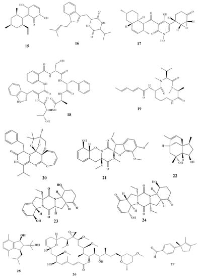
Ieodoglucomide C (
1 in
Scheme 1) and ieodoglycolipid (
2 in
Scheme 1) are two glycolipids which are both isolated from the aquatic bacterium
Bacillus licheniformis. It was found that they both have potent antifungal activity, with MIC values of 0.02–0.03 µM against the human pathogens
Candida albicans,
Colletotrichum acutatum,
Botrytis cinerea,
Rhizoctonia solani, and
Aspergillus niger [4][5][19,20].
Scheme 1.
Chemical structures of cited compounds that were isolated from marine organisms and that showed antimicrobial activities.
Hedaya48, which was synthesized by the
Aplysina fistularis sponge when subjected to various UV radiation dosages, 5,7-dimethoxy-4-p-methoxyl phenyl coumarin (
3 in
Scheme 1), and saadamycin (
4 in
Scheme 1) were all new antimycotic substances identified from endophytic
Streptomyces sp. The MIC value of saadamycin was reported to be 1–5.16 µg/mL, whereas 7.5–100 µg/mL was observed for 5,7-dimethoxy-4-p-methoxyl phenyl coumarin against dermatophytes as well as other fungi, including
Cryptococcus humicolus,
Fusarium oxysporum,
Aspergillus fumigatus,
A. niger, and
Microsporum gypseum [6][7][21,22].
Actinomycetes were used to create the new and superior antifungal drug caerulomycin A. (
5 in
Scheme 1). Actinomycete strain PM0525875 for extraction was obtained from a marine invertebrate. Actinomycetes extracts showed strong effectiveness against drug-resistant fungus strains in in vitro investigations. The fluconazole-resistant
Candida glabrata,
C. albicans,
C. albicans CO9, and
Candida krusei were the pathogenic fungal test strains used to determine the MIC value of caerulomycin A. The MIC values reported ranged between 0.39 and 1.56 µg/mL
[8][9][23,24].
The secondary metabolite, pedein A (
6 in
Scheme 1), was isolated from the cell mass of the myxobacterium
Chondromyces pediculatus. Pedein A inhibited the growth of a broad spectrum of yeasts and fungi, whereas Gram-positive and Gram-negative bacteria such as
Bacillus subtilis,
Brevibacterium ammoniagenes,
Corynebacterium fascians,
Micrococcus luteus,
Staphylococcus aureus,
Escherichia coli,
Enterobacter aerogenes,
Pseudomonas aeruginosa, and
Salmonella typhimurium were not sensitive to the antibiotic. MIC value for
Rhodotorula glutinis was reported to be 0.6 µg/mL, and an MIC value of 1.6 µg/mL was reported for both
Saccharomyces cerevisiae and
Candida albicans. Furthermore, pedein A showed inhibitory activity against the growth of some filamentous fungi with a zone diameter range of 22–35 mm for
Botrytis cinerea,
Gibberella fujikuroi,
Pythium debaryanum,
Rhizopus arrhizus,
Trichoderma koningii, and
Ustilago maydis [5][6][20,21].
Other important isolated antifungal compounds, their marine sources, and their activities are listed in Table 1.
Table 1.
Some antifungal compounds isolated from marine bacteria.
Lobophorolide (
26 in
Scheme 1), isolated from
Lobophora variegata (marine brown alga) of the Bahamas and Egypt, has excellent activity against the pathogenic ascomycete
Lindra thalassiae and the saprophytic deuteromycete
Dendryphiella salina, with IC
50 values of 0.135 and 0.034 µg/mL, respectively. Further, it showed antifungal activity against
C. albicans wild and amphotericin-resistant strains, with IC
50 values of 1.3 and 0.5 µg/mL
[9][25][24,40].
The isolated isolauraldehyde (
27 in
Scheme 1) showed antifungal activity against
C. albicans,
A. fumigatus, and
A. flavus with MIC values of 70, 100, and 1000 µg/mL, respectively. The organic extract of isolauraldehyde was obtained from the red alga
Laurencia obtuse [26][27][41,42].
The growth of
Mycobacterium smegmatis and
Neurospora crassa could be inhibited by the ethanolic extract of
Gracilaria domigensis [28][43].
Gracilaria sjoestedii and
Gracilaria debilis ethanolic extract had antifungal activity against
C. albicans [29][44].
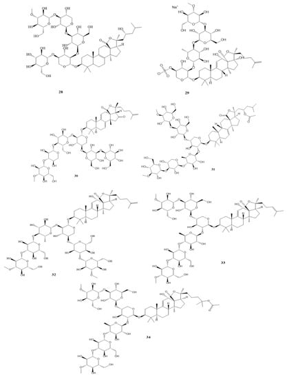
4. Antifungal Compounds Isolated from Sea Cucumbers
Sea cucumbers are animals with long bodies and leathery skin. They contain several antifungal compounds, such as variegatuside D. (
28 in
Scheme 1), which was isolated from
Stichopus variegates and which showed antifungal activity against
Microsporum gypseum,
C. albicans,
C. pseudotropicalis, and
C. parapsilosis, all of which have 3.4 µg/mL MIC
80 value
[30][31][45,46].
Scabraside A (
29 in
Scheme 1) isolated from
Holothuria scabra exhibited antifungal activities against
A. fumigatus, C. pseudotropicalis,
M. gypseum,
T. rubrum, and
C. albicans, with MIC values of 2, 4, 4, 8, and 8 µg/mL, respectively
[32][47].
Antifungal activity against
C. tropicalis and
M. gypseum 31388 with MIC
80 values of 1.4–5.7 µM were reported for holotoxin D1 (
30 in
Scheme 1) and stichloroside C1 (
31, in
Scheme 1), which were isolated from
Apostichopus japonicus Selenka
[33][48].
The growth of
Cryptococcus neoformans,
Richophyton rubrum,
C. albicans,
C. tropicalis,
A. fumigatus, and
C. krusei could be inhibited with MIC
80 values ranging from 0.7 to 2.81 µM by marmoratoside A, impatient side A, and bivittoside D (
32–
34 in
Scheme 1) isolated from
Bohadschia marmorata Jaeger
[34][35][49,50].
5. Antifungal Compounds Isolated from Sea Sponges
Sponges are elementary multi-cellular animals with dense skeleton muscles. They have a vast repertoire of antifungal compounds, which are useful in cases of resistance to amphotericin B and fluconazole
[36][51].
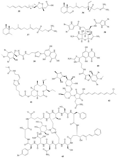
The growth of
C. albicans was inhibited by the isolated epiplakinic acid F (
35, in
Scheme 1) and agelasidine F and C (
36,
37 in
Scheme 1), which have MIC values of 3.1, 4, and 0.5 µg/mL, respectively. Epiplakinic acid F was extracted from the Seychelles sponge genus
Plakinastrella. Agelasidine F and C were obtained from
Agelas citrina (Caribbean sponge)
[36][37][38][51,52,53].
Table 2 lists other isolated compounds from sea sponges that exhibit antifungal activity against
C. albicans.
Table 2.
Compounds with antifungal activity against
C. albicans
isolated from sea sponges.
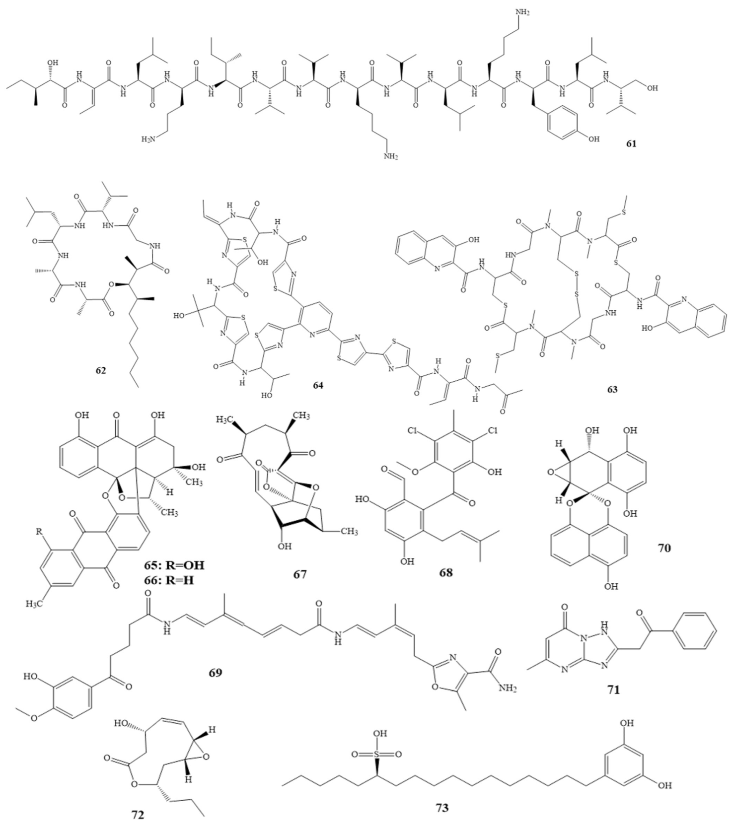
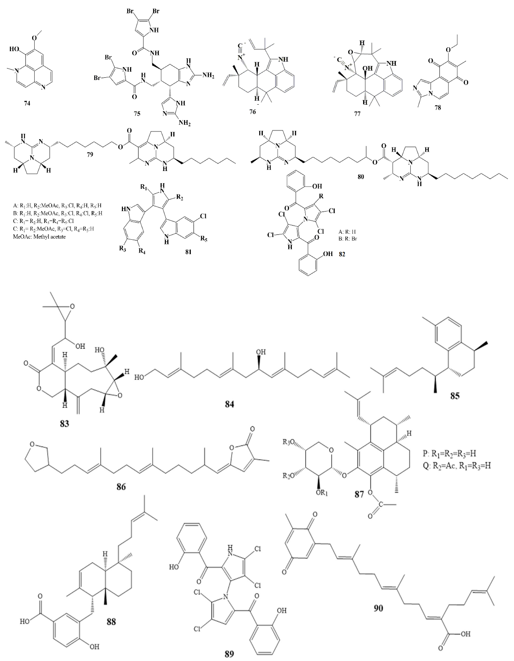
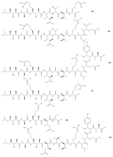
Compounds 7–14 as shown in Scheme 1
2. Antifungal Compounds Isolated from Marine Fungi
From the ocean’s surface to its deepest parts, fungi have been discovered to exist in almost every aquatic habitat studied
[10][25]. As a result of marine fungi’s superior biological characteristics to terrestrial fungi and their ability to adapt to extreme pH, temperature, and salinity, a wider range of biotechnological applications of marine fungi are possible
[11][26].
In Greenland,
Trichoderma sp. strain MF106 was the source of pyridoxatin (
15 in
Scheme 1), demonstrating antifungal activity with IC
50 values of 1.07 ± 0.34 µM against
Trichophyton rubrum and 6.9 ± 0.04 µM against
C. Albicans [12][13][27,28]. The Japanese isolated diketopiperazine (
16 in
Scheme 1) demonstrated growth inhibition against
P. oryzae. and
P. yezoensis with an IC
50 value of 350 nM
[14][15][29,30].
Didymellamide A (
17 in
Scheme 1), isolated from the fungus
Stagonosporopsis cucurbitacearum, reduced the growth of
C. albicans,
Candida glabrata, and
Cryptococcus neoformans strains at doses of 1.6–3.1 µg/mL
[16][17][31,32]. Additionally, the
Aspergillus sclerotiorum PT06-1 isolates of sclerotide B (
18 in
Scheme 1) and sclerotide B (
19 in
Scheme 1) both exhibited activity against
Candida albicans, with MIC values of 7.0 and 3.5 µM, respectively
[18][19][33,34].
The plant pathogenic fungus
Fusarium graminearum,
Alternaria brassicae, and
Colletotrichum gloeosporioides were all inhibited by varioxepine A, which has an MIC value of 4 µg/mL; peniciadametizine A, which has an MIC value of 4 µg/mL; and penicibilaenes A, which has an MIC value of 1.0 µg/mL (
20–
22 in
Scheme 1), respectively. They were extracted from
Paecilomyces variotii,
Phoma sp. Q60596, and
Penicillium bilaiae MA-267 fungus
[20][21][22][35,36,37]. On the other hand, penicibrocazines B and E (
23,
24 in
Scheme 1), which were isolated from
Penicillium brocae MA-231 (
Avicennia marina culture extract), showed activity against the plant pathogen
Gaeumannomyces graminis, with a 0.25 µg/mL MIC value for both
[23][38].
3. Antifungal Compounds Isolated from Marine Algae
Caulerprenylol B (
25 in
Scheme 1), which was obtained from Chinese alga
Caulerpa racemosa, has excellent antifungal activity against
T. rubrum fungus, which causes two of the most common fungal infections, known as ’athlete’s foot’ and ’jock itch’, with an MIC
80 value of 16 µg/mL
[24][39].
* Aurantoside K has activity against wild type C. albicans with MIC value of 1.95 μg/mL [44][59]. Compounds 39–49 as shown in Scheme 1.
The highly oxygenated alkaloid massadine (
38 in
Scheme 1), which was isolated from the marine sponge
Stylissa aff. massa, inhibited Geranylgeranyltransferase-I from
C. albicans with an IC50 value of 3.9 µM. Moreover, massadine inhibited the growth of
Cryptococcus neoformans with an MIC value of 32 µM, but it did not inhibit the growth of
C. albicans at a concentration of 64 µM
[38][53].
Haliscosamine (
50 in
Scheme 1), plakortide F acid (
51 in
Scheme 1) and simplexolide E (
52 in
Scheme 1) showed antifungal effectiveness against
C. neoformans with MIC values ranged 0.2–3.66 μg/mL, respectively.
Haliscosamine was obtained from
Haliclona viscosa (Moroccan sponge), plakortide F acid from
Plakortis halichondrioides sponge, and simplexolide E from the sponge
Plakortis simplex found in China
[47][48][49][62,63,64].
Puupehenone (
53 in
Scheme 1) (isolated from
Hyrtios sp. sponge) showed antifungal activity with MIC values of 1.25 µg/mL and 2.50 µg/mL against
C. neoformans and
C. krusei, respectively
[50][65]. The isolated Chinese Hippolachnin A (
54 in
Scheme 1) from
Hippospongia lachne sponge showed antifungal activity against
C. neoformans,
T. rubrum, and
M. gypseum, with MIC values of 0.41 μM for each fungus
[51][66]. Furthermore, with MIC values ranging between 1.9 and 7.8 µg/mL, the Brazilian batzelladine L (
55 in
Scheme 1) isolated from the
Monanchora arbuscular sponge exhibited activity against
A. flavus strains
[52][67].
A reasonably new nematicide (a substance active against nematode worms), onnamide F (
56 in
Scheme 1), which was isolated from
Trachycladus laevispirulifer, is helpful in
Saccharomyces cerevisiae or baker’s yeast infections. It has an LD
99 (dosage required to kill 99% of the fungi population) of 1.4 μg/mL
[53][68].
Fluconazole resistance has been increasing recently, specifically in immunocompromised individuals such as HIV patients prescribed fluconazole prophylactically. Because of that, other antifungal compounds have been screened for efficacy in resistant strains. Geodisterol-3-O-sulfite and 29-demethylgeodisterol-3-O-sulfite, active constituents of
Topsentia sp. extracts, have been used in fluconazole-resistant strains. Many
Saccharomyces cerevisiae strains can overexpress the MDR1 efflux pump (a pump responsible for pumping out toxic substances such as fluconazole). Hence, these two compounds have been used in reverse
[54][69].