From the Discovery of Targets to Delivery Systems: Comparison
Please note this is a comparison between Version 2 by Rita Xu and Version 1 by Ilaria Iacobucci.

Metals are indispensable for the life of all organisms, and their dysregulation leads to various disorders due to the disruption of their homeostasis. Nowadays, various transition metals are used in pharmaceutical products as diagnostic and therapeutic agents because their electronic structure allows them to adjust the properties of molecules differently from organic molecules. Therefore, interest in the study of metal–drug complexes from different aspects has been aroused, and numerous approaches have been developed to characterize, activate, deliver, and clarify molecular mechanisms. The integration of these different approaches, ranging from chemoproteomics to nanoparticle systems and various activation strategies, enables the understanding of the cellular responses to metal drugs, which may form the basis for the development of new drugs and/or the modification of currently used drugs. The purpose of this review is to briefly summarize the recent advances in this field by describing the technological platforms and their potential applications for identifying protein targets for discovering the mechanisms of action of metallodrugs and improving their efficiency during delivery.

  • metallodrugs
  • medicinal chemistry
  • chemoproteomics
  • thermal proteome profiling

1. Introduction

The involvement of metals in many cellular and subcellular functions and their role in numerous vital processes is well known. Zinc (Zn), for example, is a mineral that is involved in numerous functions of cellular metabolism by supporting the catalytic activity of over 100 enzymes [1]. Calcium (Ca) is essential for cell physiology, and its transfer across membranes serves as a signal for many cellular processes, such as muscle contraction and the transmission of nerve impulses [2]. Copper (Cu) plays a key role in the central nervous system (CNS) [3] and in hematopoiesis [4], which is the process of blood cell formation. In addition, Cu and Cu-dependent enzymes are cofactors of numerous redox reactions and are involved, for example, in neurotransmission [5]. Evolution has made metals indispensable for the life of all organisms, and their dysregulation causes various, sometimes serious, disorders, such as a number of neurodegenerative diseases caused by the disturbance of their homeostasis [6,7][6][7]. It has long been known that metals can also serve as therapeutic agents, for example, as antimicrobial and antiviral weapons [8,9,10][8][9][10]. Recently, many metals have been incorporated into pharmaceutical products as diagnostic and therapeutic agents. The electronic structure of transition metals has the advantage of being very versatile in tuning molecular properties, unlike organic molecules [11]. The use of Pt coordination compounds in cancer therapy was certainly [12] one of the most unexpected developments in the field of medicine in the last 50 years. The first representative of chemotherapeutic agents, cisplatin, was discovered and synthesized by Michele Peyrone (1813–1883) [13], and its cytotoxic activity was revealed by chance by Barnett Rosemberg (1927–2009) in 1965. The clinical success of cisplatin as an anticancer drug accelerated the search for metals in medicinal chemistry [14]. Subsequently, several other Pt drugs, including carboplatin and oxaliplatin, were developed to improve the therapeutic efficacy and reduce the systemic toxicity caused by cisplatin [15,16,17][15][16][17].
Clinical studies have revealed controversial evidence. While some patients showed very positive results, others displayed the high toxicity of the compound [18]. The spectacular results obtained in patients suffering from tumors of the genital tract, particularly testicular cancer, in which cisplatin proved to be practically curative, led to its approval by the FDA (Food and Drug Administration) in 1978. Since then, cisplatin has been one of the most widely used drugs in cancer therapy worldwide and by far one of the most successful drugs (a so-called blockbuster drug) [19,20][19][20]. The spontaneous or induced resistance of tumors to cisplatin is an extremely serious problem, as it significantly limits the use of the drug. The main cellular processes by which cisplatin attacks tumor cells are as follows: uptake and transport, the formation of adducts with DNA and their recognition by specific proteins (damage-response proteins), the “translation” [21] of the DNA damage signal, which inhibits its replication and transcription and activates numerous signal transduction pathways that can lead to cell cycle arrest and damage repair or cell death via a variety of proteins that control cell growth, differentiation, and response to stress [22]. Research soon turned to other metals with the goal of finding complexes that would be effective against tumors resistant to platinum compounds and that might also have less systemic toxicity. For this reason, other metals have been tested as potential anticancer compounds, such as those containing ruthenium (Ru) [23], arsenic (As) [24], gold (Au) [25], and osmium (Os) [26], and they are currently being developed [14,27][14][27].
Among them, Ru compounds were the first to be studied, which is still a very active branch of research today, followed by arsenic (As) compounds, the only other non-radioactive metal (at the time) approved by the FDA (in 2000) for the treatment of tumors [28]. It was brought into clinical use in the 1970s for the treatment of numerous leukemias, especially acute promyelocytic leukemia [29]. At the moment, gold compounds are receiving increasing attention, as they have fewer tolerability limitations and appear to be target-specific [21]. Recent studies show that Au(I) complexes have antitumor activity, which is caused by inducing apoptosis in various cancer cell lines in vitro, comparing the effect of such compounds on resistant cells and analyzing the mechanism of action [30]. In addition, gold compounds have been proposed as potential anti-infective and anti-tuberculosis agents [31].
Despite the great potential of metal compounds, only a few have been finally accepted and marketed by the FDA because of toxicity and resistance induction, poor biodistribution, and short- and mid-term side effects [32]. Metallodrugs’ toxicity is mainly related to renal damage and neurotoxic effects [33], whereas resistance is due to targeting individual proteins or enzymes. On the other hand, the biodistribution and side effects could be improved by the further development of drug delivery strategies, such as nanoparticles [32,34][32][34]. The steps for the clinical approval established by the FDA are as follows: (1) discovery and development, (2) preclinical research, (3) clinical research, (4) FDA drug review, and (5) FDA post-market drug safety monitoring (https://www.fda.gov/patients/learn-about-drug-and-device-approvals/drug-development-process, accessed on 12 July 2023). All of these steps aim to determine the safety and efficacy of the drug under development in humans. Specifically, the clinical trial begins with Phase 0, a preliminary study in which some healthy volunteers are studied with an administration dose of less than 1% of the therapeutic dose for a maximum of seven days, followed by Phase I. After these phases, the metal drug can be tested in affected patients, which is Phase II of the clinical trials. The efficacy of the drug is tested in patients, and a placebo is used as a control. The final step is Phase III, which accounts for the majority of the clinical trials’ cost, and aims to confirm the safety and efficacy tested in Phase I and Phase II, respectively [35]. Nowadays, there are several metallodrugs approved by the United States and/or European Union (EU) countries for the medical treatment of human diseases. The application of these approved metallodrugs range from “Anticancer Metallodrugs” and “Therapeutic Radiopharmaceuticals or Phototherapeutic Metallodrugs” to “Antimicrobial and Antiparasitic Metallodrugs”, but also “Antidiabetes” and others (see ref. [35] for more information). Prior to the entire preclinical and clinical investigation, the first step is, of course, the investigation of one or more disease targets and the discovery of the molecular mechanisms of action of the metallodrug under investigation. Functional proteomics aims to identify protein–protein (PPI) [36,37][36][37] and protein–DNA/RNA [38] interactions in vivo in order to define protein complexes and, thus, the cellular pathways involved in the biological processes of interest, as detailed in several papers and reviews [39,40][39][40]. There are many proteomic approaches used for the study of such interactions based on classical biochemical protocols adapted for the study of so-called “interactomics”, as well as coupling with advanced mass spectrometry instruments. This approach can also be used to define and identify the molecular partners of metal compounds in the context of chemical proteomics. In addition, drug delivery (i.e., the targeted administration of a compound to a tissue or cell where its controlled release ensures greater efficiency) is also an important area of study. Its application in the pharmacological field allows the transport of a molecule in our body, the selective delivery to the target tissue, and controlled release [41,42,43][41][42][43]. This method allows uresearchers to reduce the dose of the administered drug, reducing its possible side effects, and making it more bioavailable. Nanoparticles (NPs) currently represent a major advance in this field [32].

2. Protein Target Identification through Proteomic Approaches

The study of protein interactions with different classes of biomolecules has ancient roots in the field of proteomics, with many different biochemical strategies developed and advanced in conjunction with mass spectrometry techniques [39,40][39][40]. In recent years, several proteomic strategies coupled with mass spectrometry (MS) have emerged as a powerful and systematic approach for the large-scale identification of drug–protein interactions and the elucidation of their associated mechanisms. Chemical proteomics, or chemoproteomics, is primarily devoted to the study of protein–small-molecule interactions and is attracting increasing attention in drug target discovery [44,45][44][45]. Chemoproteomics is a leading tool in the field of drug discovery that relies on affinity-based or label-free approaches to identify protein targets.

2.1. Affinity-Based Strategies

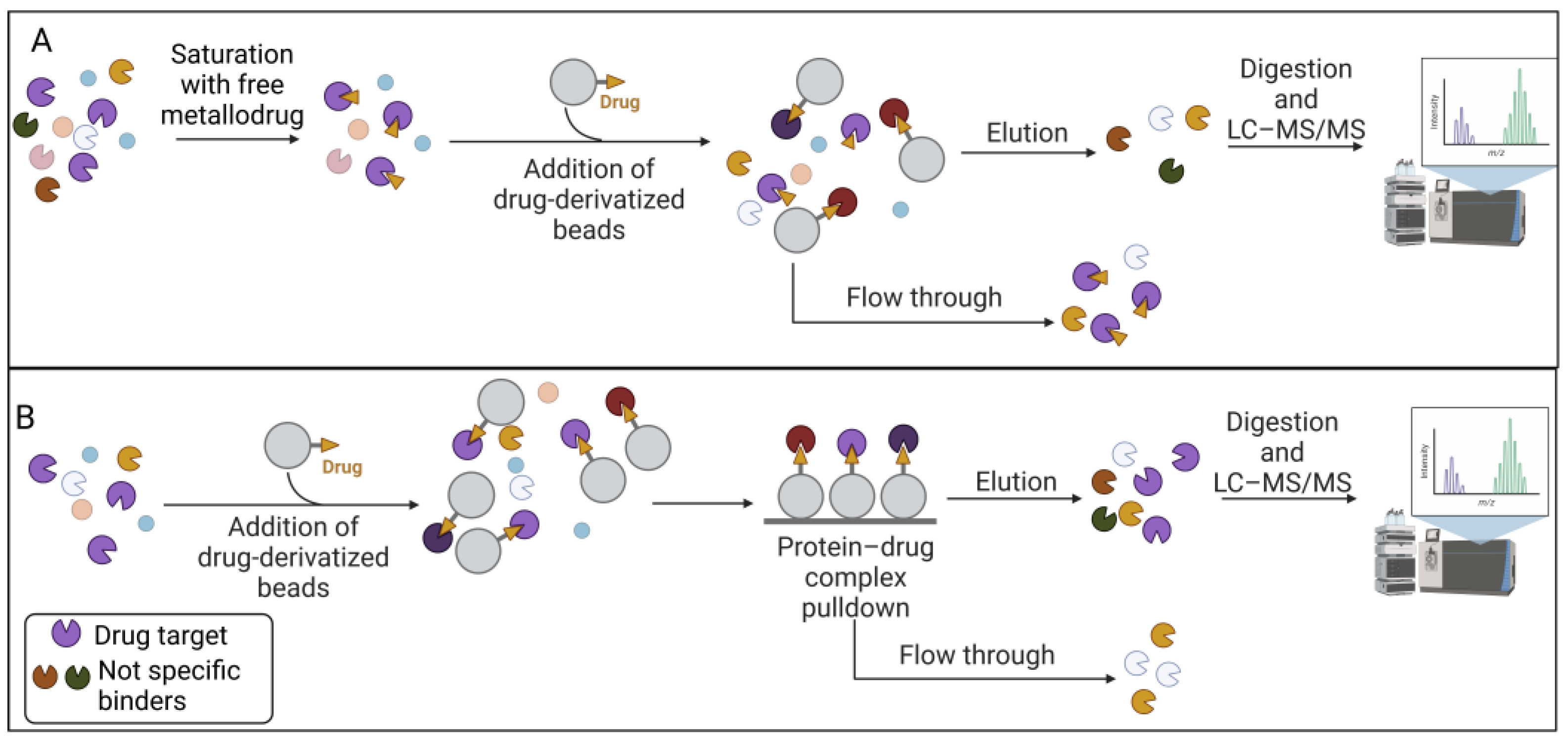
The affinity-based approach to chemical proteomics aims to immobilize the drug on a solid support and isolate the target proteins. This strategy is also referred to as drug pulldown [46,47][46][47]. Pulldown strategies and, more generally, affinity purification-mass spectrometry approaches (AP-MS), are proteomic techniques that allow uresearchers to isolate multiprotein complexes starting from a known molecule to hypothesize the intracellular processes in which it is involved [48,49,50][48][49][50]. Drug pulldown requires the following four main steps: (1) the immobilization of the pharmacophore on resin beads with affinity chromatography; (2) the isolation of the target molecules; (3) the identification of the target molecules with liquid chromatography–tandem mass spectrometry (LC–MS/MS); and (4) bioinformatics analysis to identify and quantify the proteins (Figure 1).
Figure 1. Drug pulldown consists of four steps: (1) immobilization of the pharmacophore on beads, which can be performed using different approaches (streptavidin beads and biotinylated metallodrugs or human serum albumin (HSA)–antiHSA antibody interaction); (2) target isolation, which can be preceded by a stabilization step involving UV-crosslinking between the proteic target and the drug; (3) mass spectrometry analysis of the target; and (4) bioinformatic analysis for the targets identification. Legend: M: metallodrug; B: biotin.
The advantage of pulldown techniques is that the physiological state of the proteins, the concentration levels (i.e., abundance), the post-translational modifications, and the natural binding partners are preserved by simply using non-denaturing lysis buffers. In particular, for Step 1 of the drug-pulldown workflow (Figure 1), different methods can be used to immobilize the active pharmacophore, which also depend on the stability of the organic- and inorganic-metal complexes. For this reason, Babak et al. used a biotin/streptavidin approach to immobilize a Ru(II) compound that is classified as a member of the RAPTA family [51]. The arene ligand of the complex was functionalized with a primary amine that can be biotinylated via an aminocaproic acid linker. The research results led to the identification of 15 cancer-related proteins that can explain the activity of RAPTA molecules, including MK, PTN, and FGF3 as metastasis-related effectors, but also proteins related to cell cycle regulation, i.e., GNL3, CGBP1, FAM32A, and VIR. Drug biotinylation can also be performed by using the click reaction (Cu(I)-catalyzed alkyne–azide cycloaddition (CuAAC)). CuAAC is a reaction between an alkyne and an azide group, catalyzed by Cu(I) ions [52,53][52][53]. This reaction was used by Neuditschko and colleagues to derivatize the drug with biotin and fish the targets with streptavidin beads. Another clever method for the immobilization of metal drugs was reported by the same [54] research group. A complex was formed between the Ru(III) compound (i.e., BOLD-100) and human serum albumin (HSA). Then, the authors used the anti-HSA beads utilized for the human serum depletion to form the BOLD-100-HSA-beads adduct used to isolate drug targets [55]. Because high false positive rates occur in pulldown experiments, the authors also performed a competitive assay. The cell lysate was pre-treated with free BOLD-100 before exposure to the immobilized drug on the beads. This latter strategy allowed the saturation of the selective binding sites, as shown in Figure 2, so that the resulting target profile contains only non-selective binding partners. The subtraction of these from the target profile obtained by normal pulldown deletes the non-selective interactors and provides a list of selective binding partners. The competitive pulldown experiment is an alternative way to validate and remove the possible false-positive targets from a canonical pulldown experiment. Using this approach, the authors identified ribosomal proteins and the transcription factor GTF2I as BOLD-100 targets. In particular, further transcriptome analysis validated RPL10 and/or RPL24 among the ribosomal protein BOLD-100 targets.
Figure 2. Competitive pulldown workflow. (A) The total protein extract was incubated with the free metallodrug and then subjected to the chemoproteomic strategy. Only the non-specific binders were eluted. Instead, the specific target was not retained on the beads. (B) The total protein extract was incubated with the metallodrug-derivatized beads for the chemoproteomic strategy. The non-specific and specific proteins were eluted. The comparison between the eluted proteins in the competitive and non-competitive will highlight the false-positive content.
Although the simple pulldown strategy is a straightforward approach, it suffers from the classic drawbacks that are generally associated with affinity purification strategies, such as the lack of transient interactions and the problem of false positives, as mentioned above (see [38] for specific further insights on this topic). For these reasons, the drug immobilization step is the most critical, as the final yield of target purification depends on it. In classical pulldown experiments with small organic molecules, the drug is usually covalently functionalized on an N-hydroxysuccinimidyl (NHS) sepharose carrier, then the non-functionalized portion of the carrier is blocked using several washes with ethanolamine [45]. For metal-based drugs, these types of washes are not compatible, because ethanolamine can compete with the metal complex ligands. Therefore, alternative methods have been developed, for example, using the biotin–streptavidin interaction (as described previously). The metal complex site carrying the label must be carefully selected in order to avoid interference with the biological activity of the pharmacophore. The CuAAC strategy is one of the best known for metal complex biotinylation and offers the advantage of high reaction efficiency and a favorable reaction time under mild conditions. However, a key drawback is the toxicity of copper ions to proteins. They can damage the structure and function of the target proteins and promote the formation of ROS [56]. In addition to the clickable part, i.e., the alkyne, a photo affine group can often also be produced [57]. Indeed, this strategy can be used to stabilize transient interactions, and it is also possible to stabilize the interactions in living cells, i.e., photoaffinity labeling (PAL). The photoactivatable group, usually a diazirine or benzophenone group, has the function of facilitating covalent adducts with the metal/metal-drug-interacting proteins upon light irradiation, as reported by Liu et al. [58]. The authors synthesized a photoactivatable molecule that enables the probe to form covalent adducts with the proteins interacting with the metal drug upon irradiation with UV light [59]. The authors incubated HeLa cells with a Au(III) meso-tetraphenylporphyrin (gold-1 a) compound and irradiated them at 365 nm to activate the benzophenone reaction with proteins via a radical mechanism. After cell lysis, an alkyne group on the probe was used to functionalize the protein–drug complexes with biotin in order to enrich the adducts on streptavidin beads. In their study, they identified heat shock protein 60 (Hsp60) as a target of gold-1a. These approaches can be used to identify interactions characterized by non-covalent and coordinative bonds. The covalent detection of drug–target interactions in cells can be used to identify physiologically occurring and moderately strong binding events by affinity-based chemoproteomics in vivo [60,61][60][61].

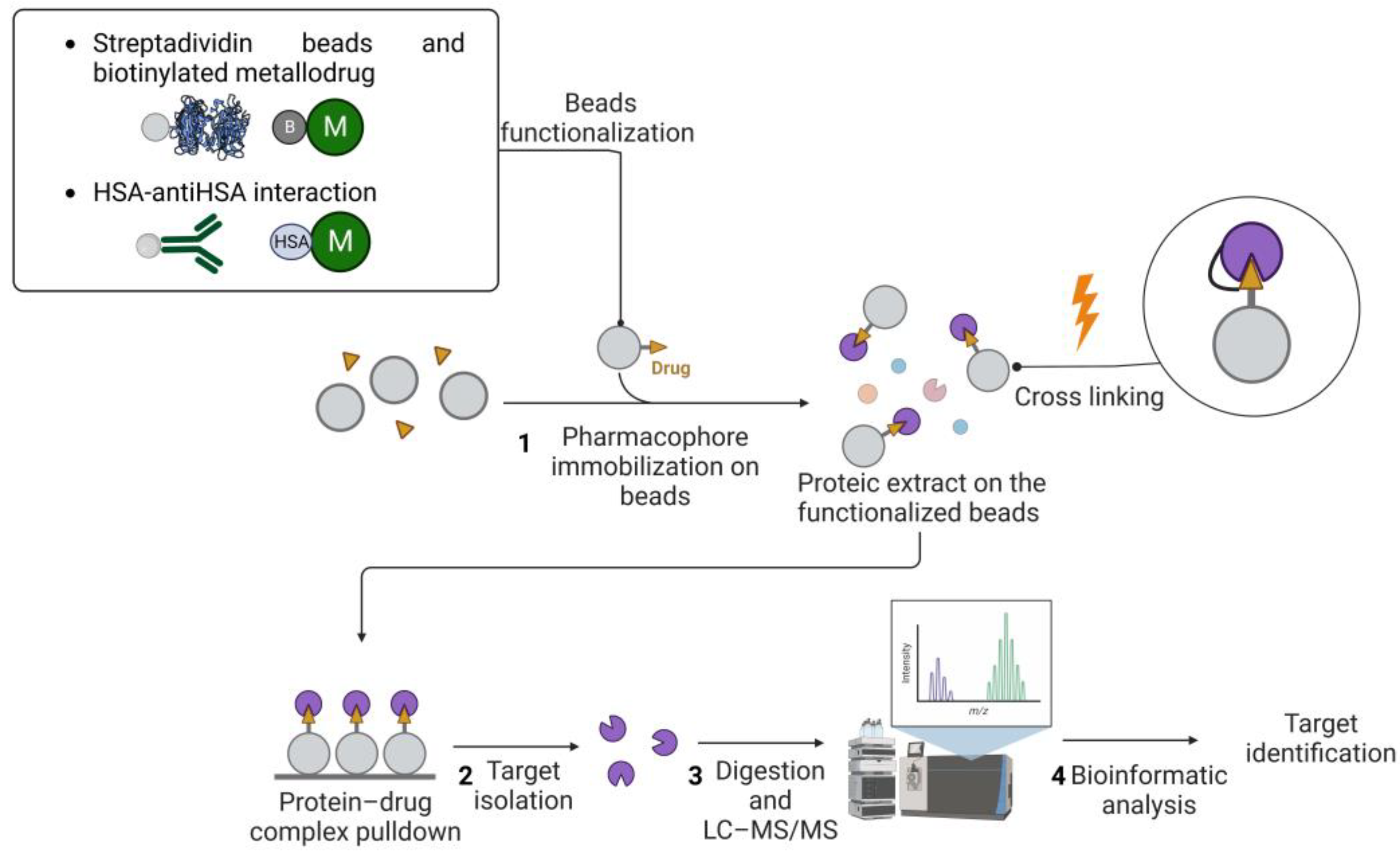
2.2. Label-Free Approaches

On the other hand, label-free approaches exploit protein stability due to the drug interaction. For example, thermal proteome profiling (TPP) [62] has been used to study metallodrug targets [63]. TPP measures the extent of the drug–target interaction by monitoring the effects of pharmacological treatment on protein denaturation/solubility as a function of a progressive increase in temperature. The extent of protein stabilization by the small molecule is proportional to the strength of the interaction and can be assessed by multiplexed mass spectrometry analysis (see [64] for a comprehensive description of all TPP applications). As shown in Figure 3, TPP can be performed by varying the temperature range (TR-TPP) (Figure 3A) or drug concentration (CR-TPP) (Figure 3B). After protein digestion and mass spectrometry analysis using LC–MS/MS, proteins are relatively quantified by the comparison between the drug-treated and drug-free conditions. The soluble fraction for each protein is reported as a function of the temperature or drug concentration.
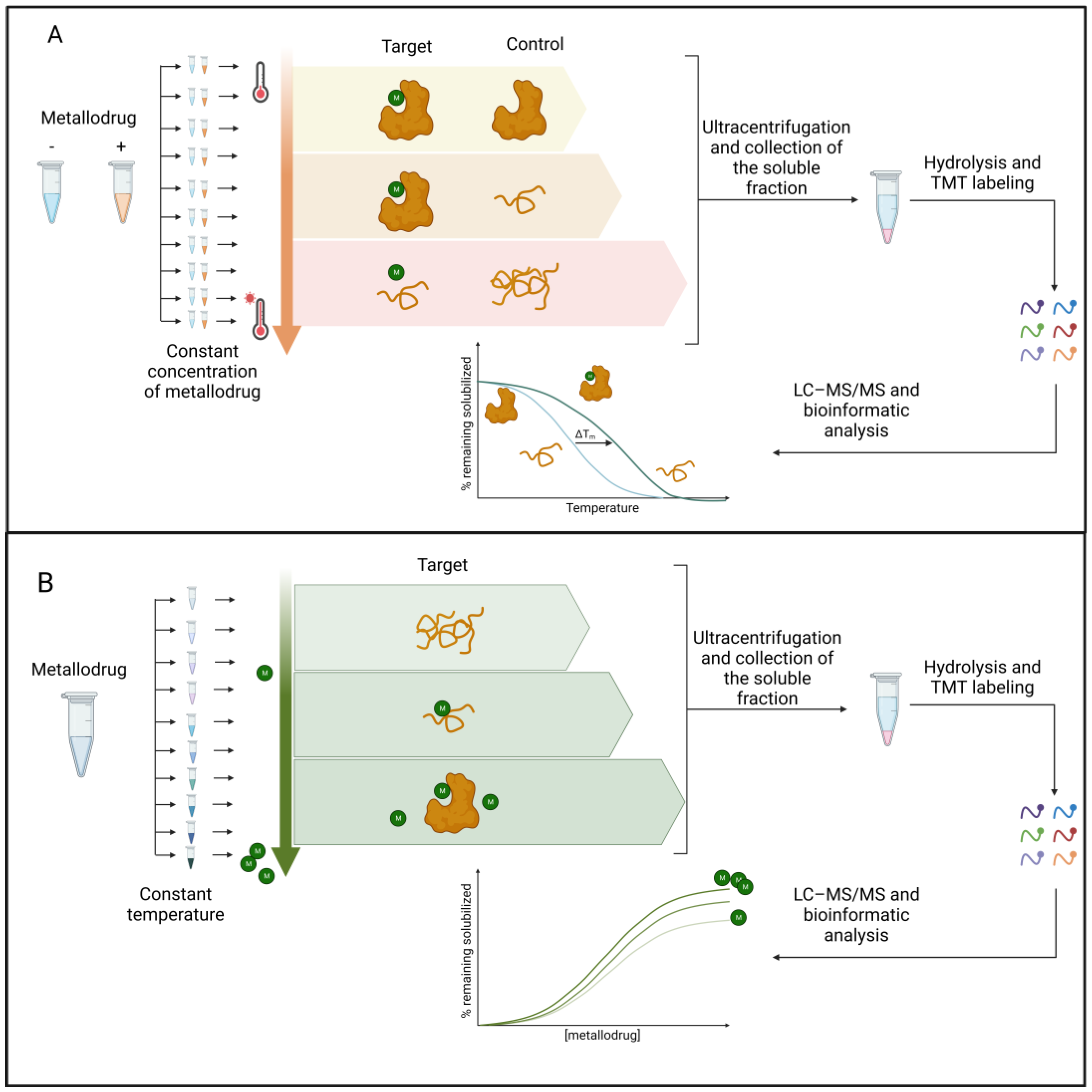
Figure 3. (A) TR-TPP experiment. The protein extract is incubated with both the vehicle and the metallodrug. Ten aliquots are prepared for each condition. Each aliquot is heated at a specific temperature, and then the soluble fractions are digested with trypsin and labeled with a TMT isotope tag. The samples are analyzed with LC–MS/MS and the protein is identified. Melting curves are fitted and melting temperatures are calculated for all proteins. (B) CR-TPP experiment. Untreated protein extract, as a control, and those treated with nine different concentrations of metallodrug, are heated to the same temperature. The soluble fractions are then digested with trypsin and labeled with a TMT isotope tag. The samples are analyzed using LC–MS/MS and proteins are identified and quantified. Dose–response curves are fitted, and thermal stability parameters are calculated. Legend: M: metallodrug.
TR-TPP was recently applied to discover the antitumor Bis(N-Heterocyclic Carbene)Pt(II) complex targets in intact cells, leading to the identification of asparagine synthetase (ASNS) [65]. Another label-free method used in the study of metallodrug targets is the functional identification of target by expression proteomic (FITExP) approach [66]. The basic principle is that protein targets and major mechanistically related proteins are modulated upon prolonged drug exposure; in particular, they are overexpressed when a lethal concentration (LC50) is administered to the cells. Lee et al. [67] applied the FITExP method to evaluate the mechanism of action of Ru(II) complexes, RAPTA-T, and RAPTA-EA. Although the approach suggested many protein targets, the best results were obtained in terms of revealing the mechanism of action of the molecules. For example, RAPTA-EA and RAPTA-T were found to induce the overexpression of several oxidative-stress-related and tumor-suppressing proteins, respectively [67]. FITExP analysis of proteins extracted from cells treated with RAPTA-T revealed, among the proteins identified, PLD3 protein, which belongs to the phospholipase D enzyme (PLD) family, with the lowest p-value. Instead, cells treated with RAPTA-EA had heat shock protein 1A/1B (HSPA 1A/1B) as the protein with the top-ranked p-value. This approach allowed the authors to hypothesize the following two different mechanisms of action for the two drugs: RAPTA-T targets are involved in multiple processes, suggesting a broader mechanism of action of RAPTA-T. In contrast, the targets of RAPTA-EA are exclusively involved in the regulation of the oxidative stress response. The combination of TPP and FITExP was performed in the work of Saei and colleagues to reduce the number of false positives and false negatives [63]. In principle, the other label-free strategies used in the study of protein–small-molecule interactions could also be applied to the field of metallodrugs. These methods encompass the protein stability based on the oxidation rate (SPROX), [68] pulse proteolysis (PP) [69], drug affinity responsive target stability (DARTS) [70], and limited proteolysis-coupled mass spectrometry (LiP-MS) [71]. However, to our knowledge, no no work has been published. Label-free techniques are capable of detecting the transient interactions of drug candidates with potential protein targets and globally assessing drug-altered temperature and the proteolytic or chemical stability of proteins, and otherwise do not require synthetic modification of the drug [72,73][72][73].

References

  1. Kambe, T.; Tsuji, T.; Hashimoto, A.; Itsumura, N. The Physiological, Biochemical, and Molecular Roles of Zinc Transporters in Zinc Homeostasis and Metabolism. Physiol. Rev. 2015, 95, 749–784.
  2. Bagheri-Mohammadi, S.; Farjami, M.; Suha, A.J.; Zarch, S.M.A.; Najafi, S.; Esmaeili, A. The Mitochondrial Calcium Signaling, Regulation, and Cellular Functions: A Novel Target for Therapeutic Medicine in Neurological Disorders. J. Cell. Biochem. 2023, 124, 635–655.
  3. An, Y.; Li, S.; Huang, X.; Chen, X.; Shan, H.; Zhang, M. The Role of Copper Homeostasis in Brain Disease. Int. J. Mol. Sci. 2022, 23, 13850.
  4. Ruiz, L.M.; Libedinsky, A.; Elorza, A.A. Role of Copper on Mitochondrial Function and Metabolism. Front. Mol. Biosci. 2021, 8, 711227.
  5. Opazo, C.M.; Greenough, M.A.; Bush, A.I. Copper: From Neurotransmission to Neuroproteostasis. Front. Aging Neurosci. 2014, 6, 143.
  6. Wang, L.; Yin, Y.-L.; Liu, X.-Z.; Shen, P.; Zheng, Y.-G.; Lan, X.-R.; Lu, C.-B.; Wang, J.-Z. Current Understanding of Metal Ions in the Pathogenesis of Alzheimer’s Disease. Transl. Neurodegener. 2020, 9, 10.
  7. Haywood, S. Brain–Barrier Regulation, Metal (Cu, Fe) Dyshomeostasis, and Neurodegenerative Disorders in Man and Animals. Inorganics 2019, 7, 108.
  8. Franz, K.J.; Metzler-Nolte, N. Introduction: Metals in Medicine. Chem. Rev. 2019, 119, 727–729.
  9. Galib; Mashru, M.; Patgiri, B.; Barve, M.; Jagtap, C.; Prajapati, P. Therapeutic Potentials of Metals in Ancient India: A Review through Charaka Samhita. J. Ayurveda Integr. Med. 2011, 2, 55.
  10. Begley, T.P. Wiley Encyclopedia of Chemical Biology; John Wiley & Sons: Hoboken, NJ, USA, 2007; ISBN 978-0-470-04867-2.
  11. Sodhi, R.K. Metal Complexes in Medicine: An Overview and Update from Drug Design Perspective. Cancer Ther. Oncol. Int. J. 2019, 14, 555883.
  12. Rosenberg, B.; Van Camp, L.; Krigas, T. Inhibition of Cell Division in Escherichia Coli by Electrolysis Products from a Platinum Electrode. Nature 1965, 205, 698–699.
  13. Kauffman, G.B.; Pentimalli, R.; Doldi, S.; Hall, M.D. Michele Peyrone (1813–1883), Discoverer of Cisplatin. Platin. Met. Rev. 2010, 54, 250–256.
  14. Sigel, A.; Sigel, H.; Freisinger, E.; Sigel, R.K.O. (Eds.) Metallo-Drugs: Development and Action of Anticancer Agents; (Metal Ions in Life Sciences); De Gruyter: Berlin, Germany; Boston, MA, USA, 2018; ISBN 978-3-11-046984-4.
  15. Khoury, A.; Deo, K.M.; Aldrich-Wright, J.R. Recent Advances in Platinum-Based Chemotherapeutics That Exhibit Inhibitory and Targeted Mechanisms of Action. J. Inorg. Biochem. 2020, 207, 111070.
  16. Zhong, T.; Yu, J.; Pan, Y.; Zhang, N.; Qi, Y.; Huang, Y. Recent Advances of Platinum-Based Anticancer Complexes in Combinational Multimodal Therapy. Adv Healthc. Mater. 2023, 2300253.
  17. Wang, X.; Wang, X.; Guo, Z. Functionalization of Platinum Complexes for Biomedical Applications. Acc. Chem. Res. 2015, 48, 2622–2631.
  18. Barabas, K.; Milner, R.; Lurie, D.; Adin, C. Cisplatin: A Review of Toxicities and Therapeutic Applications. Vet. Comp. Oncol. 2008, 6, 1–18.
  19. Rosenberg, B.; Vancamp, L.; Trosko, J.E.; Mansour, V.H. Platinum Compounds: A New Class of Potent Antitumour Agents. Nature 1969, 222, 385–386.
  20. Williams, C.J.; Whitehouse, J.M. Cis-Platinum: A New Anticancer Agent. BMJ 1979, 1, 1689–1691.
  21. Moreno-Alcántar, G.; Picchetti, P.; Casini, A. Gold Complexes in Anticancer Therapy: From New Design Principles to Particle-Based Delivery Systems. Angew. Chem. Int. Ed. 2023, 62, e202218000.
  22. Coffetti, G.; Moraschi, M.; Facchetti, G.; Rimoldi, I. The Challenging Treatment of Cisplatin-Resistant Tumors: State of the Art and Future Perspectives. Molecules 2023, 28, 3407.
  23. Florio, D.; La Manna, S.; Annunziata, A.; Iacobucci, I.; Monaco, V.; Di Natale, C.; Mollo, V.; Ruffo, F.; Monti, M.; Marasco, D. Ruthenium Complexes Bearing Glucosyl Ligands Are Able to Inhibit the Amyloid Aggregation of Short Histidine-Peptides. Dalton Trans. 2023, 52, 8549–8557.
  24. Paul, N.P.; Galván, A.E.; Yoshinaga-Sakurai, K.; Rosen, B.P.; Yoshinaga, M. Arsenic in Medicine: Past, Present and Future. Biometals 2023, 36, 283–301.
  25. Florio, D.; Iacobucci, I.; Ferraro, G.; Mansour, A.M.; Morelli, G.; Monti, M.; Merlino, A.; Marasco, D. Role of the Metal Center in the Modulation of the Aggregation Process of Amyloid Model Systems by Square Planar Complexes Bearing 2-(2’-Pyridyl)Benzimidazole Ligands. Pharmaceuticals 2019, 12, 154.
  26. Jaouen, G.; Metzler-Nolte, N.; Alberto, R. (Eds.) Medicinal Organometallic Chemistry; (Topics in Organometallic Chemistry); Springer: Berlin/Heidelberg, Germany; New York, NY, USA, 2010; ISBN 978-3-642-13184-4.
  27. Zhou, Y.; Li, H.; Sun, H. Metalloproteomics for Biomedical Research: Methodology and Applications. Annu. Rev. Biochem. 2022, 91, 449–473.
  28. Kostova, I. Ruthenium Complexes as Anticancer Agents. Curr. Med. Chem. 2006, 13, 1085–1107.
  29. Huang, H.; Cao, K.; Kong, Y.; Yuan, S.; Liu, H.; Wang, Y.; Liu, Y. A Dual Functional Ruthenium Arene Complex Induces Differentiation and Apoptosis of Acute Promyelocytic Leukemia Cells. Chem. Sci. 2019, 10, 9721–9728.
  30. Ahrweiler-Sawaryn, M.-C.; Biswas, A.; Frias, C.; Frias, J.; Wilke, N.L.; Wilke, N.; Berkessel, A.; Prokop, A. Novel Gold(I) Complexes Induce Apoptosis in Leukemia Cells via the ROS-Induced Mitochondrial Pathway with an Upregulation of Harakiri and Overcome Multi Drug Resistances in Leukemia and Lymphoma Cells and Sensitize Drug Resistant Tumor Cells to Apoptosis in Vitro. Biomed. Pharmacother. 2023, 161, 114507.
  31. Ferraro, M.G.; Piccolo, M.; Misso, G.; Santamaria, R.; Irace, C. Bioactivity and Development of Small Non-Platinum Metal-Based Chemotherapeutics. Pharmaceutics 2022, 14, 954.
  32. Peña, Q.; Wang, A.; Zaremba, O.; Shi, Y.; Scheeren, H.W.; Metselaar, J.M.; Kiessling, F.; Pallares, R.M.; Wuttke, S.; Lammers, T. Metallodrugs in Cancer Nanomedicine. Chem. Soc. Rev. 2022, 51, 2544–2582.
  33. Oun, R.; Moussa, Y.E.; Wheate, N.J. The Side Effects of Platinum-Based Chemotherapy Drugs: A Review for Chemists. Dalton Trans. 2018, 47, 6645–6653.
  34. Casini, A.; Vessieres, A.; Meier-Menches, S.M. (Eds.) Metal-Based Anticancer Agents; (Metallobiology Series); Royal Society of Chemistry: London, UK, 2019; ISBN 978-1-78801-406-9.
  35. Mjos, K.D.; Orvig, C. Metallodrugs in Medicinal Inorganic Chemistry. Chem. Rev. 2014, 114, 4540–4563.
  36. Barhamand, B.A. Difficulties Encountered in Implementing Guidelines for Handling Antineoplastics in the Physician’s Office. Cancer Nurs. 1986, 9, 138–143.
  37. Zanca, C.; Cozzolino, F.; Quintavalle, C.; Di Costanzo, S.; Ricci-Vitiani, L.; Santoriello, M.; Monti, M.; Pucci, P.; Condorelli, G. PED Interacts with Rac1 and Regulates Cell Migration/Invasion Processes in Human Non-Small Cell Lung Cancer Cells. J. Cell. Physiol. 2010, 225, 63–72.
  38. Fusco, S.; Aulitto, M.; Iacobucci, I.; Crocamo, G.; Pucci, P.; Bartolucci, S.; Monti, M.; Contursi, P. The Interaction between the F55 Virus-Encoded Transcription Regulator and the RadA Host Recombinase Reveals a Common Strategy in Archaea and Bacteria to Sense the UV-Induced Damage to the Host DNA. Biochim. Biophys. Acta (BBA)—Gene Regul. Mech. 2020, 1863, 194493.
  39. Iacobucci, I.; Monaco, V.; Cozzolino, F.; Monti, M. From Classical to New Generation Approaches: An Excursus of -Omics Methods for Investigation of Protein-Protein Interaction Networks. J. Proteom. 2021, 230, 103990.
  40. Cozzolino, F.; Iacobucci, I.; Monaco, V.; Monti, M. Protein–DNA/RNA Interactions: An Overview of Investigation Methods in the -Omics Era. J. Proteome Res. 2021, 20, 3018–3030.
  41. Kumara, B.N.; Kalimuthu, P.; Prasad, K.S. Synthesis, Properties and Potential Applications of Photoluminescent Carbon Nanoparticles: A Review. Anal. Chim. Acta 2023, 1268, 341430.
  42. Xia, Y.; Fu, S.; Ma, Q.; Liu, Y.; Zhang, N. Application of Nano-Delivery Systems in Lymph Nodes for Tumor Immunotherapy. Nano-Micro Lett. 2023, 15, 145.
  43. Kargozar, S.; Moghanian, A.; Rashvand, A.; Miri, A.K.; Hamzehlou, S.; Baino, F.; Mozafari, M.; Wang, A.Z. Nanostructured Bioactive Glasses: A Bird’s Eye View on Cancer Therapy. WIREs Nanomed. Nanobiotechnol. 2023, e1905.
  44. Fedorov, I.I.; Lineva, V.I.; Tarasova, I.A.; Gorshkov, M.V. Mass Spectrometry-Based Chemical Proteomics for Drug Target Discoveries. Biochemistry 2022, 87, 983–994.
  45. Skos, L.; Borutzki, Y.; Gerner, C.; Meier-Menches, S.M. Methods to Identify Protein Targets of Metal-Based Drugs. Curr. Opin. Chem. Biol. 2023, 73, 102257.
  46. Steel, T.R.; Hartinger, C.G. Metalloproteomics for Molecular Target Identification of Protein-Binding Anticancer Metallodrugs. Metallomics 2020, 12, 1627–1636.
  47. Ziegler, S.; Pries, V.; Hedberg, C.; Waldmann, H. Target Identification for Small Bioactive Molecules: Finding the Needle in the Haystack. Angew. Chem. Int. Ed. 2013, 52, 2744–2792.
  48. Iacobucci, I.; Monaco, V.; Canè, L.; Bibbò, F.; Cioffi, V.; Cozzolino, F.; Guarino, A.; Zollo, M.; Monti, M. Spike S1 Domain Interactome in Non-Pulmonary Systems: A Role beyond the Receptor Recognition. Front. Mol. Biosci. 2022, 9, 975570.
  49. Federico, A.; Sepe, R.; Cozzolino, F.; Piccolo, C.; Iannone, C.; Iacobucci, I.; Pucci, P.; Monti, M.; Fusco, A. The Complex CBX7-PRMT1 Has a Critical Role in Regulating E-Cadherin Gene Expression and Cell Migration. Biochim. Biophys. Acta (BBA)—Gene Regul. Mech. 2019, 1862, 509–521.
  50. Cozzolino, F.; Vezzoli, E.; Cheroni, C.; Besusso, D.; Conforti, P.; Valenza, M.; Iacobucci, I.; Monaco, V.; Birolini, G.; Bombaci, M.; et al. ADAM10 Hyperactivation Acts on Piccolo to Deplete Synaptic Vesicle Stores in Huntington’s Disease. Hum. Mol. Genet. 2021, 30, 1175–1187.
  51. Babak, M.V.; Meier, S.M.; Huber, K.V.M.; Reynisson, J.; Legin, A.A.; Jakupec, M.A.; Roller, A.; Stukalov, A.; Gridling, M.; Bennett, K.L.; et al. Target Profiling of an Antimetastatic RAPTA Agent by Chemical Proteomics: Relevance to the Mode of Action. Chem. Sci. 2015, 6, 2449–2456.
  52. Wang, X.; Zhu, M.; Gao, F.; Wei, W.; Qian, Y.; Liu, H.-K.; Zhao, J. Imaging of a Clickable Anticancer Iridium Catalyst. J. Inorg. Biochem. 2018, 180, 179–185.
  53. Wang, X.; Zhang, J.; Zhao, X.; Wei, W.; Zhao, J. Imaging and Proteomic Study of a Clickable Iridium Complex. Metallomics 2019, 11, 1344–1352.
  54. Neuditschko, B.; King, A.P.; Huang, Z.; Janker, L.; Bileck, A.; Borutzki, Y.; Marker, S.C.; Gerner, C.; Wilson, J.J.; Meier-Menches, S.M. An Anticancer Rhenium Tricarbonyl Targets Fe−S Cluster Biogenesis in Ovarian Cancer Cells. Angew. Chem. Int. Ed. 2022, 61, e202209136.
  55. Neuditschko, B.; Legin, A.A.; Baier, D.; Schintlmeister, A.; Reipert, S.; Wagner, M.; Keppler, B.K.; Berger, W.; Meier-Menches, S.M.; Gerner, C. Interaction with Ribosomal Proteins Accompanies Stress Induction of the Anticancer Metallodrug BOLD-100/KP1339 in the Endoplasmic Reticulum. Angew. Chem. Int. Ed. 2021, 60, 5063–5068.
  56. Li, L.; Zhang, Z. Development and Applications of the Copper-Catalyzed Azide-Alkyne Cycloaddition (CuAAC) as a Bioorthogonal Reaction. Molecules 2016, 21, 1393.
  57. Wang, Y.; Li, H.; Sun, H. Metalloproteomics for Unveiling the Mechanism of Action of Metallodrugs. Inorg. Chem. 2019, 58, 13673–13685.
  58. Hu, D.; Liu, Y.; Lai, Y.-T.; Tong, K.-C.; Fung, Y.-M.; Lok, C.-N.; Che, C.-M. Anticancer Gold(III) Porphyrins Target Mitochondrial Chaperone Hsp60. Angew. Chem. Int. Ed. 2016, 55, 1387–1391.
  59. Wang, S.; Tian, Y.; Wang, M.; Wang, M.; Sun, G.; Sun, X. Advanced Activity-Based Protein Profiling Application Strategies for Drug Development. Front. Pharmacol. 2018, 9, 353.
  60. Lu, K. Chemoproteomics: Towards Global Drug Target Profiling. ChemBioChem 2020, 21, 3189–3191.
  61. Plowright, A.T. Target Discovery and Validation: Methods and Strategies for Drug Discovery; (Methods and Principles in Medicinal Chemistry); Wiley-VCH: Weinheim, Germany, 2020; ISBN 978-3-527-34529-8.
  62. Savitski, M.M.; Reinhard, F.B.M.; Franken, H.; Werner, T.; Savitski, M.F.; Eberhard, D.; Molina, D.M.; Jafari, R.; Dovega, R.B.; Klaeger, S.; et al. Tracking Cancer Drugs in Living Cells by Thermal Profiling of the Proteome. Science 2014, 346, 1255784.
  63. Saei, A.A.; Gullberg, H.; Sabatier, P.; Beusch, C.M.; Johansson, K.; Lundgren, B.; Arvidsson, P.I.; Arnér, E.S.J.; Zubarev, R.A. Comprehensive Chemical Proteomics for Target Deconvolution of the Redox Active Drug Auranofin. Redox Biol. 2020, 32, 101491.
  64. Mateus, A.; Kurzawa, N.; Becher, I.; Sridharan, S.; Helm, D.; Stein, F.; Typas, A.; Savitski, M.M. Thermal Proteome Profiling for Interrogating Protein Interactions. Mol. Syst. Biol. 2020, 16, e9232.
  65. Hu, D.; Yang, C.; Lok, C.; Xing, F.; Lee, P.; Fung, Y.M.E.; Jiang, H.; Che, C. An Antitumor Bis(N-Heterocyclic Carbene)Platinum(II) Complex That Engages Asparagine Synthetase as an Anticancer Target. Angew. Chem. Int. Ed. 2019, 58, 10914–10918.
  66. Chernobrovkin, A.; Marin-Vicente, C.; Visa, N.; Zubarev, R.A. Functional Identification of Target by Expression Proteomics (FITExP) Reveals Protein Targets and Highlights Mechanisms of Action of Small Molecule Drugs. Sci. Rep. 2015, 5, 11176.
  67. Lee, R.F.S.; Chernobrovkin, A.; Rutishauser, D.; Allardyce, C.S.; Hacker, D.; Johnsson, K.; Zubarev, R.A.; Dyson, P.J. Expression Proteomics Study to Determine Metallodrug Targets and Optimal Drug Combinations. Sci. Rep. 2017, 7, 1590.
  68. Strickland, E.C.; Geer, M.A.; Tran, D.T.; Adhikari, J.; West, G.M.; DeArmond, P.D.; Xu, Y.; Fitzgerald, M.C. Thermodynamic Analysis of Protein-Ligand Binding Interactions in Complex Biological Mixtures Using the Stability of Proteins from Rates of Oxidation. Nat. Protoc. 2013, 8, 148–161.
  69. Park, C.; Marqusee, S. Pulse Proteolysis: A Simple Method for Quantitative Determination of Protein Stability and Ligand Binding. Nat. Methods 2005, 2, 207–212.
  70. Lomenick, B.; Jung, G.; Wohlschlegel, J.A.; Huang, J. Target Identification Using Drug Affinity Responsive Target Stability (DARTS). Curr. Protoc. Chem. Biol. 2011, 3, 163–180.
  71. Feng, F.; Zhang, W.; Chai, Y.; Guo, D.; Chen, X. Label-Free Target Protein Characterization for Small Molecule Drugs: Recent Advances in Methods and Applications. J. Pharm. Biomed. Anal. 2023, 223, 115107.
  72. Jia, S.; Wang, R.; Wu, K.; Jiang, H.; Du, Z. Elucidation of the Mechanism of Action for Metal Based Anticancer Drugs by Mass Spectrometry-Based Quantitative Proteomics. Molecules 2019, 24, 581.
  73. Roberts, E.A.; Sarkar, B. Metalloproteomics: Focus on Metabolic Issues Relating to Metals. Curr. Opin. Clin. Nutr. Metab. Care 2014, 17, 425–430.
More
Video Production Service