2. Biomimetic Cell-Derived Nanoparticles
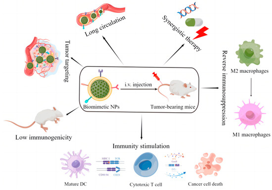
Biomimetic cell-derived nanoparticles are synthetic nanoparticles camouflaged with natural or engineered cell membrane materials to trick the immune system and enhance tumor targeting
[21][22][21,22]. The structure of biomimetic cell-derived nanoparticles is a core–shell structure, where the core is the nanoparticles delivering the therapeutic agents to the target site and the shell is the membrane materials extracted from different cells. These resulting biomimetic nanoparticles combine the physical and chemical properties of nanoparticles with the intrinsic properties of natural cell membranes, possessing the capacity to evade the immune system, prolong blood circulation, and actively target diagnostic and therapeutic agents to the targeted sites
[17][18][19][20][17,18,19,20]. In 2011, Hu et al. constructed erythrocyte membrane-camouflaged nanoparticles by co-extruding PLGA polymeric nanoparticles and erythrocyte membrane-derived vesicles
[23]. This is the first report of nanoparticles derived from cell membranes. Since then, researchers have designed various biomimetic cell-derived nanoparticles to their meet desired functions, and flexibly combined different types of nanoparticles with different sources of biomimetic membrane materials
[24][25][26][27][24,25,26,27], such as cancer cell membranes, white blood cell or leukocyte membranes, stem cell membranes, platelet membranes, and bacteria membranes. The biomimetic cell-derived nanoparticles have been widely recognized for a variety of applications in drug delivery, disease diagnosis, immune modulation, and disease treatment
[28][29][30][28,29,30].
2.1. Unique Function of Biomimetic Cell-Derived Nanoparticles
Biomimetic cell-derived nanoparticles inherit abundant important biological functions related to their source cells, including “self” labeling, biological targeting, cross-talk with the immune system, and region-specific homing. Most of these important functions are attributed to specific proteins or molecules on the surface of cell membranes.
2.1.1. Erythrocyte Membranes
Erythrocytes can prolong the systemic circulation time of wrapped nanoparticles due to some special membrane proteins
[31], and the most important biomarker is CD47. Briefly, signal-regulatory protein alpha (SIRPα) on the surface of phagocytes interacts with CD47 expressed by erythrocytes, which disguises the immune cells as self and prevents immune phagocytes from phagocytizing erythrocytes
[32]. In addition, CD59, C8 binding protein (C8bp), and complement receptor 1 (CR1) on the surface of erythrocytes have a role in defending against complement system attack
[33]. Therefore, nanoparticles coated with erythrocyte membranes have a prolonged circulation half-life and are less immunogenic. Nevertheless, erythrocyte membranes have no tumor-targeting properties. To remedy this deficiency, researchers further utilize tumor-targeting ligands/peptides to modify erythrocyte membranes or construct erythrocyte membrane-based hybrid membranes to realize tumor targeting
[34][35][36][37][34,35,36,37].
2.1.2. Immune Cell Membranes
Tumors are chronic inflammatory tissues that attract and recruit immune cells by secreting a variety of chemokines and cytokines
[38][39][38,39]. Therefore, the adhesive property of immune cells can be exploited to actively target drugs for cancer treatment
[40].
Dendritic cells (DCs) are professional antigen presenting cells (APCs). Thus, mature DC membrane-wrapped nanoparticles can thoroughly inherit the antigen-presenting function of DCs
[41]. With this advantage, mature dendritic cell membrane-wrapped nanoparticles can specifically activate T cells due to the peptide/major histocompatibility complex (MHC) complexes on the surface of biomimetic nanoparticles
[42]. In addition, costimulatory molecules and adhesion molecules on DC membranes, including integrins, CD44, CD40, and ICAM-3, can facilitate the interaction between T cells and DCs
[43].
Macrophages can accumulate in the tumor microenvironment owing to some adhesion molecules and specific receptors, such as intercellular adhesion molecule-1, C-C chemokine receptor 2, and vascular cell adhesion molecule-1 (VCAM-1)
[43]. In addition, macrophages can achieve active tumor targeting owing to the interaction between α4 integrins on macrophage membranes and VCAM-1 on tumor cell membranes
[44].
T cells or cytotoxic T cells are also a subtype of leukocytes that can kill tumor cells. T cells have various properties, such as searching for antigens in the systemic circulation, activating cytolysis by recognizing a single peptide, and producing interferon-γ (IFN-γ, a cytokine with multiple antitumor properties), which make them attractive mediators of antitumor immunity
[45][46][45,46]. Additionally, cytotoxic T cells can promote tumor cell apoptosis mediated by granule and receptors
[47]. In addition, TCR complexes expressed on T cells can specifically bind to tumor-associated antigens with high affinity, which makes T cells more specific in tumor-targeting
[48][49][48,49].
Natural killer (NK) cells play an important role in the recognition and killing of tumor cells owing to their innate capacity to monitor the abnormal expression of stress proteins and MHC-I
[50]. Although NK cells lack tumor antigen-specific receptors on their surface, they have some alternative receptors that can recognize tumor cells, such as DNAM-1, NKG2D, and NKp46
[51][52][51,52]. Therefore, NK cell membrane-wrapped nanoparticles possess good tumor-targeting ability.
2.1.3. Platelet Membranes
Similar to erythrocytes, platelet cell membrane-wrapped nanoparticles also have a prolonged blood circulation time and are less immunogenic due to their reduced immunogenicity. Firstly, CD47 expressed on the surface of platelets can inhibit the uptake of platelet cell membrane-wrapped nanoparticles by macrophages
[53]. Moreover, CD55 and CD59 expressed on the surface of platelet membranes, along with CD47, can avoid immune complement system attack
[54][55][54,55]. In addition, P-selectin is overexpressed on the surface of platelets, which allows platelet membrane-derived nanoparticles to specifically bind to the CD44 receptors expressed on tumor cells
[56]. Thus, nanoparticles can achieve aggressive tumor targeting and long circulation capability through platelet membrane coating.
2.1.4. Cancer Cell Membranes
Cancer cell membrane-camouflaged nanoparticles can enable immune escaping, prolong systemic circulation, and target homotypic tumors owing to a series of membrane proteins expressed by cancer cells
[57][58][57,58]. CD47 on the tumor cell surface plays an important role in immune escape, especially in 4T1, MDA-MB-231, and MCF-7
[59][60][61][62][59,60,61,62]. Thomsen–Friedenreich antigen, E-cadherin, Galectin-3, N-cadherin, and epithelial cell adhesion molecules on the surface of tumor cells are essential for homologous targeting and adhesion
[63][64][65][63,64,65]. Therefore, the application of cancer cell membranes for nanoparticle surface coating has the unique advantages of inherent immune escaping and homologous targeting and adhesion.
2.1.5. Stem Cell Membranes
Stem cell membranes are easy to isolate and have a various of molecular recognition sites, which can be used in biomimetic nanoparticles
[66][67][66,67]. Chemokine receptors on mesenchymal stem cell membranes respond to ligand molecules on tumor cells, promoting the migration of mesenchymal stem cells to the tumor
[68]. Moreover, P-selectin, E-selectin, and TGF-β expressed on the membrane of mesenchymal stem cells also influence the tumor tropism of mesenchymal stem cells
[68][69][68,69]. Therefore, mesenchymal stem cell membrane-wrapped nanoparticles have also attracted increasing attention owing to their good biocompatibility, prolonged circulation time, and tumor targeting.
2.1.6. Bacteria Membranes
In the 1890s, William Coley used toxins made from attenuated bacteria to activate the anti-tumor immune system
[70]. This is the first report of bacteria being used in cancer immunotherapy. Outer membrane vesicles (OMVs) produced by Gram-negative bacteria are composed of lipid bilayers and inherit various parent bacteria-derived components, such as enzymes, bacteria antigens, adhesins, and a variety of pathogen-associated molecular patterns (PAMPs)
[71][72][73][71,72,73]. Among them, bacteria-derived antigens and PAMPs play a vital role in inducing the humoral and cellular anti-tumor immune responses
[74][75][74,75]. Taking the advantages of OMVs, researchers have attempted to construct bacterial membrane-derived nanoparticles for cancer immunotherapy
[76].
2.1.7. Extracellular Vesicles
Extracellular vesicles (EVs) are a group of nanoscale membrane-bound vesicles secreted by almost all eukaryotic cells
[77][78][77,78]. The surface of EVs is rich in a variety of transmembrane proteins, such as ICAM-1, integrin, and tetraspanin, which give EVs the ability to target specific tissues or cells
[79]. In addition, compared with traditional drug delivery platforms, EVs have excellent biocompatibility, low immunogenicity, phagocytosis avoidance, controllable biological characteristics, and the potential to cross natural barriers such as the blood–brain barrier
[80]. Especially, EVs with immunomodulatory capacities, such as DC-derived EVs, NK-derived EVs, and T cell-derived EVs, can be used as effective therapeutics for cancer immunotherapy
[81][82][81,82].
2.1.8. Hybrid Cell Membranes
Compared with single-cell membranes, hybrid cell membranes not only retain the physical and chemical characteristics of nanoparticles, but also endow nanoparticles with the biological functions of two or more derived cells, which makes biomimetic cell-derived nanoparticles more attractive
[83]. Hybrid cell membrane-camouflaged nanoparticles can enhance the flexibility of nanoparticle functionality and thereby achieve better anti-tumor effects
[84][85][86][87][84,85,86,87].
2.2. Fabrication of Biomimetic Cell-Derived Nanoparticles
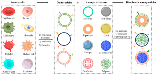
The fabrication of biomimetic cell-derived nanoparticles mainly includes: (1) isolation and construction of parent cell membrane-derived vesicles and (2) fusion of the parent cell membrane with nanoparticle cores (Figure 2).
Figure 2. The fabrication of biomimetic cell-derived nanoparticles.
2.2.1. Isolation and Preparation of Parent Cell Membrane-Derived Vesicles
The isolation of cell membranes should be gentle in order to obtain biologically active membrane vesicles and usually involves cell lysis and membrane purification. Anucleated cells, such as platelets and erythrocytes, are isolated from whole blood by centrifugation. After that, the collected cells are lysed via repeated freeze–thaw or hypotonic treatment. Purified membranes are obtained by removing soluble proteins by centrifugation. Finally, the purified membranes are extruded through a polycarbonate porous membrane to gain parent cell membrane-derived vesicles
[23][54][55][23,54,55]. Bacteria are coated with cell membranes and peptidoglycans, making membrane extraction more difficult. Luckily, Gram-negative bacteria can naturally produce OMVs, which can be directly separated from their culture via ultrafiltration
[88][89][88,89]. Compared with anucleated cells, the extraction and purification of parent membranes from eukaryotic cells, such as stem cells and leukocytes, is more complicated. First,
reswe
archers have to collect source cells from the tissue, blood, or culture medium. Then, a variety of methods are used for cytolysis, including hypotonic solution treatment, repeated freeze–thaw, and/or mechanical rupture. Additionally, the obtained membranes are purified by discontinuous sucrose gradient centrifugation to remove nuclei, intracellular vesicles, and intracellular biomacromolecules
[90][91][90,91].
2.2.2. Fusion of Parent Cell Membrane-Derived Vesicles with Nanoparticle Cores
Membrane extrusion and ultrasonic and microfluidic electroporation are commonly used methods to facilitate the coating of parent cell membrane-derived vesicles onto nanoparticle cores
[2][14][2,14]. Mechanical extrusion is one of the commonly used methods reported in the literature. Briefly, the membrane vesicles and nanoparticles are extruded through the polycarbonate porous membrane with progressively smaller pore sizes. The fusion of vesicles and particles is achieved by applying mechanical forces to facilitate the passage of nanoparticles through the lipid bilayer of the membrane vesicles
[92]. This approach can largely ensure the bioactivity of membrane proteins. Nevertheless, it is a time-consuming process
[93]. Ultrasound is another effective method. When the nanoparticles and membrane vesicles are mixed together, the membranes surrounding the nanoparticles are reassembled by sonication. Compared to the extrusion method, this method is time-saving. However, the parameters of the ultrasound apparatus, including power, frequency, and duration, need to be optimized to guarantee fusion efficiency while avoiding protein inactivation. Microfluidic electroporation can also be utilized for coating different types of nanoparticles. Electromagnetic energy forms holes in cell membranes by a microfluidic chip, thereby promoting membrane vesicles to wrap nanoparticle cores
[94]. In this process, the parameters also need to be optimized, such as pulse voltage, flow rate, and duration. The biomimetic cell-derived nanodrugs constructed by this method are completely coated, uniformly distributed, and highly reproducible. However, this technology comes at a high cost. In contrast to nanoparticle-templated membrane coating, Zhang and his colleagues reported a simple method to synthesize cell membrane-wrapped nanogels by cell membrane-templated polymerization
[95]. Unlike the previously mentioned approaches, this new strategy uses membrane vesicles to ‘guide’ the growth of nanoparticle cores, thereby surmounting the “coatability” limitation of nanoparticles, as well as adding elevated controllability for the application of biomimetic cell-derived nanoparticles. The key challenge for this technique is how to efficiently and precisely inhibit the polymerization reaction outside the vesicles while maintaining the reaction activity inside the vesicles.