Your browser does not fully support modern features. Please upgrade for a smoother experience.
Please note this is a comparison between Version 1 by Zongjie Wang and Version 2 by Sirius Huang.
Rare cells play essential roles in the initiation and progression of diseases and therefore their analysis is of great interest. The micro-magnetofluidic system is one of the emerging platforms that have been proposed for the rapid, sensitive, and cost-effective analysis of rare cells.
- microfluidics
- magnetic nanoparticles
- rare cell analysis
- cell sorting
1. Rare Cells: Definition and Examples
A cell is considered ‘rare’ when the number of cells of that subpopulation represents less than 0.01% of the total cell populations in a tissue or biofluid [1][2][43,44]. These cells, although occur as a minimal population, may play critical roles in disease progression and tissue regeneration. Below are a few examples highlighting the essentiality of rare cells in important bioprocesses.
For disease progression, one vivid instance is the initiation of cancer metastasis [3][45], where the primary tumor evolves and generates delayed secondary tumors in distal organs [4][46]. While many theories have been proposed to explain the process of cancer metastasis, the most accepted mechanism is through the formation of circulating tumor cells (CTCs) [5][6][47,48] (Figure 1A). In brief, CTCs are rare cells that emigrate from the primary tumor, enter and circulate within the peripheral blood, and eventually settle down in different organs for growing [7][49]. CTCs are generally considered as the seed of metastasis given their critical role during cancer migration [8][9][50,51] and therefore have attracted significant attention as a general biomarker for monitoring and predicting cancer progression [10][11][12][52,53,54]. However, one critical challenge of using CTCs is their rarity—the number of CTCs is extremely low, usually at the scale of a few CTCs in millions of normal cells [13][14][15][55,56,57].
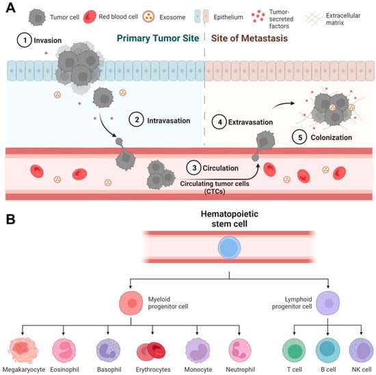
Figure 1. Rare cell-mediated bioprocesses in disease progression and tissue regeneration. (A) Circulating tumor cells (CTCs) play a critical role in cancer metastasis. In primary tumor sites, some highly invasive cancer cells undergo intravasation to enter the blood circulation and become CTCs. CTCs travel to distal organs and tissues, undergo extravasation, and eventually form deadly metastatic colonies. (B) Human hematopoietic stem/progenitor cells (HSPCs) have the potential to regenerate all types of blood cells. Under proper conditions in vitro and in vivo, HSPCs can differentiate into all myeloid (monocytes, neutrophils, etc.) and lymphoid cell lineages (T cells, B cells, etc.).
For tissue regeneration, a representative example is the use of human hematopoietic stem/progenitor cells (HSPCs) for transplantation. HSPCs only represent 0.2% to 0.5% of the leukocyte population (or 0.0003–0.0008% of the blood cell population) in peripheral blood [16][58], but have the full potency to produce all types of blood cells (e.g., lymphocytes and monocytes, Figure 1B). Therefore, the transplantation of HSPCs has been essential to the immune reconstruction post the treatment of leukemia [17][18][19][59,60,61]. Besides, rare cells may modulate the fate of a specific regenerative therapy given to a patient. It has been reported that rare undifferentiated stem cells in therapeutic cells, such as cardiomyocytes [20][62] and beta cells [21][63], may undergo uncontrolled growth in vivo in animal models [22][23][64,65] and eventually convert regenerative therapies into stem cell cancers [24][25][26][27][66,67,68,69].
Thus far, over 50 different types of microfluidic devices have been designed and validated for the analysis of rare cells, including circulating tumor cells [28][29][70,71], circulating tumor clusters [30][31][32][72,73,74], circulating cancer stem cells [33][34][35][75,76,77], circulating fetal cells [36][37][78,79], and circulating endothelial cells [38][80] in peripheral blood, as well as contaminating tumor cells [39][81] in therapeutic cell products. Multiple strategies have been proposed, such as filtration, inertial fluidics, deterministic lateral displacement (DLD), and immunomagnetic sorting [40][13].
2. Micro-Magnetic Deflection
2.1. Principles
Magnetic deflection is a main approach of magnetophoresis. It relies on the dictated movement/migration of target cells to achieve separation. The main procedures of magnetic deflection include the specific magnetic labeling of target cells, the incorporation of an external magnetic field, and on-chip magnetic sorting. The magnetic deflection of target cells is the result of interaction between magnetic force and hydrodynamic force on the cells. In the presence of an external magnetic field, magnetically labeled cells experience a magnetic force produced by the inserted magnetic gradient. In the meantime, the cells experience Stoke’s drag force from the fluid flow. The magnetic force given as2.2. Magnetic and Hydrodynamic Optimization
The design of magnetic field is an important aspect for the optimization of magnetic deflection. To achieve a good deflection, a large magnetic gradient is required. To increase the magnetic gradient, researchers have tried different types of permanent magnets and different ways to place the magnets. The easiest strategy is to apply a macroscale permanent magnet on the side or at the bottom of the microfluidic device to produce magnetic gradient [41][42][43][107,108,109]. Target cells will be deflected due to the non-uniform magnetic field produced by the permanent magnet. However, the magnetic gradient may not be strong enough to achieve high-resolution magnetic deflection when target cells have low magnetic susceptibility. Alternatively, magnetic blocks arranged with opposing poles were used for a larger magnetic field gradient [44][45][110,111]. Micro-magnet arrays have also been applied to create strong local magnetic gradients to increase magnetic deflection sensitivity [46][47][112,113]. Insertion of paramagnetic materials has also been used to further enhance local magnetic gradients in the microfluidic device. Soft magnetic materials such as nickel and ion oxide micro- or nanostructures were integrated in microfluidic devices to create strong local magnetic gradients in the microchannel [48][49][50][51][105,114,115,116]. Tom Soh et al. have designed nickel microchannel strips to deflect magnetic labeled rare cells [52][117]. The magnetic ratcheting cytometry system developed by the Di Carlo group also applied soft magnetic materials for magnetic field enhancement and adjustment. In this system, micropillars made of soft materials were arranged at various distances to create multiple magnetic ratcheting zones in the sorting chamber. Periodically switching magnetic fields were produced under the microchip through rotating permanent magnets. The micropillars activated by the switching magnetic field drove magnetically labeled cells across the sorting chamber, and cells with different magnetic loading levels would be stabilized at different ratcheting zones [53][54][118,119]. In recent years, the Kelley group has developed a magnetic deflection-based microfluidic chip-termed PRISM [55][56][57][106,120,121]. This system made use of the superb ferromagnetic property of an amorphous metal alloy ribbon called metglas to produce an extremely strong local magnetic field when external magnets were introduced. Through lithography and wet etching, metglas-based magnetic guide could be patterned on a glass substrate. SU8-based microchannels were then generated on top of the magnetic guide for sample processing and magnetic deflection. The magnetic guides embedded under the microchannel were designed to branch out from the middle to the sidewall of the microchannel with multiple magnetic deflection angles. Magnetically labeled rare cells were thus segregated into different subpopulations when following the magnetic guides. Making use of hydrodynamic force is another way to dictate magnetic deflection. For example, designing microstructures in the microchannel to produce a low drag force zone can be used to isolate magnetically labeled cells for single-cell analysis [58][122]. Using different materials for channel substrates and optimized channel shapes to obtain uniform flow profiles is another way for drag force manipulation [59][123]. A third way is to introduce ‘flow pockets’ on the side the microchannel and apply permanent magnets along the ‘flow pockets’ [60][61][124,125]. The flow velocity is close to zero in the ‘flow pockets’ and non-target cells flowing along the fluid flow will not enter the ‘flow pockets’, while magnetic labeled target cells will be deflected and trapped in the ‘flow pockets’. Table 1 summarizes different magnetic sorting methods and their relevant performance including throughput, target cell isolation efficiency, purity, sensitivity and their potential limitations. Due to the miniaturized channel scale and strong inserted external magnetic field, microfluidics-based magnetic deflection can bring high deflection efficiency of target cells with good purity [62][63][64][65][14,126,127,128]. Additionally, since magnetically labeled cells move continuously along the sorting chamber, deflected target cells can be easily collected in the designated outlet with few cell loss. However, this method solely depends on magnetic force added on the target cells, which may not be enough to address the heterogeneity of target cells. Rare cells such as CTCs are known to be heterogenous in both biological and physical properties [66][67][129,130]. For example, CTCs undergoing epithelial to mesenchymal transition (EMT) will lose the expression of EpCAM [68][69][70][71][131,132,133,134]. Targeting EpCAM alone cannot isolate CTCs with low EpCAM expression. To address this problem, researchers introduced multiplex marker targeting [72][73][74][135,136,137]. In addition to EpCAM, biomarkers such N-cadherin, vimentin were used to differentiate CTCs under EMT from blood cells [75][76][138,139]. Alternatively, magnetic deflection can be combined with physical-property based cell separation methods to achieve superb CTC isolation efficiency.Table 1.
Comparison of different magnetic deflection-based cell sorting strategies.
| Sorting Strategy | Application | Throughput | Efficiency | Purity | Sensitivity | Limitation | Reference |
|---|---|---|---|---|---|---|---|
| Magnetic guided deflection | CTC and CTC cluster isolation | 0.5 mL/h | ~90% | 5.7 log white blood cell depletion | 10 targe cell per mL | Strong cell–cell interaction at high cell concentration can affect its performance | [56][120] |
| Modified magnetic beads | Separation of cancer cells | NA | ~90% | >80% | 106 cells /mL | Large bead size reduce sensitivity | [77][140] |
| Captured magnetic bead-bounded target cells with dead-ended side chambers near the a permanent magnet | CTC isolation from whole blood | 1.2 mL/h | >90% | <0.4% white blood cell capture | 2–80 target cells spiked in 1 mL of blood | Lack of clinical sample processing | [42][108] |
| Magnetophoresis assisted cell capture activated by magnet arrays | Isolation of cancer cells spiked in human blood | 7.2 mL/h | >60% | ~30% | 3.5 × 10⁴ cancer cells spiked in 1 mL of blood | Release of the rare cells would be challenging | [44][110] |
| Two-stage magnetic separation | White blood cell sorting from whole blood | 1.2 mL/h | 93% | >70% | 103 cells/min | Require further optimization of rare cell separation | [49][114] |
| Ferromagnetic guide based deflection in a large scale 3D printed system | Isolation of mature natural killer cells from blood | >18 mL/h | >50% | ~90% | 107 cells per mL | Complicated fabrication and assembly process | [57][121] |
| Magnetic deflection facilitated single-cell capture | Capture of rare tumor cells from mouse blood | 2 mL/h | ~90% | >90% | <100 CTCs from 1 mL of whole blood | The system can be saturated due to the limited number of trap units | [58][122] |
| Magnetic deflection and capture on wavy-herringbone structures | Capture CTCs from whole blood | 0.54 mL/h | 92% | 91% | 100 target cells per mL blood | Lack of clinical processing | [61][125] |
| Magnetic ratcheting | Profiling of magnetically labeled immune cells in spiked samples | NA | 87% | 95% | 107 cells per mL | The throughput was limited as no in-flow allowed during magnetic ratcheting | [53][118] |