Treatment Drugs for Silicosis: Comparison
Please note this is a comparison between Version 1 by Shi Chen and Version 3 by Lindsay Dong.

Silicosis, characterized by irreversible pulmonary fibrosis, remains a major global public health problem. CNowadays, cumulative studies are focusing on elucidating the pathogenesis of silicosis in order to identify preventive or therapeutic antifibrotic agents. However, the existing research on the mechanism of silica-dust-induced pulmonary fibrosis is only the tip of the iceberg and lags far behind clinical needs. Idiopathic pulmonary fibrosis (IPF), as a pulmonary fibrosis disease, also has the same problem.

  • silicosis
  • IPF
  • pulmonary fibrosis
  • treatment drugs

1. Introduction

The prevalence of crystalline silicon dioxide dust is widespread in many areas [1]. As a result of the continuous inhalation of silica particles, many workers develop silicosis, an irreversible and incurable disease [2]. Silicosis is a chronic interstitial lung disease characterized by fibrosis, inflammation, and destruction of the pulmonary structures. It causes pulmonary hypertension, progressive dyspnoea and death from respiratory insufficiency [3]. Many measures have been taken in recent decades to protect workers in the workplace, but millions of workers still suffer from silicosis [4].
Silicosis is a severe concern in construction and mining workers, particularly young workers, who are exposed to quartz conglomerates during sandblasting, bolting, cutting, shaping, and installing kitchen countertops [5][6][7][5,6,7]. Recent studies have also indicated that exposure to nanosilica can cause inflammation and fibrosis in the lungs. The risk of nanosilica exposure in this emerging industry is noteworthy, despite the lack of reported cases [8]. The increasing number of silicosis cases worldwide presents new challenges for prevention in many countries [4]. The present circumstance underscores the significance of exercising caution in authorizing development of emerging industries and implementing early identification and control measures from a public health perspective. Furthermore, it is crucial to accelerate the discovery of remedies for silicosis from a clinical treatment standpoint.
The pathogenesis of silicosis is not fully understood, and the disease is complex [9][12]. Although further research is required to clarify the role of intricate signaling pathways [10][13], multiple pathways are thought to be involved in the development of silicosis (Figure 1). Silica-induced lung injury is characterized by various mechanisms, including direct cytotoxic effects on macrophages, activation of macrophage surface receptors, lysosomal rupture, reactive oxygen species (ROS) production, inflammasome activation, cytokine and chemokine production, apoptosis/softening, and lung fibrosis [11][14].
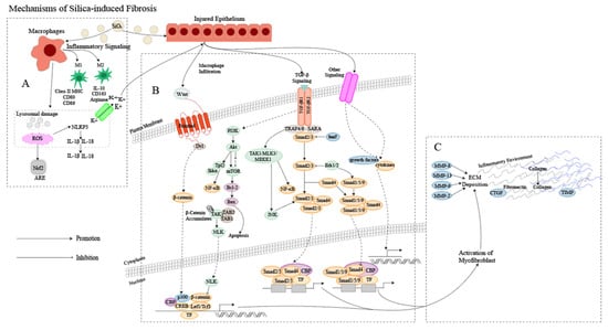
Figure 1. Mechanism of silica-induced fibrosis. (A) Alveolar macrophages (AMs) engulf silica dust, causing them to turn into dust cells. Subsequently, AMs may synergize with alveolar epithelial cells (AECs) to release a large amount of ROS to participate in oxidative stress reactions, activate NOD-like receptor thermal protein domain associated protein 3 (NLRP3) inflammatory bodies through lysosomal damage and potassium outflux, and activate the release of inflammatory mediator interleukin (IL) -1β, IL-18 and other cytokines inducing epithelial–mesenchymal transition (EMT). Meanwhile, AMs can polarize into M1 and M2 types, playing a role in promoting inflammation, fibrosis, and antigen presentation, increasing the proliferation of lung fibroblasts and collagen synthesis and secretion, and promoting the formation of fibrosis through apoptosis and autophagy. (B) Ongoing damage and damage to lung cells by silica lead to pathological overdeposition of extracellular matrix (ECM) proteins accompanied by upregulation of myofibroblast activity, resulting in a chronic inflammatory environment of macrophage and immune cell infiltration. In this cellular environment, cytokines and growth factors are released in large quantities, activating many signaling cascades, including members of the transforming growth factor-beta (TGF-β) family and Wingless/Int (Wnt) 1, the phosphatidylinositol 3-kinase (PI3K)/protein kinase B (AKT)/mammalian target of rapamycin (mTOR) pathway and other pathways. (C) Fibroblasts then aggregate in the area of injury, and the combination of ECM degradation by MMPs and excessive collagen deposition leads to granuloma formation and lung tissue remodeling.

2. Characteristics of Pulmonary Fibrosis

2.1. Characteristics of IPF

The origin of idiopathic pulmonary fibrosis (IPF), much like silicosis, remains unclear, with a convoluted pathogenesis likely involving multiple interconnected signaling pathways, including the TGF-β/Smad signaling pathway, Wnt/β-catenin signaling pathway, platelet-derived growth factor (PDGF) signaling pathway, PI3K/AKT signaling pathway and other signaling pathways [12][16]. IPF is thought to be a consequence of damage to the alveolar epithelium and abnormal wound healing, but it has also been shown that both genetic factors and environmental conditions can significantly contribute to the development of the disease [13][14][17,18]. The condition is characterized by subpleural basal fibrosis, honeycomb changes, and collagen and ECM deposition, which ultimately result in life-threatening structural changes in lung tissue and loss of pulmonary ventilation and diffusion function.
Alveolar epithelial damage, caused by external factors (infection, toxins, smoke) or internal factors (inflammation, oxidative stress, abnormal immune response), leads to the release of fibrogenic cytokines, including TGF and tumor necrosis factor (TNF), as well as growth factors such as PDGF and connective tissue growth factor (CTGF) [15][19]. Elevated levels of these fibrogenic cytokines and growth factors, both locally and systemically, stimulate the activation and proliferation of lung fibroblasts to some extent. Upon activation, fibroblasts differentiate into pulmonary myofibroblasts, which are responsible for the excessive production of ECM proteins in fibrotic lung tissue. These myofibroblasts also regulate the balance between MMP and tissue inhibitors of metalloproteinases (TIMPs), thereby facilitating the process of IPF [16][20].

2.2. The Relationship between Silicosis and IPF

2.2.1. Cause of Disease

The inhalation of free silica dust in the air is the main cause of silicosis. The emergence of silicosis is closely linked to the volume, structure and dimensions of the silica particles inhaled [17][21], whereas IPF is a particular type of interstitial pneumonia that is fibrosing, chronic and progressive, but for which the origin is yet to be determined [18][22].
Despite the fact that occupational exposure can induce IPF to some extent, a plethora of epidemiological investigations have demonstrated a significant link between smoking, chronic viral infections, and the genetics of IPF [18][19][20][22,23,24].

2.2.2. Pathogenesis

Extensive research has revealed that the development of silicosis fibrosis is not solely due to one factor, but rather a complex outcome resulting from various factors and links [21][25]. The primary pathogenic mechanisms of silicosis involve direct cytotoxic effects, the generation of ROS and reactive nitrogen radicals, the release of inflammatory chemokines, the initiation of fibrotic pathways and cell death [4]. The cellular molecule and gene transcription regulation fields are also being explored in relation to silicosis [22][26].
Cellular senescence, which includes molecular changes such as telomere shortening, is involved in the pathogenesis of various chronic diseases, including lung diseases [23][28]. Telomere shortening may be a common causative feature of the development of IPF and silicosis [24][25][29,30].
Silicosis and IPF are respiratory diseases that cause damage to the lungs. In response to the inflammatory process, fibroblasts proliferate and produce excessive collagen fibers, leading to the deposition of ECM lung tissue remodeling, ultimately resulting in impaired lung function [26][27][33,34]. Moreover, the two diseases share similarities in the upregulation of TGF-β and extracellular signal-regulated kinase (ERK) signaling pathways in cytokine and growth factor pathways, and a relationship with autophagy [28][35]. While the immediate causes of silicosis and IPF may differ, the overall mechanisms of the subsequent profibrotic reaction are comparable, being characterized by ECM deposition and fibroblast proliferation [29][36]. Therefore, potential therapeutic drugs for the treatment of silicosis may be sought from IPF.

2.2.3. Symptoms and Complications

Silicosis and IPF share certain symptoms, such as dyspnoea and loss of appetite. However, patients with silicosis may experience additional manifestations, such as chest pain, pulmonary dysfunction, low-grade fever, night sweats, and active shortness of breath, while IPF typically presents as cough and sputum production [30][37]. In some severe cases, patients with IPF may also exhibit general discomfort, including weakness and joint pain.
The development of silicosis can result in various complications, including tuberculosis, chronic obstructive pneumonia, and rheumatoid arthritis [31][38]. Similarly, individuals with IPF may experience pulmonary hypertension, acute exacerbation of pulmonary fibrosis, respiratory tract infections, acute coronary syndrome, and thromboembolic disease [32][39]. However, as the diseases advance, both silicosis and IPF can increase the likelihood of developing lung cancer [33][34][35][40,41,42].

3. Treatment of Silicosis and IPF

3.1. Drug Research Targeting Mechanisms of Silicosis

The study findings have demonstrated the considerable scope of pharmaceuticals in the treatment of silicosis fibrosis. These drugs can address one or more fundamental mechanisms of silicosis, mitigating the inflammation and/or fibrosis triggered by silica.

3.1.1. Oxidative Stress Response

The oxidative stress encountered during pulmonary fibrosis is closely associated with both the nuclear factor kappa-B (NF-κB) signaling pathway, the kelch-like ECH-associated protein 1 (Keap1)/NF-E2 p45-related factor 2 (Nrf2)/antioxidant-responsive element (ARE) pathway and the NADPH oxidase (NOX) 4-Nrf2 signaling cascade [36][47]. According to available evidence, it has been suggested that the administration of dioscin, dihydroquercetin, and quercetin may have a beneficial effect on pulmonary fibrosis by impeding the infiltration of macrophages, B lymphocytes, and T lymphocytes into the lung tissue [37][38][39][48,49,50]. Oleanolic acid has been found to be advantageous in the management of pulmonary fibrosis, most likely due to its capacity to diminish serum TNF-α levels, decrease collagen content in lung tissues, and hinder oxidative stress and NF-κB activation [40][51]. The fibrosis process in silicosis may be impacted by the use of traditional Chinese medicine (TCM) compound preparations that target oxidative stress. A recent study has revealed that the low-dose nebulized inhalation of Chinese herbal preparations can reduce the levels of malondialdehyde (MDA) and interferon γ, while also improving pulmonary fibrosis and inflammation in silicosis [41][60]. Other compounds, such as panicolin, bletilla striata polysaccharide, small molecule components, and N-acetylcysteine (NAC) have also been found to be effective in controlling the progression of silicosis. These drugs work by modulating the hydroxyproline levels and regulating factors such as superoxide dismutase (SOD) and MDA in the oxidation system [42][61].

3.1.2. Autophagy and Apoptosis

Currently, research has shown that apoptosis of AM induced by SiO2 can be regulated by various intracellular pathways, including the mitochondria-mediated intracellular apoptosis program [43][64], NF-κB signaling pathway [44][65], factor-related apoptosis (Fas)-mediated exogenous pathway [45][66], p53 signaling pathway [46][67], endoplasmic reticulum stress [47][68], PI3K/AKT/mTOR signaling pathway [48][69], Janus kinase (JAK)2/signal transducer and activator of transcription (STAT)3 signaling pathway [49][70], and others. Studies have demonstrated that emodin can inhibit silica-induced apoptosis and exert antifibrotic effects by increasing the expression of anti-apoptotic protein B-cell lymphoma-2 (BCL2) and decreasing the expression of pro-apoptotic protein-BCL2-associated X(Bax) protein [50][58]. Similarly, dioscin can promote AM autophagy, enhance the clearance of mitochondria damaged by silica dust, and reduce the activation of mitochondria-mediated apoptosis pathway, so that AM resists apoptosis caused by silica dust and reduces the secretion of pro-inflammatory and profibrotic factors [37][48].

3.1.3. Regulation of Signaling Pathways Related to EMT

TGF-β is a crucial regulator of EMT, which reduces the expression of epithelial markers such as E-cadherin and α-catenin while increasing the expression of mesenchymal markers such as N-cadherin, vimentin, and alpha-smooth muscle actin(α-SMA) [51][75]. Certain natural compounds such as emodin, quercetin, tadalafil, and sodium ferulate have been found to increase E-cadherin levels and reduce the expression of Vimentin, α-SMA, Col-I, TNF-α, IL-1β, and pro-inflammatory factors TGF-β1, thereby regulating EMT to relieve fibrosis in silicosis [52][53][76,77]. Tamoxifen, on the other hand, reduces the serum TGF-β1 content in rats in the model group in a dose-dependent manner, effectively inhibiting the process of silicosis [54][78], but also exhibits certain hepatotoxic effects. Interestingly, AKEX0011 has also been found to reduce the infiltration of neutrophils and macrophages in lung tissue and decrease the protein levels and mRNA expression of fibrosis-associated proteins [55][71].

3.1.4. Blocking Silicosis Fibrosis by Targeting Fibroblasts

Pulmonary fibrosis is characterized by excessive secretion of collagen, fibronectin, and elastin by myofibroblasts, leading to an imbalance in MMP/TIMP regulation and accumulation of the ECM [56][84]. Quercetin has been found to reduce the expression of MMP-2 and MMP-9, thereby inhibiting the formation of the ECM and reducing pulmonary fibrosis [57][58][59][85,86,87]. Schisandrin B has also been shown to inhibit the expression of MMP-2, slowing the onset of pulmonary fibrosis in rats induced by silica [60][88]. Additionally, gallus domesticus extract has been found to reduce the content of FNs, Col-I, MMP-9 and MMP-12 in bronchoalveolar lavage fluid (BALF) in rats [61][89]. Dasatinib has been found to induce macrophage bias towards the M2 macrophages phenotype, improving lung mechanics in mouse models of acute silicosis by downregulating the expression of IL-1β, TNF-α and TGF-β proteins in lung tissue and upregulating the expression of arginase and MMP-9 [62][90].

3.1.5. Other Mechanisms to Prevent and Treat Silicosis Fibrosis

In addition to the aforementioned studies, researchers have been investigating alternative mechanisms through which drugs can affect silicosis fibrosis. For instance, kaempferol has been found to restore silica-induced microtubule-associated protein 1A/1B-light chain 3 (LC3) lipidation without increasing p62 levels [63][73]. Moreover, the combination of tetrandrine tablets and matrine injection has been shown to have a low incidence of adverse reactions and to be able to improve lung ventilation function, alleviate symptoms, and exhibit significant clinical value [64][93]. The combined use of desipramine and NAC has been found to effectively suppress the inflammatory response and delay the progression of silicosis fibrosis in a synergistic manner [42][61]. This synergy has also been observed with the combination of NAC and tetrandrine [65][94]. However, drug combinations not only show synergistic effects, but also are accompanied by the side effects of multiple drugs. In summary, it has been discovered that numerous drug components and TCM compound preparations can hinder the development of silicosis fibrosis by intervening in the silicosis TGF-β/Smad signaling pathway, oxidative stress mechanism, apoptosis, and autophagy. Additionally, various pathways have been observed to interact with each other, such as emodin, tan IIA, curcumin, and other drugs, which can exert anti-inflammatory and antifibrotic effects through different intervention mechanisms [66][95].

3.2. Antifibrosis Treatment Drugs for IPF

Numerous studies have been found that by inhibiting the NF-κB signaling pathway, parthenolide, hesperidin and dehydrocostus can inhibit the early inflammatory response, thereby exerting an antifibrotic effect [67][68][69][96,97,98]. Moreover, sulforaphane, dihydroartemisinin, melatonin and ginkgo biloba extract have been found to reduce oxidative stress to exert anti-pulmonary-fibrosis effects [70][71][72][73][74][99,100,101,102,103]. Similarly, the total extract of Yupingfeng and the combination of tan IIA and puerarin can exert antifibrotic effects by regulating the PI3K/AKT/mTOR signaling pathway and JAK/STAT signaling pathway [75][76][77][104,105,106]. As for regulating signaling pathways related to EMT, asiatic acid and oridonin inhibit the expression of TGF-β1 in lung tissue, accompanied by a decrease in Col-I, Col-III, α-SMA and TIMP-1, as well as inactivation of Smads and ERK1/2 [78][79][107,108].

3.3. Other Potential Therapies

3.3.1. Stem Cell Therapy

Mesenchymal stem cells (MSCs) are considered relatively safe due to their abundant sources, ease of isolation and culture, low immunogenicity, and secretion factors that can reduce inflammation [80][130]. Therefore, stem cell therapy has shown significant promise in the treatment of silicosis. Preclinical studies have shown that the administration of MSCs through endotracheal or tail vein effectively inhibits inflammation and fibrosis in mouse models of silicosis, leading to therapeutic benefits [81][82][131,132]. Moreover, previous research suggests that N-acetyl-seryl-aspartyl-lysyl-proline (Ac-SDKP) may alleviate the fibrotic symptoms of silicosis by regulating endoplasmic reticulum stress [83][133]. Averyanov’s study demonstrated that transplantation of bone-marrow-derived MSCs at high cumulative doses (1.6 × 10⁹/mL) is well tolerated and safe in patients with pulmonary fibrosis, with only minor side effects such as fever. These findings confirm the safety, tolerability, and potential benefits of high-dose MSCs, and may pave the way for future stem cell transplantation trials [84][134].

3.3.2. Antifibrotic Target Therapy

The advent of high-throughput omics technology and its amalgamation with bioinformatics has brought about a paradigm shift in gene editing technology, presenting a viable remedy for investigating the causation and management of silicosis. Recent genomic analyses have revealed that certain single nucleotide polymorphisms (SNPs) are connected to an increased risk of silicosis, particularly rs73329476 and rs12812500, which are linked to the development of pneumoconiosis [85][136]. By conducting weighted gene co-expression network analysis, Jiaqi Lv et al. identified silicosis-related modules and pivot genes and found that the Hippo signaling pathway plays a beneficial role in silicosis fibrosis. This discovery has helped to elucidate the precise mechanisms of silica-induced pulmonary fibrosis and identify the molecular initiation events and adverse outcome pathways of silicosis [86][137]. In the field of epigenomics, research has demonstrated that certain miRNAs are significantly linked to the development and progression of pneumoconiosis and may serve as non-invasive biomarkers and prognostic indicators for early pneumoconiosis. The use of transcriptomics and proteomics has led to the identification of a variety of protein-coding genes and proteins that are differentially expressed in silicosis [87][141]. These findings suggest potential targets and signaling pathways that may play a significant role in lung disease. Bo C and Mingyao Wang et al. conducted a study using different clusters for pathway enrichment. They classified differentially significant proteins and found that although differential expression features in the omics datasets are involved in different pathways, these features are found in some key signaling pathways, such as inflammatory response and interstitial fibrosis regulation [22][88][26,142]

3.4. Clinical Drugs of Silicosis and IPF

3.4.1. Silicosis

Management of silicosis consists of using bronchodilators and cough medication. However, symptomatic treatment may only ameliorate symptoms rather than restoring health. Most drugs that have shown positive effects in animal models, especially reducing lung fibrosis, have not been yet translated into clinically approved drugs in many countries, including Europe and the USA [89][144]. Tetrandrine stands as the sole drug sanctioned for silicosis treatment in China, as per the approval of regulatory authorities [90][145]. Recent clinical trials have demonstrated that the combined administration of tetrandrine with other drugs yields more pronounced therapeutic outcomes than conventional treatment modalities [65][94]. Despite the minimal adverse effects of tetrandrine therapy for pulmonary fibrosis, it remains incapable of reversing fibrosis and curing patients afflicted with silicosis.

3.4.2. IPF

Since many of the signaling pathways of silicosis and IPF overlap, it is possible that clinical medications used in the treatment of IPF could hold promise in the treatment of silicosis. For instance, antifibrotic drugs like pirfenidone and nintedanib, which are commonly prescribed for IPF [91][146], have demonstrated efficacy in reducing lung inflammation and fibrotic changes in animal models of silicosis [92][93][94][147,148,149]. Due to the significant side effects of nintedanib, it has not been an option for silicosis treatment, but recently an experiment engineered a nanocrystal-based suspension formulation of nintedanib possessing specific physicochemical properties to enhance drug retention in the lung for localized treatment of silicosis [95][150], bringing new hope to nintedanib for treating silicosis and highlighting that overcoming the side effects of existing clinical drugs can provide a new direction for the treatment of silicosis. Therefore, insights gained from antifibrosis drugs utilized in IPF could offer potential avenues for treating silicosis.
Video Production Service