1. Introduction
Intravenous administration of image contrast agents/drugs typically results in their passage through the circulatory system, followed by renal elimination, which can affect the kinetic parameters within the body. Incorporating these agents within nanoparticles (NPs) can enhance their pharmacokinetic performance and allow for targeted delivery
[1][15]. However, several biological barriers, such as immune recognition/activation and systemic clearance, must be overcome. PEGylation, charge modulation (by attaching zwitterionic entities), or utilization of surface ligands that bypass immune surveillance are some of the available surface modification approaches to improve NP performance
[2][3][4][16,17,18]. However, in most cases, the benefits of employing such strategies are undermined by the added complexity of synthesis. Hence, the attention of the scientific fraternity has shifted toward cell membrane coating as a “bioinspired” strategy to functionalize NPs for biomedical applications. Depending on the membrane source, the corresponding nanosystem will typically have a unique set of properties that can be leveraged for theranostics applications.
Figure 1 represents various sources for procuring functional membranes whereas an overview of membrane sources and their attributes are enlisted in
Table 1.
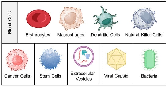
Figure 1.
Cell membrane sources.
Table 1.
Overview of membrane sources and their attributes.
|
Membrane Source
|
Surface Markers/
Proteins
|
Key Advantages
|
Limitations
|
Ref.
|
|
Erythrocytes
|
CD47, C8 binding protein
|
Prolonged circulation time; ease of isolation;
reduced susceptibility to macrophage uptake
|
Absence of tumor-specific ligands
|
[5][20]
|
|
Platelets
|
CD62p, PECAM-1, CD44, CD47
|
Prolonged circulation time; ease of isolation; robust immune evasion
|
Aggregation of coated nanoparticles; absence of tumor-specific ligands
|
[6][26]
|
|
Macrophages
|
CCR2, VCAM-1, ICAM-1
|
Facilitates immune cell trafficking towards tumor; evades reticuloendothelial system; trans-endothelial migration through intercellular adhesion
|
Limited tumor-targeting ability
|
[7][32]
|
|
Dendritic cells
|
|
|
Bacteria |
|
|
|
|
OmpA, OmpC, AcrA |
|
Immune cell activation; self-adjuvant characteristic
|
Pathogenicity needs to be adequately addressed before in vivo use
|
[14][82]
|
2. Blood Cells
Blood cells are a highly diverse and extensively utilized class of membrane source, encompassing a variety of cell types produced in the bone marrow and circulating in the bloodstream. This includes erythrocytes (red blood cells; RBCs), platelets (PLTs), and leukocytes such as macrophages, dendritic cells (DCs), and natural killer (NK) cells.
Erythrocytes are the most abundant cellular component of blood, with approximately 5 million cells per microliter. Due to their size (7–8 μm diameter with a thickness of approximately 1 μm at the center) and lack of nuclei/sub-cellular organelles, their membrane is easily extractable and purifiable. Their long retention time of up to 120 days in humans makes them ideal for conferring long-circulation properties to entrapped NPs
[15][19]. Furthermore, their surface is rich in “self-tagged” proteins such as CD47 (which protects them from phagocytosis by modulating recognition by the signal-regulatory protein alpha glycoprotein), as well as glycans and acidic sialic acid fractions. These attributes collectively provide immune evasion and deterrence from forming the protein corona
[5][20]. Other membrane proteins such as C8 binding protein, homologous restriction protein, decay accelerating factor, membrane cofactor protein, and complement receptor 1 also play a role in resistance to complement system attacks
[16][21]. It is widely accepted that Hu et al. (2011) reported the first bioinspired nanosystem comprising biodegradable polymeric NPs encapsulated by an erythrocyte membrane
[17][22]. Since then, their abundance and ease of availability have led to their widespread use
[18][23].
PLTs, which originate from mature megakaryocytes, are another type of anucleate blood cell with significant applications in developing bioinspired nanosystems
[19][24]. They are abundant in the blood (approximately 150,000–350,000 cells/mL) and have a circulatory lifespan of 7–10 days. PLTs play a vital role in generating an inflammatory response and hemostasis, especially after thrombosis. Many studies have established a correlation between platelet-induced hemostatic properties and cancer progression/metastasis. PLTs can recognize, interact with, and cover circulating tumor cells (CTCs), allowing them to evade immune clearance and spread to new tissues
[20][25]. The surface marker proteins of PLTs, including P-Selectin (CD62p), PECAM-1, CD47, and CD44 receptors, play a crucial role in this function. The unique properties of these surface marker proteins make PLT membranes an attractive coating material for NPs
[6][26]. Coating with PLT membranes can confer beneficial characteristics such as immune evasion and specific adhesion to injured vessels and tumor tissues. Furthermore, the inflammatory characteristics of tumors can “recall” PLTs to accumulate passively at the site of cancerous growth
[21][27].
Leukocytes are derived from multipotent hematopoietic stem cells in the bone marrow, where they differentiate and mature before entering the bloodstream. As immune cells, they play a vital role in protecting the body from infections, repairing tissue injuries, and resisting diseases by engulfing foreign invaders. Leukocytes have migration properties similar to CTCs and share adhesion molecules with the vascular endothelium, allowing for interaction with activated endothelial cells
[22][28]. Leukocytes accumulate near the endothelial cell wall of blood vessels due to rheological differences with erythrocytes. To enter the target site, leukocytes undergo a “rolling adhesion phenomenon,” where weak interactions are mediated by selectins (on endothelial cells) and their ligands (on leukocytes), such as P-selectin glycoprotein ligand-1 and L-selectin. Later, they induce strong/firm adhesion mediated by the binding of the intercellular ICAM-1 adhesion molecule expressed on the vascular endothelium to leukocytes’ β2 integrins (e.g., lymphocyte function-associated antigen-1, macrophage-1 antigen)
[23][29]. Additionally, several chemoattractants present in the tumor microenvironment (in the form of surface receptors of cancer cells or soluble chemokines) are imperative for leukocytes’ movement toward the tumor
[24][30]. Unique features such as adhesive interactions with vascular walls and their active recruitment at tumor/inflammatory locations make them lucrative membrane sources. Some important receptors/adhesion molecules and their tumor-associated roles are discussed below:
-
Macrophage: C-C chemokine receptor 2 (CCR2), vascular cell adhesion molecule-1 (VCAM-1), and intercellular adhesion molecule-1 (ICAM-1) facilitate the movement towards inflammatory tumor sites. α4 and β1 integrins interact with VCAM-1 on cancer cell membranes, allowing selective interaction with target cancer cells
[25][31]. In addition, CD45, CD11a, and glycans act as functional molecules that aid in tumor localization by preventing internalization by phagocytes
[7][32].
-
DCs: They activate T cells by presenting antigens through their broad spectrum of membrane peptide/MHC complexes. ICAM-3, CD40, CD44, and integrins are a few molecules that aid in the adhesion and interaction of DCs
[26][33]. DCs, as a membrane source, provide the advantage of lymph node targeting via the CCR7 receptor
[27][34].
|
|
Peptide/MHC Complex, ICAM-3, CD40, CD44, CCR7 |
|
|
|
| Upregulated co-stimulatory molecules; antigen-specific T-cell activation |
|
Poor sensitivity and specificity of peptide/MHC complex to bind to CD8+ cells
|
[8][89]
|
|
Natural killer cells
|
NKG2D, NKp44, NKp46, NKp30, DNAM-1
|
Tumor recognition, a wide range of tumor targeting
|
Restricted proliferation of primary NK cells; lower infiltration in solid tumors
|
[9][90]
|
|
Cancer cells
|
CCAM, CD44, IG-SF
|
Strong adhesion among homotypic tumor cells, source of tumor antigens
|
Laborious isolation process; cell culture conditions, passage number, and genetic drift can induce variability in surface markers
|
[10][42]
|
|
Mesenchymal stem cells
|
CXCR4, PDGFR, VEGFR, E-selectin, P-selectin
|
Natural affinity toward tumor cells
|
Varying composition of the cell membrane may lead to ineffective therapeutic response
|
[11][48]
|
|
Exosomes
|
CD9, CD63, Alix, EP-CAM, CD55, CD59, MHC
|
Low immunogenicity; efficient cellular uptake; intrinsic tumor targeting
|
Laborious isolation process; presence of inherent biological cargo can cause unwanted biological effects
|
[12][57]
|
|
Viral Capsids
|
AAV-Rep78, Parvovirus NS1
|
Activates host immune system; selective apoptosis of tumor cells
|
Non-specific binding to healthy cells may lead to immunogenic responses
|
[13][65]
|
-
-
NK cells: They play a crucial role in cancer elimination by monitoring the atypical expression of MHC-I and stress proteins on the cell surface. Despite the absence of tumor antigen-specific cell surface receptors, membranes sourced from NK cells possess several alternative receptors (such as NKG2D, NKp44, NKp46, NKp30, and DNAM-1) that enable them to recognize cancer cells, enhancing biocompatibility and tumor homing ability
[28][29][35,36].
3. Cancer Cells
Cancer cells have the unique advantage of indefinite propagation, which means that they can continue to divide and proliferate indefinitely under certain conditions. This property is known as immortalization and is a hallmark of cancer cells. Cancer cells achieve immortalization through various genetic and epigenetic changes that allow them to bypass the normal regulatory mechanisms that control cell division and death
[30][37]. For example, mutations in genes that regulate cell cycle checkpoints, such as p53 or RB1, can lead to uncontrolled cell proliferation and evasion of apoptosis, which are characteristic of cancer cells
[31][38]. This indefinite propagation ability makes cancer cells an excellent source for cell membrane isolation because it allows for the continuous production of large quantities of cells for experiments. This is especially important for the isolation of cell membranes, which can be a labor-intensive and low-yield process. By using cancer cells, researchers can generate large amounts of membrane material from a single source, ensuring the consistency and reproducibility of their experiments
[32][39].
The membrane properties of cancer cells are often distinct from those of healthy cells. These unique properties can include the overexpression of specific receptors or antigens as well as changes in lipid composition. These membrane features are significant contributors to the development, progression, and metastasis of tumors. CD47 overexpression on the cell membrane is known to contribute to immune evasion
[33][40]. The mechanism of homotypic response in cancer cells heavily relies on cancer cell adhesion molecules (CCAMs). CCAMs comprise membrane receptors such as selectins, cadherins, integrins, the immunoglobulin superfamily (Ig-SF), and lymphocyte-homing receptors (e.g., CD44). Cadherins significantly impact cell–cell adhesion, signaling, migration, and gene regulation. Integrins, on the other hand, play a crucial role in cell–cell and cell-extracellular membrane interactions, which are essential for cell proliferation, differentiation, and migration
[34][41]. In addition to these CCAM proteins, the Thomsen–Friedenreich glycoantigen (TF-Ag) associated with tumors, along with galectin-3, can mediate metastatic cell homotypic aggregation
[10][42].
The presence of the abovementioned surface markers makes them an excellent choice for developing tumor-targeted nanosystems. Fang et al. were the first to report the functionalization of polymeric NPs with a layer of membrane coating obtained from cancer cells. The coating resulted in a 20-fold increase in particle uptake compared to non-coated systems due to homotypic binding mechanisms
[35][43]. This discovery has led to extensive exploration of cancer cells for localized tumor theranostics. In addition to cancer cell lines, cancer cells can be sourced directly from a patient’s tumor biopsy or indirectly through patient-derived xenografts. This allows for the isolation of membrane coatings with patient-specific surface markers and tumor-associated antigens, which offer unique immunotherapeutic advantages
[36][44].
4. Stem Cells
Stem cells are a type of progenitor cell that possess self-renewal and multi-lineage differentiation capabilities. Among them, mesenchymal stem cells (MSCs) are the most extensively studied for the functional coating of nanosystems
[37][45]. These adult stem cells can be found in various tissues and organs, such as bone marrow, adipose tissue, and umbilical cord tissue, and have the capacity to differentiate into multiple cell types, such as osteoblasts, chondrocytes, adipocytes, and myocytes. Additionally, they interact with the immune system to regulate inflammation and immune responses. In the context of cancer, MSCs can directly influence tumor cells and promote the formation of tumor vasculature
[38][46]. They may also differentiate into other types of cells in the tumor stroma, such as tumor-associated fibroblasts. Exogenous MSCs have an inherent tendency to migrate toward the microenvironment of developing tumors, attributed to their homing effect. The mechanism of MSCs’ homing to tumors is similar to the chemotaxis of immune cells to the site of inflammation
[39][47]. During tumorigenesis, adhesion molecules, chemokines, and growth factors are overexpressed, inducing MSCs to become integral components of the tumor stroma. Ligands and corresponding receptors, such as SDF-1/CXCR4, PDGF/PDGFR, and VEGF/VEGFR, play important roles in this migration by binding to surface proteins on MSCs. The tumor tropism of MSCs is also influenced by the presence of membrane proteins such as TGF-β, E-selectins, and P-selectins
[11][48]. Since MSCs hardly express MHC molecules, their targeting mechanism is tumor-specific rather than species-specific. This characteristic allows the use of MSCs from other species and expands the sources of cells for cancer targeting
[40][49].
5. Extracellular Vesicles
Extracellular vesicles (EVs) are small, anucleated, and dynamic membranous particles present in the extracellular space, blood, and different body fluids. The primary structural components of the EVs are lipids, nucleic acids, and proteins associated with the plasma membrane and cytosol. EVs are mainly involved in intracellular communication as well as modulating important cellular processes such as homeostasis, regulation of inflammation, and promotion of tissue repair
[41][42][50,51]. EVs have been successfully isolated from diverse sources, encompassing mammalian and prokaryotic cell cultures, blood plasma, bovine milk, and plants
[43][52]. EVs are formed as a result of a complex cellular process that begins with the inward folding of the endosomal limiting membrane, forming the intraluminal vesicles (ILV), which then form a unique cellular compartment called multivesicular bodies (MVBs). MVBs later merge with the plasma membrane and are eventually secreted as exosomes. Based on the route of formation, structure, size, cargo profile, membrane compositions, and functions, EVs can be further divided into three different types, viz., exosomes (30–100 nm), microvesicles (MVs, 100–1000 nm), and apoptotic bodies (>1000 nm)
[44][53]. Apoptotic bodies are non-living fragments released as a consequence of the rupture of cells due to apoptosis, containing histones and genomic DNA
[45][54]. The process of microvesicle biogenesis entails the vertical transport of molecular cargo to the plasma membrane, a reorganization of membrane lipids, and the utilization of contractile machinery on the cell surface to facilitate vesicle formation through pinching
[46][55]. Upon their release into the extracellular space and entry into circulation, these vesicles have the capacity to transfer their cargo to adjacent or distant cells, leading to phenotypical and functional alterations
[47][56]. Among them, exosomes are extensively studied for their biological attributes and therapeutic applications.
Exosomes are commonly detected in a diverse range of cell types, including tumor cells, mesenchymal stem cells, fibroblasts, neurons, endothelial cells (ECs), and epithelial cells, as reported in the scientific literature. Exosomes are able to interact with their intended targets because their membranes are enriched with transmembrane proteins such as tetraspanins (CD9, CD63, and CD81), antigen-presenting molecules (tumor-associated antigens), glycoproteins and adhesion molecules, ligands, and receptors. Due to the nature of their membrane, exosomes are not immunogenic, which prevents the immune system from recognizing them and lengthens the circulation half-life of the drug carrier system they are encapsulating
[12][57]. The exosomes derived from mesenchymal stem cells (MSCs-EXs) express various markers such as CD9, CD63, CD55, CD59, CD81, Alix, and EP-CAM on their surface. CD9, CD63, Alix, and EP-CAM are the markers used for the isolation of exosomes. Owing to the presence of opsonin and coagulating factor-inhibiting markers (CD55 and CD59), the MSCs-EXs are uniformly distributed in the bloodstream. MSCs-EXs have shown the potential to inhibit tumor growth and immunomodulation. When the exosomal membrane-coated drug carriers are administered to the host, the circulation half-life is increased
[48][58].
Tumor cells secrete a higher number of exosomes than healthy body cells. Exosomes from tumor cells contain similar membrane proteins to the tumor cells themselves, which makes them more likely to home in on the tumor. In addition to immunosuppressive proteins such as PD-L1 and death receptor ligands such as FasL and TRAIL, tumor-derived exosomes express tumor-associated antigens and MHC components that can help modulate the immune system and have anticancer effects. The transmembrane proteins on the surface of exosomes can bind directly to receptors on tumor cells, leading to the activation of apoptotic signaling pathways
[49][59].
6. Viral Capsids
Viruses are tiny parasites that can only survive within host cells and are thought to have evolved alongside human genetic blueprints
[50][60]. They consist of genetic material, capsid, envelope (in some viruses), and matrix proteins. The genetic material of viruses is delicate, contains either DNA or RNA but not both, and is surrounded by a shell made up of repeating protein units called capsids. The capsid, or envelope, of viruses contains various attachment proteins that help the virus invade the host cell
[51][52][61,62]. There are two types of viral capsids: helical and icosahedral. The helical capsid forms when capsid proteins coil around the virus’s helical genome. The icosahedral capsid is made up of three identical or different proteins and is the structural unit of the capsid. The nucleocapsid, which is composed of only one type of protein along with the viral genetic material, requires less energy to assemble
[53][54][63,64].
Various viral proteins are reported to be involved in altering and inhibiting tumor growth
[13][65]. Viral capsids derived from cowpea mosaic virus, cowpea chlorotic mosaic virus, and MS2 bacteriophage are a few examples of commonly used viral capsids for tumor targeting. The MS2 capsid was used to deliver molecules to hepatic carcinoma cells upon modification with an HCC targeting peptide, which showed 104-fold higher affinity towards HCC than endothelial cells and hepatocytes
[55][66]. Another protein, Rep6/U94, is a single-stranded DNA-binding, helicase-ATPase, helicase protein that is expressed during the replication of the virus and is involved in DNA replication. This protein has been evaluated for its antitumor effect in prostate cancer. It causes the upregulation of FN-1, which eventually reduces tumorigenesis
[56][67]. Human viral proteins, such as Parvovirus NS1, are 672 amino acids (aa). When Thr-435 and Ser-473 residues are modified, this leads to tumor cell death via mitochondrial outer membrane permeabilization, DNA damage, cell cycle arrest, and caspase activation
[57][68]. Similarly, in a protein, Rep78 from the Adeno-Associated Virus, the function of p5 is enhanced to block the cell cycle and cause DNA damage
[58][69]. Plant viruses are non-infectious and safe without causing any biological response in humans as compared to most mammalian viruses. The icosahedral capsid of the plant virus JgCSMV was modified with folic acid to deliver an anticancer drug
[59][70]. Virus-like NPs have received extensive attention because of their higher encapsulation efficiency, increased circulation time in the bloodstream, biocompatibility, and controlled release. Though immunogenicity is an important property when it comes to cancer therapy, using virus-like NPs as carriers for diagnostic molecules is required to protect them from the immune system. This can be achieved by attaching polyethylene glycol (PEG), which is famous for increasing circulation time and stability in plasma
[60][61][71,72]. The viral capsids can also be easily modified, conjugated with other cell membranes, and complexed with a myriad of chemotherapeutic agents to enhance the anticancer effect
[62][63][73,74].
7. Bacteria
The bacterial envelope is an essential organelle that surrounds the cytoplasm and maintains the shape and integrity of the cell while also regulating the exchange of nutrients, metabolites, and signaling molecules. It is composed of three main components: the cell wall, cell membrane, and associated proteins
[64][75]. In gram-negative bacteria, the envelope consists of an outer membrane of lipopolysaccharides, an inner membrane of phospholipids, and a periplasmic space containing a peptidoglycan cell wall. Outer membrane vesicles (OMVs), small spherical structures ranging from 20–400 nm, are produced by all gram-negative bacteria. These vesicles form as a result of bulges in the outer membrane of the envelope that detach from the cell and are released into the surrounding environment
[65][66][67][76,77,78]. OMVs can participate in horizontal gene transfer, cell-to-cell communication, and metabolite exchange, as well as cause infections. Unlike intact bacteria, OMVs are non-replicating but immunogenic, making them a safer option for use and direct administration
[68][79].
The structural components of the OMVs are similar to the cell membrane and include nucleic acids, lipids (phosphatidylglycerol and phosphatidylethanolamine), proteins (membrane proteins: OmpA, OmpC, and OmpF; periplasmic proteins: AcrA), virulence factors, adhesion proteins, and proteins needed for host cell invasion
[69][70][80,81]. The bacterial membrane, as well as OMVs, both show the presence of antigens specific to their origin and various pathogen-associated molecular patterns (PAMPs), which stimulate the immune system through uptake by DCs and activation of T cells
[14][82]. The presence of a wide variety of PAMPs, higher immunogenicity, efficient lymphatic drainage, enhanced tumor retention, activation of immune cells at the tumor site, ease of production, and the flexibility to genetically modify the structure have attracted researchers’ attention toward the use of bacterial membranes and OMVs as cancer vaccines. The OMVs derived from Salmonella exerted antitumor activity by increasing the production of IFN-γ, IL-12, and TNF-α
[71][83]. To enhance the therapeutic potential of cancer therapy, various chemotherapeutic, radioactive, and photoactive molecules can be attached physically or chemically to OMVs for a robust antitumor treatment regimen
[72][73][84,85].
Recently, it has been reported that gram-positive bacteria also release a vesicle-like structure called membrane vesicles (MVs). The MV formation is quite challenging due to the presence of a thick peptidoglycan wall. A study conducted to examine the vesicle formation of S. aureus concluded that cytoplasmic vesicle formation occurs in response to elevated levels of phenol-soluble modulins, which alter membrane fluidity
[74][86]. Unlike gram-negative bacteria, not all gram-positive bacteria secrete membrane vesicles. The components of MVs include lipopolysaccharides, nucleic acids, and proteins. The lipid profile of the MVs is not similar to that of its cell of origin and has different aggregation patterns. These MVs are involved in cellular processes such as biofilm formation, immune regulation, stress response, and communication. The bacterial membrane and OMVs’ coating help reduce off-target therapeutic effects, improve cell specificity, and increase cellular uptake. The fundamental concept of utilizing bacteria to enhance antitumor immunity has been established for over a century
[75][87]. However, ongoing research in this field needs to focus on addressing challenges such as minimizing batch-to-batch variation, the presence of immunogens, and specific cell targeting approaches
[76][88].