Pulmonary Fibrosis: Comparison
Please note this is a comparison between Version 2 by Sirius Huang and Version 1 by Guoying Yu.

Pulmonary fibrosis (PF) is the end-stage consequence of various interstitial lung diseases (ILD). It is a chronic progressive disease with an average survival of 3–5 years after diagnosis. The pathological features of PF are the abnormal activation and proliferation of myofibroblasts and the extraordinary deposition of the extracellular matrix (ECM). 

  • pulmonary fibrosis
  • endothelial cells
  • myofibroblasts

1. Introduction

Pulmonary fibrosis (PF) is the end-stage consequence of various interstitial lung diseases (ILD). It is a chronic progressive disease with an average survival of 3–5 years after diagnosis [1]. PF results from the dysregulation of alveolar epithelial cell (AECs) repair in response to alveolar and vascular damage, which leads to the excessive accumulation of ECM, proliferation of myofibroblasts, distortion of pulmonary architecture, and loss of pulmonary tissue function [2]. Studies have reported that circulating fibrocytes, pulmonary alveolar epithelial cells, fibroblasts, pericytes, macrophages, and endothelial cells were the progenitors of myofibroblasts and contributed to the development of PF.

More than 200 causes are known to induce PF, including genetic factors, autoimmune deficiency, environmental exposure, viral infection, etc. Pulmonary fibrosis caused by injuries can lead to different degrees of impaired gas exchange, inducing dyspnea, coughing, fatigue, and significant deterioration in quality of life, even resulting in death [7][3]. PF affects an increasing number of people and has become one of the fastest-growing global healthcare concerns.

2. Etiology of Common Pulmonary Fibrosis

According to the etiology, PF was divided into secondary pulmonary fibrosis with known etiologies, PF caused by genetic mutation, and idiopathic pulmonary fibrosis (IPF) with unknown etiology. At present, anti-inflammatory drugs/immunosuppressants, anti-fibrosis drugs, anticoagulant drugs, mesenchymal stem cells, non-coding RNA, and lung transplantation have been used in treating pulmonary fibrosis; however, there is still no effective remedy for this disease [8][4] (Figure 1).
Figure 1.
Etiology and therapeutic drugs of pulmonary fibrosis.

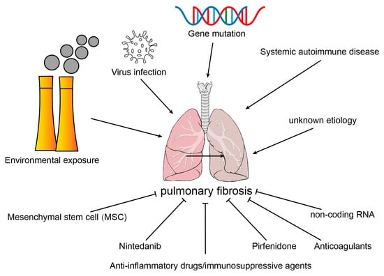
2.1. Environmental Exposure

PM2.5 (particulate matter less than 2.5 μm in aerodynamic diameter), asbestos, silica, and fumes could induce the accumulation of reactive oxygen stress (ROS) in type II alveolar epithelial cells (AECII) of lung tissue, which leads to oxidative stress and inflammatory damage in lung tissue [9][5]. A study reported that PM2.5 instillation-induced lung inflammation and fibrosis was associated with aberrant epithelial–mesenchymal transition (EMT), ROS, mitochondrial injury, and mitophagy [10][6]. Compared with WT, AECII from Ogg1−/− mice presented increased mtDNA damage and reduced mitochondrial aconitase expression, and these changes were intensified at 3 weeks after crocidolite exposure [11][7]. Environmental exposure-induced PF may be closely related to oxidative stress, mitochondrial damage, and mitophagy in epithelial cells.

2.2. Virus Infection

Human immunodeficiency virus (HIV), cytomegalovirus (CMV), murine gamma-herpes virus 68 (MHV-68), avian influenza virus, and other viruses may result in PF [12][8]. Viruses may induce pulmonary fibrosis through two pathways. First, the virus induces persistent damage and abnormal damage repair of alveolar epithelial cells, leading to the development of PF. Second, lung injury induced inflammatory infiltration and the activation of the immune system. Immune cells aggregate in the injured lung tissue and release many proinflammatory and profibrotic factors, including TGF-β, TNF-α, MMPs, and interleukin. This induces sustained lung damage and PF [13][9].

2.3. Systemic Autoimmune Disease

Systemic sclerosis (SSc), rheumatoid arthritis (RA), polymyositis/dermatomyositis (PM/DM), and Sjögren’s syndrome (SS) may promote inflammatory responses in lung inflammation through TGF-β, TGF-β-mediated pulmonary fibrosis, by mediating myofibroblast activation, proliferation, and ECM protein deposition [14,15,16,17][10][11][12][13].

2.4. Gene Mutation

Cystic fibrosis (CF) is a monogenetic disease induced by genovariation in the cystic fibrosis transmembrane conductance regulator (CFTR). Mutation in the CFTR gene affects the rheology of CF mucus, which impairs the mucociliary clearance of the respiratory tract. CF is related to the interaction of multiple intrinsic and extrinsic factors, such as genotype, mucus composition and motility abnormalities, chronic inflammation, and chronic airway infections [18][14].

2.5. Idiopathic Pulmonary Fibrosis

IPF is the consequence of reiterative epithelial cell injury and severe abnormal wound healing, and it is characterized by AEC injury, activation and proliferation of fibroblasts, and accumulation of ECM. The progression of IPF is influenced by many factors, including genetic predisposition, aging, and environmental factors. However, the magnitude of the contribution and the sequence of the pathogenic causes are indeterminate [19][15].

3. Source of Myofibroblasts in Fibrotic Lung Tissues

Studies have shown that myofibroblasts in fibrotic lung tissue exhibit a high heterogeneity. Early studies have found that myofibroblasts were mainly derived from interstitial fibroblasts, epithelial cells, and fibroblasts in peripheral blood circulation. Recent studies indicated that endothelial cells, microvascular pericytes, and macrophages could also transform into myofibroblasts in fibrotic lung tissue (Figure 2).
Figure 2.
Source of myofibroblasts in fibrotic lung tissues.

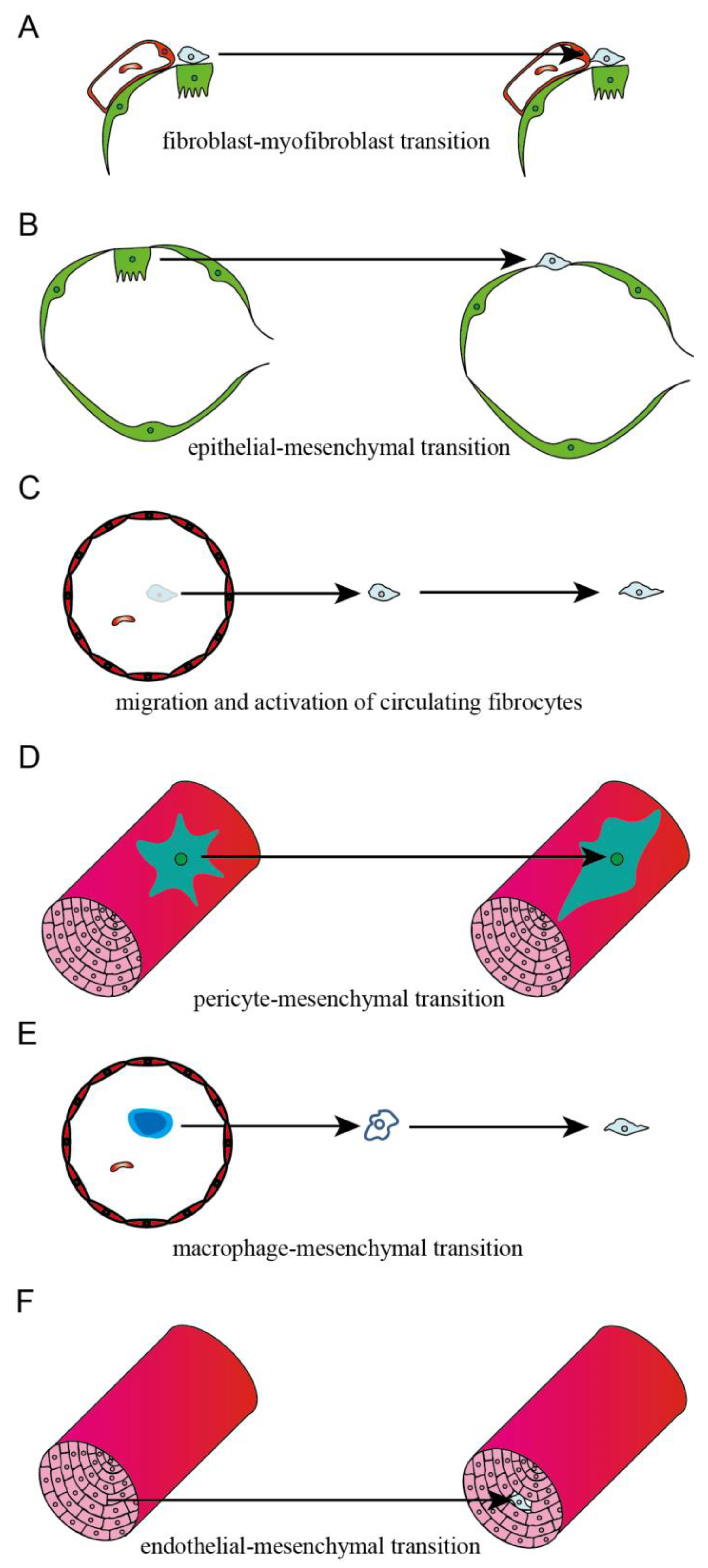
3.1. Interstitial Fibroblast

The damage of lung tissue in the process of pulmonary fibrosis induces the infiltration of immune cells, and then TGF-β derives from immune cells leads to the activation and differentiation of lung fibroblasts into myofibroblasts. In addition, TGF-β activates the cytoplasmic SMAD-2/3 complexes and stimulates the nuclear transportation of SMAD-2/3/4 complexes, which binds to the binding elements in the promoters of α-SMA, collagen, and other related genes, and leads to the synthesis and remodeling of ECM [20][16] (Figure 2A).

3.2. Epithelial Cell

Epithelial and mesenchymal cell markers were co-located in IPF lung tissue; studies have indicated that TGF-β was a primary contributor of EMT. TGF-β decreased the expression of epithelial cell marker gene E-cadherin through transcription factors, including Snail, Slug, Twist, Zeb1, SIP1, and E12, while it induced the expression of mesenchymal cell marker gene N-cadherin, vimentin, and α-SMA [21][17]. Rock JR et al. used the SFTPC-CreERT2 knock-in allele to track the fate of AECII in vivo and found that mesenchymal cell marker genes in epithelial cells do not upregulate after the BLM challenge, and labeled epithelial cells have not been found to transdifferentiate into a myofibroblast [22][18]. It was suggested that there were less epithelial-derived fibroblasts in animal models of PF or that AECII cells are not major contributors to myofibroblasts in PF (Figure 2B).

3.3. Circulating Fibrocyte

Fibrocytes are bone marrow-derived leukocytes, which infiltrate into lung tissues with the induction of CXCL12 and differentiate into fibroblasts or myofibroblasts, leading to the excessive accumulation of ECM during PF [23][19]. A platelet-derived growth factor (PDGF), such as Cxcl12, is a powerful chemoattractant of fibroblasts and directly contributes to fibrocyte infiltration into damaged lungs. Imatinib (PDGFR-blocking antibody) significantly reduces fibrocyte migration in vitro and decreases the number of fibrocytes in the lungs after a BLM challenge [24][20]. These studies demonstrated that circulating fibrocytes presented a crucial role in the development of PF by transdifferentiating into a myofibroblast (Figure 2C).

3.4. Microvascular Pericyte

Pericytes are mesenchymal cells located at the abluminal surface of the endothelium of microvessels and help to keep the homeostasis of microvascular and participate in angiogenesis [25][21]. Sava et al. reported that the pericytes marker NG2 co-localized with α-SMA in IPF, and α-SMA+ pericytes accumulated with the treatment of IPF lung ECM [26][22]. Wang et al. reported that miR-107 was significantly decreased in ECs of fibrotic lung tissue, which induced the activation of the HIF-1α/Notch1/PDGFRβ/YAP1/Twist1 axis and promoted the expression of α-SMA and Coll1A, thus contributing to the fibrotic transdifferentiation of pericytes [27][23] (Figure 2D).

3.5. Macrophage

The macrophage–myofibroblast transition (MMT), which is found in kidney fibrosis, is a recent term. It indicates that macrophages, derived from circulating monocytes originating in the bone marrow, could transform into myofibroblasts and contribute to kidney fibrosis [28][24]. Yang et al. found that most of the myofibroblasts in PF were derived from macrophages; in addition, the majority of MMT cells in the fibrotic lungs presented the M2 phenotype [29][25] (Figure 2E).

3.6. Endothelial Cell

Hashimoto et al. established BLM-induced lung fibrosis in Tie2-Cre/CAG-CAT-LacZ mice, in which LacZ was stably expressed in pan-ECs, and they found that X-gal-positive cells were presented in a lung fibroblast from BLM-challenged mice. This indicated that lung capillary ECs could be active and transdifferentiate to fibroblasts via the E(nd)MT in BLM-challenged mice. Furthermore, 16% of fibroblasts were derived from the ECs in BLM-induced PF [30][26]. Arterial layers’ structural changes and the deposition of collagen and elastin were presented at the adventitia of IPF patients. Compared to NC, the expression of mesenchymal biomarkers N-cadherin, S100A4, and vimentin was significantly increased in the arterial layers of IPF patients, indicating that resident ECs could transdifferentiate to mesenchymal cells in PF [31][27] (Figure 2F).

References

  1. Cheng, L.; Wang, D.; Deng, B.; Li, J.; Zhang, J.; Guo, X.; Yan, T.; Yue, X.; An, Y.; Zhang, B.; et al. DR7dA, a Novel Antioxidant Peptide Analog, Demonstrates Antifibrotic Activity in Pulmonary Fibrosis In Vivo and In Vitro. J. Pharmacol. Exp. Ther. 2022, 382, 100–112.
  2. Marchioni, A.; Tonelli, R.; Cerri, S.; Castaniere, I.; Andrisani, D.; Gozzi, F.; Bruzzi, G.; Manicardi, L.; Moretti, A.; Demurtas, J.; et al. Pulmonary Stretch and Lung Mechanotransduction: Implications for Progression in the Fibrotic Lung. Int. J. Mol. Sci. 2021, 22, 6443.
  3. Fujimoto, H.; Kobayashi, T.; Azuma, A. Idiopathic Pulmonary Fibrosis: Treatment and Prognosis. Clin. Med. Insights Circ. Respir. Pulm. Med. 2015, 9, 179–185.
  4. Somogyi, V.; Chaudhuri, N.; Torrisi, S.E.; Kahn, N.; Muller, V.; Kreuter, M. The therapy of idiopathic pulmonary fibrosis: What is next? Eur. Respir. Rev. 2019, 28, 153.
  5. Gao, Y.; Sun, J.; Dong, C.; Zhao, M.; Hu, Y.; Jin, F. Extracellular Vesicles Derived from Adipose Mesenchymal Stem Cells Alleviate PM2.5-Induced Lung Injury and Pulmonary Fibrosis. Med. Sci. Monit. 2020, 26, e922782.
  6. Xu, M.; Wang, X.; Xu, L.; Zhang, H.; Li, C.; Liu, Q.; Chen, Y.; Chung, K.F.; Adcock, I.M.; Li, F. Chronic lung inflammation and pulmonary fibrosis after multiple intranasal instillation of PM2.5 in mice. Environ. Toxicol. 2021, 36, 1434–1446.
  7. Cheresh, P.; Morales-Nebreda, L.; Kim, S.J.; Yeldandi, A.; Williams, D.B.; Cheng, Y.; Mutlu, G.M.; Budinger, G.R.; Ridge, K.; Schumacker, P.T.; et al. Asbestos-induced pulmonary fibrosis is augmented in 8-oxoguanine DNA glycosylase knockout mice. Am. J. Respir. Cell Mol. Biol. 2015, 52, 25–36.
  8. Sheng, G.; Chen, P.; Wei, Y.; Yue, H.; Chu, J.; Zhao, J.; Wang, Y.; Zhang, W.; Zhang, H.L. Viral Infection Increases the Risk of Idiopathic Pulmonary Fibrosis: A Meta-Analysis. Chest 2020, 157, 1175–1187.
  9. Huang, W.J.; Tang, X.X. Virus infection induced pulmonary fibrosis. J. Transl. Med. 2021, 19, 496.
  10. Fukui, Y.; Nakamura, K.; Hirabayashi, M.; Miyagawa, T.; Toyama, S.; Omatsu, J.; Awaji, K.; Ikawa, T.; Norimatsu, Y.; Yoshizaki, A.; et al. Serum vasohibin-1 levels: A potential marker of dermal and pulmonary fibrosis in systemic sclerosis. Exp. Dermatol. 2021, 30, 951–958.
  11. Wang, S.; Liu, M.; Li, X.; Zhang, J.; Wang, F.; Zhang, C.; Roden, A.; Ryu, J.H.; Warrington, K.J.; Sun, J.; et al. Canonical and noncanonical regulatory roles for JAK2 in the pathogenesis of rheumatoid arthritis-associated interstitial lung disease and idiopathic pulmonary fibrosis. FASEB J. 2022, 36, e22336.
  12. Shen, N.; Zhou, X.; Jin, X.; Lu, C.; Hu, X.; Zhang, Y.; Jiang, Y.; Xu, Q.; Xu, X.; Liu, M.; et al. MDA5 expression is associated with TGF-beta-induced fibrosis: Potential mechanism of interstitial lung disease in anti-MDA5 dermatomyositis. Rheumatology 2022, 62, 373–383.
  13. Shi, L.; Fu, Q.; Chen, N.; Liu, R.; Zheng, Y. Angiopoietin-like protein 2 as a novel marker for patients with primary Sjogren’s syndrome-related interstitial lung disease. Clin. Exp. Med. 2020, 20, 393–399.
  14. Caballero, I.; Ringot-Destrez, B.; Si-Tahar, M.; Barbry, P.; Guillon, A.; Lantier, I.; Berri, M.; Chevaleyre, C.; Fleurot, I.; Barc, C.; et al. Evidence of early increased sialylation of airway mucins and defective mucociliary clearance in CFTR-deficient piglets. J. Cyst. Fibros. 2021, 20, 173–182.
  15. Pardo, A.; Selman, M. The Interplay of the Genetic Architecture, Aging, and Environmental Factors in the Pathogenesis of Idiopathic Pulmonary Fibrosis. Am. J. Respir. Cell Mol. Biol. 2021, 64, 163–172.
  16. Lim, M.J.; Ahn, J.; Yi, J.Y.; Kim, M.H.; Son, A.R.; Lee, S.L.; Lim, D.S.; Kim, S.S.; Kang, M.A.; Han, Y.; et al. Induction of galectin-1 by TGF-beta1 accelerates fibrosis through enhancing nuclear retention of Smad2. Exp. Cell Res. 2014, 326, 125–135.
  17. Yao, L.; Conforti, F.; Hill, C.; Bell, J.; Drawater, L.; Li, J.; Liu, D.; Xiong, H.; Alzetani, A.; Chee, S.J.; et al. Paracrine signalling during ZEB1-mediated epithelial-mesenchymal transition augments local myofibroblast differentiation in lung fibrosis. Cell Death Differ. 2019, 26, 943–957.
  18. Rock, J.R.; Barkauskas, C.E.; Cronce, M.J.; Xue, Y.; Harris, J.R.; Liang, J.; Noble, P.W.; Hogan, B.L. Multiple stromal populations contribute to pulmonary fibrosis without evidence for epithelial to mesenchymal transition. Proc. Natl. Acad. Sci. USA 2011, 108, E1475–E1483.
  19. Chiang, H.Y.; Chu, P.H.; Lee, T.H. R1R2 peptide ameliorates pulmonary fibrosis in mice through fibrocyte migration and differentiation. PLoS ONE 2017, 12, e0185811.
  20. Aono, Y.; Kishi, M.; Yokota, Y.; Azuma, M.; Kinoshita, K.; Takezaki, A.; Sato, S.; Kawano, H.; Kishi, J.; Goto, H.; et al. Role of platelet-derived growth factor/platelet-derived growth factor receptor axis in the trafficking of circulating fibrocytes in pulmonary fibrosis. Am. J. Respir. Cell Mol. Biol. 2014, 51, 793–801.
  21. Rowley, J.E.; Johnson, J.R. Pericytes in chronic lung disease. Int. Arch. Allergy Immunol. 2014, 164, 178–188.
  22. Sava, P.; Ramanathan, A.; Dobronyi, A.; Peng, X.; Sun, H.; Ledesma-Mendoza, A.; Herzog, E.L.; Gonzalez, A.L. Human pericytes adopt myofibroblast properties in the microenvironment of the IPF lung. JCI Insight 2017, 2, 24.
  23. Wang, Y.C.; Xie, H.; Zhang, Y.C.; Meng, Q.H.; Xiong, M.M.; Jia, M.W.; Peng, F.; Tang, D.L. Exosomal miR-107 antagonizes profibrotic phenotypes of pericytes by targeting a pathway involving HIF-1alpha/Notch1/PDGFRbeta/YAP1/Twist1 axis in vitro. Am. J. Physiol. Heart Circ. Physiol. 2021, 320, H520–H534.
  24. Nikolic-Paterson, D.J.; Wang, S.; Lan, H.Y. Macrophages promote renal fibrosis through direct and indirect mechanisms. Kidney Int. Suppl. 2014, 4, 34–38.
  25. Yang, F.; Chang, Y.; Zhang, C.; Xiong, Y.; Wang, X.; Ma, X.; Wang, Z.; Li, H.; Shimosawa, T.; Pei, L.; et al. UUO induces lung fibrosis with macrophage-myofibroblast transition in rats. Int. Immunopharmacol. 2021, 93, 107396.
  26. Hashimoto, N.; Phan, S.H.; Imaizumi, K.; Matsuo, M.; Nakashima, H.; Kawabe, T.; Shimokata, K.; Hasegawa, Y. Endothelial-mesenchymal transition in bleomycin-induced pulmonary fibrosis. Am. J. Respir. Cell Mol. Biol. 2010, 43, 161–172.
  27. Gaikwad, A.V.; Lu, W.; Dey, S.; Bhattarai, P.; Chia, C.; Larby, J.; Haug, G.; Myers, S.; Jaffar, J.; Westall, G.; et al. Vascular remodelling in idiopathic pulmonary fibrosis patients and its detrimental effect on lung physiology: Potential role of endothelial-to-mesenchymal transition. ERJ Open Res. 2022, 8, 1.
More
Video Production Service