Meta-Inflammation, Diabetes and Cardiovascular Disease: Comparison
Please note this is a comparison between Version 2 by Catherine Yang and Version 1 by Alessia d'Aiello.

Type 2 diabetes mellitus (DM) represents, with its macro and microvascular complications, one of the most critical healthcare issues. The pathogenesis of DM shares with cardiovascular diseases (CAD) a complex landscape of risk factors, including genetic predisposition and various environmental factors like a high-fat diet, sedentary lifestyle, and chronic stress. In particular, CAD is the leading cause of morbidity and mortality in diabetic patients, determining a significant impact on life expectancy. Notably, DM is equivalent to established ischemic CAD risk, and patients with diabetes have a two- to four-fold greater risk of developing CAD than non-diabetic patients. Myocardial infarction, ischemic ictus, and peripheral arterial disease are the main expression of DM progression and, often, the first event in diabetic patients. CAD and DM strictly depend on various inflammatory pathways that are able to promote the onset and development of insulin resistance, atherosclerotic plaque, and heart failure (HF).

  • type 2 diabetes mellitus
  • meta-inflammation
  • cardiovascular diseases

1. Meta-Inflammation and Metabolic Endotoxemia

Dietary fats can boost levels of circulating bacterial endotoxins, contributing to metabolic endotoxemia, a chronic low-grade elevation of the bacterial component lipopolysaccharide (LPS) with content 10–50-times lower than in septic conditions, which can lead to leukocyte activation, local and systemic inflammation [16][1]. Recent evidence has proved that metabolic endotoxemia plays a critical role in the inflammatory setting milieu, thereby predisposing to metabolic diseases via pattern recognition receptor engagement, primarily through the toll-like receptor/nod-like receptor (TLR4/NLR) signaling pathway [17,18][2][3]. Following a similar pathophysiological framework, both DM and CAD are associated with significant alterations of the gut microbiota, responsible for the increased intestinal permeability and the consequent efflux of LPS into the bloodstream, thus altering the systemic metabolic response [19][4]. Indeed, already in 1990, it has been demonstrated that patients with end-stage HF show elevated levels of circulating tumor necrosis factor (TNF), which could explain the cachectic condition typical of this syndrome as a consequence of innate immune dysregulation operated by endotoxin-activated monocytes [20,21][5][6].

2. Meta-Inflammation and the “Unfolded Protein Response” (UPR)

The liver and pancreas are the classical target organs in the physiopathology of DM, but adipose tissue is probably at the core of meta-inflammation. Nutrient excess increases adipocyte size up to critical conditions, resulting in reduced vascularization and a hypoxic environment [22][7], finally precipitating the inflammatory cascade.
In obese mice, adipocytes participate in endoplasmic reticulum stress, a condition accelerated by the accumulation of unfolded/misfolded proteins and able to trigger the protective unfolded protein response (UPR). This process promotes both the synthesis of endoplasmic reticulum-resident chaperone proteins, which encourages protein folding and the protein-degradation mechanism components to achieve new endoplasmic reticulum homeostasis [23][8]. In addition to these defensive responses, the UPR may also encourage important inflammatory signals, triggering apoptotic and cell death pathways [24][9]. UPR is significantly enhanced by exposure to nutrient excess, as revealed by experiments in obese and diabetic mice, and its activation promotes inflammation through several mechanisms, i.e., inducing the expression of pro-inflammatory genes by directly acting on the transcription factor activator protein 1 [25][10]; abnormal nutrient intake pledges the NF-κB signaling pathway [26][11], promoting the downstream cleavage and activation of the transcription factor cyclic-AMP responsive- element-binding protein H (CREBH), which in turn induces the production of two acute phase proteins, C-reactive protein (CRP) and serum amyloid P-component (SAP) [27][12]. A similar process has been recorded in HF patients and represents the ground of cardiac hypertrophy, further exacerbated by the acquired immune response.

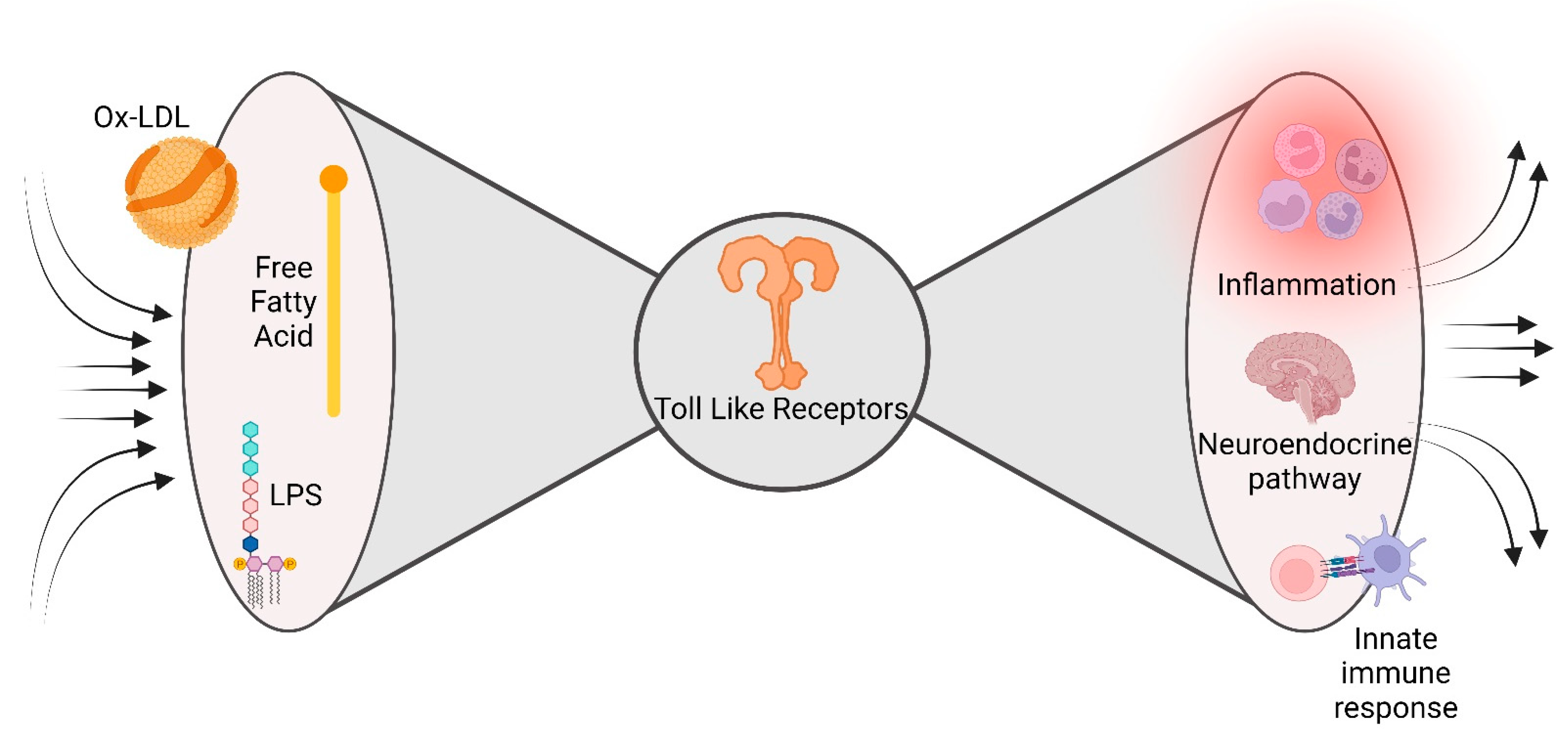
3. The Innate Immune Response

An innate immune response is an essential mechanism in triggering meta-inflammation associated with DM and CAD. Pattern recognition receptors can boost meta-inflammation following recognition of both pathogens-associated molecular patterns (PAMPs) and damage-associated molecular patterns (DAMPs). Viral nucleic acids, endotoxins, and peptidoglycans are some of the PAMPs circulating in the bloodstream because of metabolic endotoxemia, while FFAs and self-nucleoproteins are some of the endogenous ligands starring as DAMPs [28][13]. Pattern recognition receptor activation represents the main sensor to start the inflammation cascade via NF-𝜅B and some interferon regulatory factors (IRFs), which in return trigger pro-inflammatory cytokines and type-1 interferons signaling cascade [29][14]. These mechanisms could lead to insulin resistance and precipitate in DM via the mediation of macrophage polarization towards the M1 phenotype [30][15].
In HF, adverse left ventricular (LV) remodeling and LV dysfunction are structural progressions that parallel the activity of innate immunity actors and mediators such as monocyte chemoattractant protein 1 (MCP-1) and progressive monocyte-derived macrophage generation [31][16].
The innate immune response could be dysregulated at multiple levels, becoming the basement of a meta-inflammatory environment that, in turn, involves multiple cellular compartments.

4. The Adaptive Immune Response

As far as adaptive immunity is concerned, most evidence proves that CD4+T-helper (Th) cells are crucial elements in propagating meta-inflammation [32,33][17][18]. The response started by pattern recognition receptors is transferred to T cells by the antigen-presenting cell (APC) and T cell cooperation, which determines the enrolment of these activated cells into the pancreas, adipose tissue, and other target organs, consolidating the meta-inflammation mounted by the native response. The well-known Th polarization, which shapes the adaptive arm of the immune response, is also maintained in DM patients. In particular, in the murine diet-induced obesity model, Kitschier et al. demonstrated early recruitment of Th1 cells into adipose tissue, paving the way for macrophage infiltration and insulin resistance [34][19]. Notably, several studies on serum cytokine profiling in DM patients reported robust Th1 polarization during the transition from DM to macrovascular complications, especially atherosclerotic coronaropathy, highlighting the role of Th1 polarization in the physiopathology of the disease [35][20]. Furthermore, a significant action in interfering with insulin signaling and insulin-stimulated glucose uptake is pursued by interferon-gamma (IFN-𝛾), eventually leading to insulin resistance and DM [36,37,38][21][22][23]. Polarized Th1 and Th17 T-cells also play a key role in cardiac fibrosis and adverse cardiac remodeling, sustaining and amplifying the local chronic inflammation leading to HF. In vivo, experimental models demonstrated that CD4+ T-cells were expanded in the failing heart, with a Th1/Th2 ratio significantly decreased, whereas the Th17/Treg ratio was increased, underling the loss of anti-inflammatory properties and the gain of the pro-inflammatory counterpart [39][24].

5. The Bow Tie Model

Metabolic and inflammatory pathways can converge at many levels, including cell-surface receptors, intracellular chaperones, or nuclear receptors. This molecular army allows robust cooperation between the nutrient-sensing pathways and the immune response aimed at maintaining homeostasis in opposite metabolic and immune circumstances. Unfortunately, this molecular rendezvous may be one of the crucial moments in DM and CAD pathophysiology. Meta-inflammation may be considered the unpredicted consequence of the evolution-driven degeneracy of damage sensors, recently described as a ‘bow tie’ architecture [40][25]. The main feature of bow tie architecture is the possibility to converge a vast range of inputs (fan in) on an evolutionarily reduced core of components (core), able to translate the inputs into a broad spectrum of outputs (fan out) (Figure 21). Interestingly, the inputs are represented by an extensive range of self and non-self-stimuli, i.e., free fatty acid, LPS, able to bind a restricted number of evolutionarily conserved innate immunity sensors (the bow tie core), whose activation triggers a large number of inflammatory elements [41][26]. A fascinating example of this promiscuity in immune response receptors is the capacity of saturated fatty acids to turn on both TLR2 and TLR4, important innate immune response receptors involved in pathogen recognition, and trigger the release of pro-inflammatory mediators [42][27].
Figure 21. Bow tie model. The characteristic of bow tie architecture is the ability to draw in a wide variety of inputs, such as self- and non-self-stimuli (free fatty acids, LPS, and oxidized-LDL) into the core of components (toll-like receptors), which can convert the inputs into a wide range of outputs, such as a variety of inflammatory components.
This bidirectional cross-talk between metabolic alterations and immune dysregulation is also emerging as a critical component of the pathogenesis of HF, especially for the one with preserved ejection fraction (HfpEF). Indeed, all the alterations mentioned above and driven by metabolic alterations, such as macrophage polarization, accumulation of misfolded proteins, and metabolic reprogramming, contribute to structural and functional remodeling determining HF [5][28].
The exploration of the meta-inflammation route as a common denominator for DM and CAD might expand the knowledge of molecular pathways underpinning its pathophysiology, thus clearing the unpredicted effects of new anti-diabetic drugs on cardiovascular outcomes.

References

  1. Erridge, C.; Attina, T.; Spickett, C.M.; Webb, D.J. A high-fat meal induces low-grade endotoxemia: Evidence of a novel mechanism of postprandial inflammation. Am. J. Clin. Nutr. 2007, 86, 1286–1292.
  2. Pahwa, R.; Devaraj, S.; Jialal, I. The effect of the accessory proteins, soluble CD14 and lipopolysaccharide-binding protein on Toll-like receptor 4 activity in human monocytes and adipocytes. Int. J. Obes. 2016, 40, 907–911.
  3. Vandanmagsar, B.; Youm, Y.-H.; Ravussin, A.; Galgani, J.E.; Stadler, K.; Mynatt, R.L.; Ravussin, E.; Stephens, J.M.; Dixit, V.D. The NLRP3 inflammasome instigates obesity-induced inflammation and insulin resistance. Nat. Med. 2011, 17, 179–188.
  4. Biasucci, L.M.; La Rosa, G.; Pedicino, D.; D’aiello, A.; Galli, M.; Liuzzo, G. Where Does Inflammation Fit? Curr. Cardiol. Rep. 2017, 19, 84.
  5. Levine, B.; Kalman, J.; Mayer, L.; Fillit, H.M.; Packer, M. Elevated Circulating Levels of Tumor Necrosis Factor in Severe Chronic Heart Failure. N. Engl. J. Med. 1990, 323, 236–241.
  6. Kunkel, S.L.; Spengler, M.; May, M.A.; Spengler, R.; Larrick, J.; Remick, D. Prostaglandin E2 regulates macrophage-derived tumor necrosis factor gene expression. J. Biol. Chem. 1988, 263, 5380–5384.
  7. Giordano, A.; Murano, I.; Mondini, E.; Perugini, J.; Smorlesi, A.; Severi, I.; Barazzoni, R.; Scherer, P.E.; Cinti, S. Obese adipocytes show ultrastructural features of stressed cells and die of pyroptosis. J. Lipid Res. 2013, 54, 2423–2436.
  8. Gregor, M.F.; Hotamisligil, G.S. Thematic review series: Adipocyte Biology. Adipocyte stress: The endoplasmic reticulum and metabolic disease. J. Lipid Res. 2007, 48, 1905–1914.
  9. Ron, D.; Walter, P. Signal integration in the endoplasmic reticulum unfolded protein response. Nat. Rev. Mol. Cell Biol. 2007, 8, 519–529.
  10. Todd, D.J.; Lee, A.-H.; Glimcher, L.H. The endoplasmic reticulum stress response in immunity and autoimmunity. Nat. Rev. Immunol. 2008, 8, 663–674.
  11. Hu, P.; Han, Z.; Couvillon, A.D.; Kaufman, R.J.; Exton, J.H. Autocrine Tumor Necrosis Factor Alpha Links Endoplasmic Reticulum Stress to the Membrane Death Receptor Pathway through IRE1α-Mediated NF-κB Activation and Down-Regulation of TRAF2 Expression. Mol. Cell. Biol. 2006, 26, 3071–3084.
  12. Zhang, K.; Shen, X.; Wu, J.; Sakaki, K.; Saunders, T.; Rutkowski, D.T.; Back, S.H.; Kaufman, R.J. Endoplasmic Reticulum Stress Activates Cleavage of CREBH to Induce a Systemic Inflammatory Response. Cell 2006, 124, 587–599.
  13. Kawai, T.; Akira, S. The roles of TLRs, RLRs and NLRs in pathogen recognition. Int. Immunol. 2009, 21, 317–337.
  14. Pedicino, D.; Giglio, A.F.; Galiffa, V.A.; Cialdella, P.; Trotta, F.; Graziani, F.; Liuzzo, G. Infections, immunity and atherosclerosis: Pathogenic mechanisms and unsolved questions. Int. J. Cardiol. 2013, 166, 572–583.
  15. Eguchi, K.; Nagai, R. Islet inflammation in type 2 diabetes and physiology. J. Clin. Investig. 2017, 127, 14–23.
  16. Shioi, T.; Matsumori, A.; Kihara, Y.; Inoko, M.; Ono, K.; Iwanaga, Y.; Yamada, T.; Iwasaki, A.; Matsushima, K.; Sasayama, S. Increased Expression of Interleukin-1β and Monocyte Chemotactic and Activating Factor/Monocyte Chemoattractant Protein-1 in the Hypertrophied and Failing Heart With Pressure Overload. Circ. Res. 1997, 81, 664–671.
  17. Pedicino, D.; Severino, A.; Ucci, S.; Bugli, F.; Flego, D.; Giglio, A.F.; Trotta, F.; Ruggio, A.; Lucci, C.; Iaconelli, A.; et al. Epicardial adipose tissue microbial colonization and inflammasome activation in acute coronary syndrome. Int. J. Cardiol. 2017, 236, 95–99.
  18. Liuzzo, G.; Montone, R.A.; Gabriele, M.; Pedicino, D.; Giglio, A.F.; Trotta, F.; Galiffa, V.A.; Previtero, M.; Severino, A.; Biasucci, L.M.; et al. Identification of unique adaptive immune system signature in acute coronary syndromes. Int. J. Cardiol. 2013, 168, 564–567.
  19. Kintscher, U.; Hartge, M.; Hess, K.; Foryst-Ludwig, A.; Clemenz, M.; Wabitsch, M.; Fischer-Posovszky, P.; Barth, T.F.; Dragun, D.; Skurk, T.; et al. T-lymphocyte infiltration in visceral adipose tissue: A primary event in adipose tissue inflammation and the development of obesity-mediated insulin resistance. Arter. Thromb. Vasc. Biol. 2008, 28, 1304–1310.
  20. Madhumitha, H.; Mohan, V.; Deepa, M.; Babu, S.; Aravindhan, V. Increased Th1 and suppressed Th2 serum cytokine levels in subjects with diabetic coronary artery disease. Cardiovasc. Diabetol. 2014, 13, 1.
  21. McGillicuddy, F.C.; Chiquoine, E.H.; Hinkle, C.C.; Kim, R.J.; Shah, R.; Roche, H.M.; Smyth, E.M.; Reilly, M.P. Interferon γ Attenuates Insulin Signaling, Lipid Storage, and Differentiation in Human Adipocytes via Activation of the JAK/STAT Pathway. J. Biol. Chem. 2009, 284, 31936–31944.
  22. Ruggio, A.; Pedicino, D.; Flego, D.; Vergallo, R.; Severino, A.; Lucci, C.; Niccoli, G.; Trani, C.; Burzotta, F.; Aurigemma, C.; et al. Correlation between CD4+CD28null T lymphocytes, regulatory T cells and plaque rupture: An Optical Coherence Tomography study in Acute Coronary Syndromes. Int. J. Cardiol. 2019, 276, 289–292.
  23. Canonico, F.; Pedicino, D.; Severino, A.; Vinci, R.; Flego, D.; Pisano, E.; D’aiello, A.; Ciampi, P.; Ponzo, M.; Bonanni, A.; et al. GLUT-1/PKM2 loop dysregulation in patients with non-ST-segment elevation myocardial infarction promotes metainflammation. Cardiovasc. Res. 2022; Online ahead of print.
  24. Bansal, S.S.; Ismahil, M.A.; Goel, M.; Patel, B.; Hamid, T.; Rokosh, G.; Prabhu, S.D. Activated T Lymphocytes are Essential Drivers of Pathological Remodeling in Ischemic Heart Failure. Circ. Heart Fail. 2017, 10, e003688.
  25. Tieri, P.; Grignolio, A.; Zaikin, A.; Mishto, M.; Remondini, D.; Castellani, G.C.; Franceschi, C. Network, degeneracy and bow tie. Integrating paradigms and architectures to grasp the complexity of the immune system. Theor. Biol. Med. Model. 2010, 7, 32.
  26. Franceschi, C.; Garagnani, P.; Parini, P.; Giuliani, C.; Santoro, A. Inflammaging: A new immune–metabolic viewpoint for age-related diseases. Nat. Rev. Endocrinol. 2018, 14, 576–590.
  27. Lee, J.Y.; Zhao, L.; Youn, H.S.; Weatherill, A.R.; Tapping, R.; Feng, L.; Lee, W.H.; Fitzgerald, K.A.; Hwang, D.H. Saturated Fatty Acid Activates but Polyunsaturated Fatty Acid Inhibits Toll-like Receptor 2 Dimerized with Toll-like Receptor 6 or 1. J. Biol. Chem. 2004, 279, 16971–16979.
  28. Schiattarella, G.G.; Rodolico, D.; A Hill, J. Metabolic inflammation in heart failure with preserved ejection fraction. Cardiovasc. Res. 2021, 117, 423–434.
More
Video Production Service