Understanding Antimicrobial Resistance Using Genome-Scale Metabolic Modeling: Comparison
Please note this is a comparison between Version 2 by Dean Liu and Version 1 by Elena Perrin.

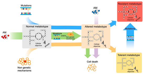
The urgent necessity to fight antimicrobial resistance is universally recognized. In the search of new targets and strategies to face this global challenge, a promising approach resides in the study of the cellular response to antimicrobial exposure and on the impact of global cellular reprogramming on antimicrobial drugs’ efficacy. The metabolic state of microbial cells has been shown to undergo several antimicrobial-induced modifications and, at the same time, to be a good predictor of the outcome of an antimicrobial treatment. Metabolism is a promising reservoir of potential drug targets/adjuvants that has not been fully exploited to date. One of the main problems in unraveling the metabolic response of cells to the environment resides in the complexity of such metabolic networks. To solve this problem, modeling approaches have been developed, and they are progressively gaining in popularity due to the huge availability of genomic information and the ease at which a genome sequence can be converted into models to run basic phenotype predictions.

  • metabolic modeling
  • antimicrobial resistance
  • bacterial metabolism

1. Introduction

Antimicrobial resistance (AMR), the ability of a microorganism to resist the action of one or more antimicrobial agents, is one of the major public health problems of this century [1]. Antimicrobial-resistant microbes are currently estimated to claim ~700,000 deaths per year, and this mortality rate is predicted to increase to 10 million per year by 2050 [1]. Consequently, antimicrobial resistance has been identified as one of the most important challenges to human health by several national and international bodies.
For bacteria, the current antimicrobials inhibit a narrow spectrum of cellular processes (e.g., DNA replication, transcription, protein synthesis, and cell wall biosynthesis) [2]. Their action is traditionally seen as a linear process in which the antimicrobial enters the cell, reaches and interacts with its target, and stops the growth or kills the bacteria [3]. Accordingly, it is generally assumed that antimicrobial resistance relies on a few specific microbial genes [4]. However, this description represents only the immediate effects of the antimicrobial. In many cases, what is not known and is still the subject of debate, is how antimicrobials actually kill bacterial cells [5]. Microbial metabolism is impacted by antimicrobials [4], and in recent years, the close link between bacterial metabolism, antimicrobial action, and antimicrobial resistance has increasingly emerged. Indeed, on the one hand, antimicrobials alter the metabolic phenotype (metabotype [6]) of bacterial cells, giving rise to an altered metabotype. On the other hand, the metabotype of bacteria (normal or altered) influences their susceptibility to antimicrobials (Figure 1) [2]. Consequently, the antimicrobial efficacy can be enhanced by altering the metabolic state of bacteria. Approaches that target the bacterial central metabolism for drug development or that use the control of nutrients’ availability as a mechanism for the selection of antimicrobial-tolerant strains (for a definition of the differences between tolerance and resistance, see below) are gaining more and more attention [2].
Figure 1. Possible paths to resistant and tolerant metabotypes. Here, researchers show the hypothetical effects of mutations, antimicrobials’ exposure, and non-genetic mechanisms on microbial metabotypes, including the effects on the single reactions (thicker links among the nodes) and the most likely objective function (“growth” or “?”).
Overall, antimicrobials can affect bacterial metabolism in three ways. First, they have a direct effect on metabolism that affects their efficacy, which seems to be different depending on whether the antimicrobial is bactericidal or bacteriostatic (even though it appears that all antimicrobials are ultimately bactericidal, and the difference between the two types of antimicrobials is only the rates at which they kill bacteria) [2][5]. The action of bactericidal antimicrobials on their primary targets causes damage to other essential macromolecules (nucleic acids, proteins, and membrane lipids) within the cell, resulting in the induction of stress response pathways. Stress responses increase metabolic activity to meet the corresponding energy demands. This results in the production of toxic metabolic byproducts such as reactive species, which damage macromolecules, and leads to the induction of additional stress response pathways. Once again, the overall cellular metabolic load results increased. The alteration of the metabotype, therefore, creates a cyclical process that ends with cell death [7][8][9][10][11][12][13][14][15]. Wong et al. [16] recently suggested that the interactions of the toxic metabolic bioproducts with the membrane induce a loss of membrane integrity, which results in cytoplasmic condensation through the leakage of cytoplasmic contents, and then in cell death. Alternative cell death pathways may involve cellular damage to nucleic acids and proteins resulting from both the primary drug–target interaction and from the subsequent generation of reactive metabolic byproducts [16]. A similar process, involving reactive metabolic byproducts, seems to contribute to antimicrobial lethality, also under anaerobic conditions [17]. Bacteriostatic antimicrobials, instead, inhibit protein biosynthesis or transcription in certain contexts, leading to a decrease in metabolic activity and subsequent cell stasis, thus, again resulting in an altered metabotype [14][18]. As mentioned before, however, bacteriostatic antimicrobials can also kill bacteria, probably depending, among other things, on the number of ribosomes. Indeed, when the number of ribosomes is reduced, ribosome-targeting antimicrobials seem to become increasingly bactericidal, suggesting a kind of lethal protein synthesis threshold. However, this could not be the ultimate mechanism by which these ribosome-targeting ‘bacteriostatic’ antimicrobials kill bacteria [5]. Therefore, the altered metabotypes resulting from antimicrobial treatment contribute to its final outcomes. The second and the third ways in which antimicrobials can influence bacterial metabolism are indirect. First, antimicrobial treatments generally involve the acquisition of resistance, through mutations or horizontal gene transfer, this often come with a fitness cost for the bacterial cell (Figure 1) [19][20]. Consequently, the acquisition of compensatory mutations that counterbalance the decreased fitness is a crucial step for the success of resistant strains [21][22]. These mutations generally restore normal growth, preserving resistance, and their number and type varies with the organism and the particular environmental conditions under which compensation occurs, indicating that fitness costs are dependent on the habitat and on the metabolic adaptation required for colonizing such a habitat [23]. Regarding the third mode, it has recently been demonstrated that, in addition to the acquisition of the classical mechanisms of resistance (target modification, drug inactivation, and drug transport), antimicrobials can also induce mutations in metabolic genes [24]. These metabolic mutations can confer resistance and are prevalent in clinical pathogens, suggesting that metabolic adaptation may represent a mechanism of resistance that confers tolerance, but may also mitigate the downstream toxic aspects of antimicrobials (Figure 1) [24]. On the other hand, the metabolic state of the bacterial cells can affect the antimicrobials’ efficacy in various ways. It must be stated that, in addition to the classical molecular mechanisms of antimicrobial resistance, bacterial cells can counteract antimicrobials in several ways, each of which relies on a general metabolic downregulation. Indeed, the term resistance is usually used to “describe the inherited ability of microorganisms to grow at high concentrations of an antimicrobial, irrespective of the duration of the treatment, and is quantified by the minimum inhibitory concentration (MIC) of the particular antimicrobial” [25]. If this resistant phenotype is observed only in a subpopulation of cells, it is known as hetero-resistance [26]. The term tolerance, instead, is used to describe “the ability, whether inherited or not, of microorganisms to survive transient exposure to high concentrations of an antimicrobial without a change in the MIC, which is often achieved by slowing down an essential bacterial process” [25]. It has been demonstrated that tolerance often evolves during frequent and intermittent antimicrobial treatments, and that its emergence often promotes the development of resistance [27]. Finally, the term persistence is used when only a subpopulation of a clonal bacterial population is able to survive exposure to high concentrations of an antimicrobial, without any genetic mutations [25]. Indeed, if persisters are isolated and regrown in the presence of antimicrobials, they display the same pattern of susceptibility as the original population [28]. The metabolic state of bacterial cells (normal or altered metabotypes, Figure 1) is mainly involved in mechanisms of tolerance to the antimicrobials’ action. Indeed, most of the so-called “phenotypic resistance”, in which metabolism has a fundamental role, is mechanisms of tolerance rather than resistance. Indeed, this term refers to all the transient situations in which a bacterial population, susceptible to an antimicrobial, becomes resistant without any genetic change taking place; therefore, resulting as not inheritable [29]. For example, the growth rate is an important parameter that affects the susceptibility to antimicrobials of bacterial populations. Resting cells are less susceptible to antimicrobials than metabolically active cells, especially to the action of bactericidal antimicrobials [4]. However, a recent study showed that the metabotype of the cell, instead of growth, better correlates with antimicrobial lethality, suggesting that antimicrobials should also be able to kill non-growing bacteria if metabolism is active, and that the metabolic response, following the initial interaction of an antimicrobial with its target, influences the bacterial response to the antimicrobial action [30]. Consequently, in all the conditions of an altered metabotype, in which the metabolism is poor or not active, bacterial cells are less susceptible to antimicrobials’ action (Figure 1). For example, during a stringent response, the accumulation of (p)ppGpp determines a global switch on bacterial metabolism that also modulates their response to antimicrobials [31][32][33][34], while bacterial persisters (whose formation also involved (p)ppGpp), as mentioned above, are subpopulations of metabolically repressed cells, which survive antimicrobial treatment, although lacking genetically encoded antimicrobial resistance determinants [35][36][37][38]. Additionally, during swarming motility, bacterial cells undergo a metabolic shift which makes them less susceptible to antimicrobials, through a mechanism that is poorly understood but that might be related to changes in the cell envelope [39][40][41][42][43]. Another example of phenotypic resistance is growth in biofilms, microbial communities embedded in an extracellular polymeric matrix [44][45][46]. Bacterial cells grown in biofilm are less susceptible to antimicrobials through a combination of resistance and tolerance mechanisms. Indeed, bacterial cells in biofilm are more resistant to antimicrobials than planktonic cells due to higher levels of both spontaneous and stress-induced mutagenesis, but also of horizontal gene transfer [45]. In addition, biofilm cells are also more tolerant to antimicrobials than their planktonic counterparts through a combination of different known mechanisms, in which the metabotypes of the cells play a key role. Indeed, biofilms are characterized by a gradient of nutrients and oxygen which decrease passing from the outermost to the innermost layers [46]. Consequently, they are composed of subpopulations with different metabotypes: metabolically active populations are located on the oxygenated and richer in nutrients surface of the biofilm, while non-growing subpopulations reside in the central anoxic and low in nutrient zones [45][46]. This stratified bacterial physiology corresponds to stratified layers of susceptibility to antimicrobials, with internal cells generally more tolerant than external ones [45]. In addition, the metabolic active cells at the surface exhibit increased expression of antimicrobial resistance genes, while the metabolically inactive subpopulation exhibits reduced or negligible expression of the antimicrobial targets and a reduced antimicrobial uptake [45]. Moreover, the gradient of nutrients and oxygen also induces a progressive activation of stringent and SOS stress responses that impairs the efficacy of antimicrobials, contributing to the antimicrobial tolerance of biofilms [45]. Finally, the presence of the biofilm matrix slows antimicrobials’ penetration and also favors their degradation due to the presence in the matrix of antimicrobial-modifying enzymes [45]. However, besides tolerance, bacterial metabolism is also involved in antimicrobial resistance. For example, classical elements involved in intrinsic resistance to antimicrobials, such as chromosomally encoded β-lactamases or multi-drug efflux pumps, play an important role in bacterial physiology, and they are not just an adaptive response to the presence of antimicrobials [4]. Furthermore, mutations in genes involved in cellular metabolism can contribute to intrinsic resistance [47]. Since, as stated before, bacterial metabolism is part of the antimicrobial-induced cell death process, mutations in the genes involved in these pathways (both their impairment and their overproduction) can influence the bacterial susceptibility to antimicrobials [4]. Moreover, all the regulatory genes involved in the control of metabolism, global regulators [48], signal transduction pathways controlled by two-component systems [49], and regulatory RNA [50] can influence bacterial susceptibility to antimicrobials. Finally, the metabolic adaptation that bacteria undergo during the colonization of a new environment (for example, during the early stages of host infection) might select antimicrobial-resistant bacteria (even in the absence of selective pressure with antimicrobials), thus further highlighting the existence of a tight link between bacterial metabolism and their susceptibility to antimicrobials [4]. In contrast to, for example, single mutations on specific genes that lead to antimicrobial resistance phenotypes, a causal relationship and/or mechanistic understanding of metabolism-dependent resistance/tolerance is much harder to achieve. Essentially, this is due to the inherent complexity of metabolic networks in which thousands of elements (reactions and metabolites) give rise to an intricate set of interconnections. One of the most powerful tools to study cellular metabolism at the system level is represented by genome-scale metabolic models (GSMMs) [51]. The starting point to work with GSMMs is the reconstruction of the metabolic network of the organism of interest, starting from its genome annotation. The methods that can be implemented to reconstruct metabolic networks include KBase server, Model SEED, or CarveMe [52][53][54]. The resulting models are then validated using several databases (such as KEGG, PubMed, and BiGG, the more the better [55][56][57]), genomes from related microorganisms, and further manual curation using experimental data as potential benchmarks. Ultimately, GSMMs are formal representations of cellular metabolism that include genes, enzymes, reactions, and metabolites, describing the associated gene–protein reaction (GPR) rules [58]. This facilitates computation and prediction of phenotypes through techniques such as Flux Balance Analysis (FBA) [59]. Using the optimization of an objective function of interest (usually cellular growth), these models are capable of predicting the activity (rate) of each reaction in the model. Despite being far from flawless, these simulations permit speculation on the role of specific reactions/pathways in the simulated conditions. Indeed, one of the most promising applications of GSMMs is the possibility, by key interventions on the model itself, to include environmental conditions in the modeling framework, i.e., researchers can opportunely tune some model parameters to represent the actual (experimental) intracellular and extracellular contexts in their GSMMs. The two most valuable examples in this context are represented by: (i) the possibility to define the nutritional landscape of the model by setting the boundaries of uptake reactions to mimic experimental nutrients’ availability, and (ii) the use of transcriptomic (as well as other -omics) data to define the set of enzymes that the cell is actually expressing in vivo. Both these methodologies constrain the model and create context-specific representations of the bacterial metabolism, according to the conditions used to obtain such extra information (nutrients’ availability and gene/protein/metabolite abundance). Since the first model ever developed, for Haemophilus influenzae [60], several groups have continued with this task, constructing models for other bacteria. Originally, GSMM have been exploited in metabolic engineering of biotechnologically relevant strains, as the optimization of natural products encoded by biosynthetic gene clusters (BGCs) [61][62]. GSMMs, however, have been progressively applied to many other areas of research. Recently, sSeveral works have illustrated the tools for the development of metabolic networks of Gram-negative pathogens and the metabolism of priority pathogens reported by the World Health Organization, the possible drug targets for them (antimicrobial pharmacology), and the awareness of the spread of antimicrobial resistance pathogens [63][64][65].

References

  1. Murray, C.J.; Ikuta, K.S.; Sharara, F.; Swetschinski, L.; Aguilar, G.R.; Gray, A.; Han, C.; Bisignano, C.; Rao, P.; Wool, E.; et al. Global Burden of Bacterial Antimicrobial Resistance in 2019: A Systematic Analysis. Lancet 2022, 399, 629–655.
  2. Stokes, J.M.; Lopatkin, A.J.; Lobritz, M.A.; Collins, J.J. Bacterial Metabolism and Antibiotic Efficacy. Cell Metab. 2019, 30, 251–259.
  3. Roemhild, R.; Bollenbach, T.; Andersson, D.I. The Physiology and Genetics of Bacterial Responses to Antibiotic Combinations. Nat. Rev. Microbiol. 2022, 20, 478–490.
  4. Martínez, J.L.; Rojo, F. Metabolic Regulation of Antibiotic Resistance. FEMS Microbiol. Rev. 2011, 35, 768–789.
  5. Baquero, F.; Levin, B.R. Proximate and Ultimate Causes of the Bactericidal Action of Antibiotics. Nat. Rev. Microbiol. 2021, 19, 123–132.
  6. Gavaghan, C.L.; Holmes, E.; Lenz, E.; Wilson, I.D.; Nicholson, J.K. An NMR-Based Metabonomic Approach to Investigate the Biochemical Consequences of Genetic Strain Differences: Application to the C57BL10J and Alpk:ApfCD Mouse. FEBS Lett. 2000, 484, 169–174.
  7. Adolfsen, K.J.; Brynildsen, M.P. Futile Cycling Increases Sensitivity toward Oxidative Stress in Escherichia coli. Metab. Eng. 2015, 29, 26–35.
  8. Belenky, P.; Ye, J.D.; Porter, C.B.M.; Cohen, N.R.; Lobritz, M.A.; Ferrante, T.; Jain, S.; Korry, B.J.; Schwarz, E.G.; Walker, G.C.; et al. Bactericidal Antibiotics Induce Toxic Metabolic Perturbations That Lead to Cellular Damage. Cell Rep. 2015, 13, 968–980.
  9. Cho, H.; Uehara, T.; Bernhardt, T.G. Beta-Lactam Antibiotics Induce a Lethal Malfunctioning of the Bacterial Cell Wall Synthesis Machinery. Cell 2014, 159, 1300–1311.
  10. Dwyer, D.J.; Belenky, P.A.; Yang, J.H.; MacDonald, I.C.; Martell, J.D.; Takahashi, N.; Chan, C.T.Y.; Lobritz, M.A.; Braff, D.; Schwarz, E.G.; et al. Antibiotics Induce Redox-Related Physiological Alterations as Part of Their Lethality. Proc. Natl. Acad. Sci. USA 2014, 111, E2100–E2109.
  11. Foti, J.J.; Devadoss, B.; Winkler, J.A.; Collins, J.J.; Walker, G.C. Oxidation of the Guanine Nucleotide Pool Underlies Cell Death by Bactericidal Antibiotics. Science 2012, 336, 315–319.
  12. Hong, Y.; Zeng, J.; Wang, X.; Drlica, K.; Zhao, X. Post-Stress Bacterial Cell Death Mediated by Reactive Oxygen Species. Proc. Natl. Acad. Sci. USA 2019, 116, 10064–10071.
  13. Kohanski, M.A.; Dwyer, D.J.; Hayete, B.; Lawrence, C.A.; Collins, J.J. A Common Mechanism of Cellular Death Induced by Bactericidal Antibiotics. Cell 2007, 130, 797–810.
  14. Lobritz, M.A.; Belenky, P.; Porter, C.B.M.; Gutierrez, A.; Yang, J.H.; Schwarz, E.G.; Dwyer, D.J.; Khalil, A.S.; Collins, J.J. Antibiotic Efficacy Is Linked to Bacterial Cellular Respiration. Proc. Natl. Acad. Sci. USA 2015, 112, 8173–8180.
  15. Vatansever, F.; de Melo, W.C.M.A.; Avci, P.; Vecchio, D.; Sadasivam, M.; Gupta, A.; Chandran, R.; Karimi, M.; Parizotto, N.A.; Yin, R.; et al. Antimicrobial Strategies Centered around Reactive Oxygen Species—Bactericidal Antibiotics, Photodynamic Therapy, and Beyond. FEMS Microbiol. Rev. 2013, 37, 955–989.
  16. Wong, F.; Stokes, J.M.; Cervantes, B.; Penkov, S.; Friedrichs, J.; Renner, L.D.; Collins, J.J. Cytoplasmic Condensation Induced by Membrane Damage Is Associated with Antibiotic Lethality. Nat. Commun. 2021, 12, 2321.
  17. Wong, F.; Stokes, J.M.; Bening, S.C.; Vidoudez, C.; Trauger, S.A.; Collins, J.J. Reactive Metabolic Byproducts Contribute to Antibiotic Lethality under Anaerobic Conditions. Mol. Cell 2022, 82, 3499–3512.e10.
  18. Lin, X.; Kang, L.; Li, H.; Peng, X. Fluctuation of Multiple Metabolic Pathways Is Required for Escherichia coli in Response to Chlortetracycline Stress. Mol. Biosyst. 2014, 10, 901–908.
  19. Dahlberg, C.; Chao, L. Amelioration of the Cost of Conjugative Plasmid Carriage in Eschericha coli K12. Genetics 2003, 165, 1641–1649.
  20. Melnyk, A.H.; Wong, A.; Kassen, R. The Fitness Costs of Antibiotic Resistance Mutations. Evol. Appl. 2015, 8, 273–283.
  21. Levin, B.R.; Lipsitch, M.; Perrot, V.; Schrag, S.; Antia, R.; Simonsen, L.; Walker, N.M.; Stewart, F.M. The Population Genetics of Antibiotic Resistance. Clin. Infect. Dis. Off. Publ. Infect. Dis. Soc. Am. 1997, 24 (Suppl. S1), S9–S16.
  22. Marciano, D.C.; Karkouti, O.Y.; Palzkill, T. A Fitness Cost Associated with the Antibiotic Resistance Enzyme SME-1 Beta-Lactamase. Genetics 2007, 176, 2381–2392.
  23. Zampieri, M.; Enke, T.; Chubukov, V.; Ricci, V.; Piddock, L.; Sauer, U. Metabolic Constraints on the Evolution of Antibiotic Resistance. Mol. Syst. Biol. 2017, 13, 917.
  24. Lopatkin, A.J.; Bening, S.C.; Manson, A.L.; Stokes, J.M.; Kohanski, M.A.; Badran, A.H.; Earl, A.M.; Cheney, N.J.; Yang, J.H.; Collins, J.J. Clinically Relevant Mutations in Core Metabolic Genes Confer Antibiotic Resistance. Science 2021, 371, eaba0862.
  25. Brauner, A.; Fridman, O.; Gefen, O.; Balaban, N.Q. Distinguishing between Resistance, Tolerance and Persistence to Antibiotic Treatment. Nat. Rev. Microbiol. 2016, 14, 320–330.
  26. Andersson, D.I.; Nicoloff, H.; Hjort, K. Mechanisms and Clinical Relevance of Bacterial Heteroresistance. Nat. Rev. Microbiol. 2019, 17, 479–496.
  27. Sulaiman, J.E.; Lam, H. Evolution of Bacterial Tolerance under Antibiotic Treatment and Its Implications on the Development of Resistance. Front. Microbiol. 2021, 12, 617412.
  28. Balaban, N.Q.; Helaine, S.; Lewis, K.; Ackermann, M.; Aldridge, B.; Andersson, D.I.; Brynildsen, M.P.; Bumann, D.; Camilli, A.; Collins, J.J.; et al. Definitions and Guidelines for Research on Antibiotic Persistence. Nat. Rev. Microbiol. 2019, 17, 441–448.
  29. Levin, B.R.; Rozen, D.E. Non-Inherited Antibiotic Resistance. Nat. Rev. Microbiol. 2006, 4, 556–562.
  30. Lopatkin, A.J.; Stokes, J.M.; Zheng, E.J.; Yang, J.H.; Takahashi, M.K.; You, L.; Collins, J.J. Bacterial Metabolic State More Accurately Predicts Antibiotic Lethality than Growth Rate. Nat. Microbiol. 2019, 4, 2109–2117.
  31. Srivatsan, A.; Wang, J.D. Control of Bacterial Transcription, Translation and Replication by (p)PpGpp. Curr. Opin. Microbiol. 2008, 11, 100–105.
  32. Pesavento, C.; Hengge, R. Bacterial Nucleotide-Based Second Messengers. Curr. Opin. Microbiol. 2009, 12, 170–176.
  33. Wu, J.; Long, Q.; Xie, J. (P)PpGpp and Drug Resistance. J. Cell. Physiol. 2010, 224, 300–304.
  34. Jain, V.; Kumar, M.; Chatterji, D. PpGpp: Stringent Response and Survival. J. Microbiol. Seoul Korea 2006, 44, 1–10.
  35. Lewis, K. Persister Cells. Annu. Rev. Microbiol. 2010, 64, 357–372.
  36. Balaban, N.Q.; Merrin, J.; Chait, R.; Kowalik, L.; Leibler, S. Bacterial Persistence as a Phenotypic Switch. Science 2004, 305, 1622–1625.
  37. Shah, D.; Zhang, Z.; Khodursky, A.; Kaldalu, N.; Kurg, K.; Lewis, K. Persisters: A Distinct Physiological State of E. Coli. BMC Microbiol. 2006, 6, 53.
  38. Prax, M.; Bertram, R. Metabolic Aspects of Bacterial Persisters. Front. Cell. Infect. Microbiol. 2014, 4, 148.
  39. Kim, W.; Killam, T.; Sood, V.; Surette, M.G. Swarm-Cell Differentiation in Salmonella enterica Serovar Typhimurium Results in Elevated Resistance to Multiple Antibiotics. J. Bacteriol. 2003, 185, 3111–3117.
  40. Overhage, J.; Bains, M.; Brazas, M.D.; Hancock, R.E.W. Swarming of Pseudomonas aeruginosa Is a Complex Adaptation Leading to Increased Production of Virulence Factors and Antibiotic Resistance. J. Bacteriol. 2008, 190, 2671–2679.
  41. Yeung, A.T.; Torfs, E.C.; Jamshidi, F.; Bains, M.; Wiegand, I.; Hancock, R.E.; Overhage, J. Swarming of Pseudomonas aeruginosa Is Controlled by a Broad Spectrum of Transcriptional Regulators, Including MetR. J. Bacteriol. 2009, 191, 5592–5602.
  42. Kim, W.; Surette, M.G. Swarming Populations of Salmonella Represent a Unique Physiological State Coupled to Multiple Mechanisms of Antibiotic Resistance. Biol. Proced. Online 2003, 5, 189–196.
  43. Irazoki, O.; Campoy, S.; Barbé, J. The Transient Multidrug Resistance Phenotype of Salmonella enterica Swarming Cells Is Abolished by Sub-Inhibitory Concentrations of Antimicrobial Compounds. Front. Microbiol. 2017, 8, 1360.
  44. Sauer, K.; Stoodley, P.; Goeres, D.M.; Hall-Stoodley, L.; Burmølle, M.; Stewart, P.S.; Bjarnsholt, T. The Biofilm Life Cycle: Expanding the Conceptual Model of Biofilm Formation. Nat. Rev. Microbiol. 2022, 20, 608–620.
  45. Ciofu, O.; Moser, C.; Jensen, P.Ø.; Høiby, N. Tolerance and Resistance of Microbial Biofilms. Nat. Rev. Microbiol. 2022, 20, 621–635.
  46. Jo, J.; Price-Whelan, A.; Dietrich, L.E.P. Gradients and Consequences of Heterogeneity in Biofilms. Nat. Rev. Microbiol. 2022, 20, 593–607.
  47. Fajardo, A.; Linares, J.F.; Martínez, J.L. Towards an Ecological Approach to Antibiotics and Antibiotic Resistance Genes. Clin. Microbiol. Infect. Off. Publ. Eur. Soc. Clin. Microbiol. Infect. Dis. 2009, 15 (Suppl. S1), 14–16.
  48. Corona, F.; Martinez, J.L. Phenotypic Resistance to Antibiotics. Antibiotics 2013, 2, 237–255.
  49. Bhagirath, A.Y.; Li, Y.; Patidar, R.; Yerex, K.; Ma, X.; Kumar, A.; Duan, K. Two Component Regulatory Systems and Antibiotic Resistance in Gram-Negative Pathogens. Int. J. Mol. Sci. 2019, 20, 1781.
  50. Dersch, P.; Khan, M.A.; Mühlen, S.; Görke, B. Roles of Regulatory RNAs for Antibiotic Resistance in Bacteria and Their Potential Value as Novel Drug Targets. Front. Microbiol. 2017, 8, 803.
  51. Kim, W.J.; Kim, H.U.; Lee, S.Y. Current State and Applications of Microbial Genome-Scale Metabolic Models. Curr. Opin. Syst. Biol. 2017, 2, 10–18.
  52. Arkin, A.P.; Cottingham, R.W.; Henry, C.S.; Harris, N.L.; Stevens, R.L.; Maslov, S.; Dehal, P.; Ware, D.; Perez, F.; Canon, S.; et al. KBase: The United States Department of Energy Systems Biology Knowledgebase. Nat. Biotechnol. 2018, 36, 566–569.
  53. Henry, C.S.; DeJongh, M.; Best, A.A.; Frybarger, P.M.; Linsay, B.; Stevens, R.L. High-Throughput Generation, Optimization and Analysis of Genome-Scale Metabolic Models. Nat. Biotechnol. 2010, 28, 977–982.
  54. Machado, D.; Andrejev, S.; Tramontano, M.; Patil, K.R. Fast Automated Reconstruction of Genome-Scale Metabolic Models for Microbial Species and Communities. Nucleic Acids Res. 2018, 46, 7542–7553.
  55. Kanehisa, M.; Goto, S. KEGG: Kyoto Encyclopedia of Genes and Genomes. Nucleic Acids Res. 2000, 28, 27–30.
  56. Sayers, E.W.; Bolton, E.E.; Brister, J.R.; Canese, K.; Chan, J.; Comeau, D.C.; Connor, R.; Funk, K.; Kelly, C.; Kim, S.; et al. Database Resources of the National Center for Biotechnology Information. Nucleic Acids Res. 2022, 50, D20–D26.
  57. King, Z.A.; Lu, J.; Dräger, A.; Miller, P.; Federowicz, S.; Lerman, J.A.; Ebrahim, A.; Palsson, B.O.; Lewis, N.E. BiGG Models: A Platform for Integrating, Standardizing and Sharing Genome-Scale Models. Nucleic Acids Res. 2016, 44, D515–D522.
  58. Passi, A.; Tibocha-Bonilla, J.D.; Kumar, M.; Tec-Campos, D.; Zengler, K.; Zuniga, C. Genome-Scale Metabolic Modeling Enables In-Depth Understanding of Big Data. Metabolites 2022, 12, 14.
  59. Orth, J.D.; Thiele, I.; Palsson, B.Ø. What Is Flux Balance Analysis? Nat. Biotechnol. 2010, 28, 245–248.
  60. Edwards, J.S.; Palsson, B.O. Systems Properties of the Haemophilus InfluenzaeRd Metabolic Genotype. J. Biol. Chem. 1999, 274, 17410–17416.
  61. Saini, D.K.; Rai, A.; Devi, A.; Pabbi, S.; Chhabra, D.; Chang, J.-S.; Shukla, P. A Multi-Objective Hybrid Machine Learning Approach-Based Optimization for Enhanced Biomass and Bioactive Phycobiliproteins Production in Nostoc sp. CCC-403. Bioresour. Technol. 2021, 329, 124908.
  62. Swayambhu, G.; Moscatello, N.; Atilla-Gokcumen, G.E.; Pfeifer, B.A. Flux Balance Analysis for Media Optimization and Genetic Targets to Improve Heterologous Siderophore Production. iScience 2020, 23, 101016.
  63. Chung, W.Y.; Zhu, Y.; Mahamad Maifiah, M.H.; Shivashekaregowda, N.K.H.; Wong, E.H.; Abdul Rahim, N. Novel Antimicrobial Development Using Genome-Scale Metabolic Model of Gram-Negative Pathogens: A Review. J. Antibiot. 2021, 74, 95–104.
  64. Zhu, Y.; Zhao, J.; Li, J. Genome-Scale Metabolic Modeling in Antimicrobial Pharmacology. Eng. Microbiol. 2022, 2, 100021.
  65. Sertbas, M.; Ulgen, K.O. Genome-Scale Metabolic Modeling for Unraveling Molecular Mechanisms of High Threat Pathogens. Front. Cell Dev. Biol. 2020, 8, 566702.
More
Video Production Service