Your browser does not fully support modern features. Please upgrade for a smoother experience.
Please note this is a comparison between Version 1 by Dr. Subrata Das and Version 3 by Camila Xu.
The diterpene lactone andrographolide, isolated from Andrographis paniculata (Burm.f.) Wall. ex Nees that showed shows a plethora of biological activities, including not only anti-cancer activity, but also anti-inflammatory, anti-viral, anti-bacterial, neuroprotective, hepatoprotective, hypoglycemic, and immunomodulatory properties. Andrographolide has been shown to act as an anti-tumor drug by affecting specific molecular targets that play a part in the development and progression of several cancer types including breast, lung, colon, renal, and cervical cancer, as well as leukemia and hepatocarcinoma.
- cancer
- mechanism of action
- apoptosis
- autophagy
1. Introduction
In spite of the progress of treatment strategies, cancer remains a major cause of death worldwide [1]. The conventional cancer treatments include chemotherapy, radiotherapy, and surgical removal. However, in some cases the resistance of cells to these therapies reduces their effectiveness. The incidence of cancer and the mortality rate have risen exponentially, with about 19.3 million new cancer cases in 2020 [2]. Despite the advances of cancer treatments, that include surgery, conventional chemotherapy, radiation therapy, hormone therapy, and immunotherapy, the overall disease-free survival rate is still inadequate. Additionally, the toxicity often associated with anti-cancer drug therapy poses additional challenges. Therefore, the search for non-toxic alternative therapies, including the use of non-toxic natural compounds of plant origin, for the prevention and treatment of cancer is drawing increasing attention. Due to their availability and wide margin of safety, plant-derived products have made a great impact on drug discovery and are gaining increasing attention for both cancer prevention and treatment [1][3][1,3].
Generally, natural compounds display multi-targeted effects, affecting various molecular targets including cytokines, transcription factors, growth-factor receptors, adhesion molecules, and inflammatory enzymes. Moreover, the combination of natural compounds with standard chemotherapeutic drugs, namely doxorubicin, cisplatin, and fluorouracil, has considerably improved patient survival by making cancer cells more sensitive to radiotherapy and chemotherapy [3][4][5][6][7][8][3,4,5,6,7,8]. Paclitaxel, vincristine, and etoposide are just a few examples of plant-derived compounds used in therapy. To date, new generations of compounds have been developed and some of these are in clinical use, whereas others are in clinical trials. In recent years, some classes of diterpenes have been investigated for their potential role as anti-cancer agents [9]. In particular, the diterpenoid andrographolide has attracted interest in the medicinal chemistry research community with its potential multiple pharmacological activities such as antioxidant [10][11][10,11], anti-inflammatory [12], immuno-regulatory [13], hypoglycemic [14], and antimicrobial [15][16][17][18][15,16,17,18] properties, and its role in improving memory impairment [19], regulating blood lipids levels, and mitigating cartilage damage [20].
2. Occurrence and Chemistry of the Labdane Diterpenoid Andrographolide
From long ago, the herbaceous plant species Andrographis paniculata (Burm.f.) Nees. which belongs to the family Acanthaceae, has been extensively used in the Chinese, Indian, and South-East Asian countries such as Thailand and Vietnam, in the traditional and clinical system of medicine for the treatment of bacterial and viral infections such as cough, sore throat, cold, carbuncle, fever, and sores [21][22][21,26]. It has several local names in different countries such as kalmegh in India, boner kalomegh in Bangladesh, Chuan-Xin-Lian in China, hempedu bumi in Malaysia, and fah talai in Thailand [22][23][26,27]. It is also known as the known as the “king of bitters”. A. paniculata has been used for centuries in traditional Asian medicines for the treatment of diarrhea, malaria, flu, leptospirosis, leprosy, rabies, syphilis, upper respiratory infections, sinusitis, HIV infection, and tuberculosis [24][25][26][27][28,29,30,31]. Since 1911, many types of compounds, such as flavonoids, lactones, terpenoids, and diterpenoids, have been identified from different parts of the plant species [28][29][32,33]. However, the most common bioactive compound from this plant species is the labdane diterpenoid andrographolides, 14-deoxyandrographolide and 14-Deoxy-11,12-dehydroandrographolide [21][22][30][21,26,34]. As reported by Gorter [28][32], andrographolide mostly accumulates in the leaves of the plant rather than in other plant parts. Recently, the diterpenoid was also isolated from the leaves of Andrographis lineata Wall. ex Nees var.l awii C.B. Clarke [31][35]. However, A. paniculata remains the main source of this promising anti-cancer diterpene lactone. Andrographolide (chemical formula: C20H30O5; PubChem CID: 5318517; CAS No. 5508-58-7) is a naturally occurring labdane diterpenoid (Figure 12). It is sparingly soluble in water.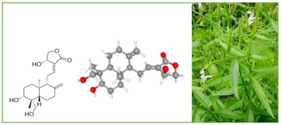
Figure 12.
A. paniculata
and its main compound andrographolide.
4. Pharmacological Importance of Andrographolide and Its Derivatives
During modern times, andrographolide and several its derivatives have been reported to possess several pharmacological properties, including anti-inflammatory, hepatoprotective, anti-viral, neuroprotective, antioxidant, anti-fibrosis, anti-hyperglycemic, anti-tumor, anti-atherosclerosis, antimicrobial, and cardiovascular protective activities. These properties have been discussed in detail in several outstanding review articles [21][29][32][33][34][35][21,33,36,37,38,39]. Andrographolide exhibits free-radical-scavenging activity and anti-inflammatory effects by inhibition of lipopolysaccharide-induced nitric oxide (NO) production and inducible NO synthase (iNOS) expression, and by suppression of IL-2 production and T-cell proliferation. The lactone diterpene demonstrated its effectiveness in the treatment of Alzheimer’s disease, Parkinsonism, spatial memory deficits, depression, and neuro-inflammation [32][36]. Jayakumar et al. [33][37] reported several works that evidenced the promising hepatoprotective effects of andrographolide. The diterpene has been shown to be able to inhibit hepatocyte apoptosis, to attenuate concanavalin-A-induced liver injury, and to protect against ethanol-induced hepatotoxicity in mice with an equivalent efficacy of silymarin. Antiviral activity has been reported against Zika virus, human immunodeficiency virus (HIV), herpes simplex virus (HSV), hepatitis C virus, pestiviruses, and flaviviruses [32][33][36,37].5. Anti-Cancer Properties and Mechanism of Action of Andrographolide and Its Derivatives
Recently, much attention has been focused on the anti-tumor/anti-cancer effects of andrographolide and its derivatives and these compounds have been demonstrated to exhibit promising anti-tumor effects in terms of inhibition of the growth, propagation, and relocation of a number of cancerous cells such as prostate carcinoma cells, colon cancer cells, bladder cancer cells, chronic myeloid leukemia cell lines, colorectal cancer cell lines, breast cancer cells, murine leukemia cells, lymphoma, adenocarcinoma PC-3, and leukemic HL-60 cells and many more human cancerous cells [21][29][36][37][38][39][40][41][21,33,40,41,42,43,44,45]. Various literature reviews have shown that andrographolide and its derivatives was able to reduce cancer cell proliferation/viability and that it is cytotoxic to a broad range of cancer cell lines, but the mechanisms were different for different cell types (Figure 24). Recent published literature has confirmed the multiple anti-cancer effects of the diterpene, in particular against the breast cancer [42][46], lung cancer [43][47], colon cancer [44][45][48,49], renal carcinoma [46][50], and cervical cancer [47][51], as well as hepatoma cancer [48][52]. The principal mechanism or mode of action of these compounds is less understood and it needs to be explored further in detail. The reduced viability of cancer cells in most of the cases could be partially described by the induction of apoptosis [49][50][51][53,54,55], but in some liver cancer cells the death resulted from the diterpene and was not due to the induction of apoptosis [52][56].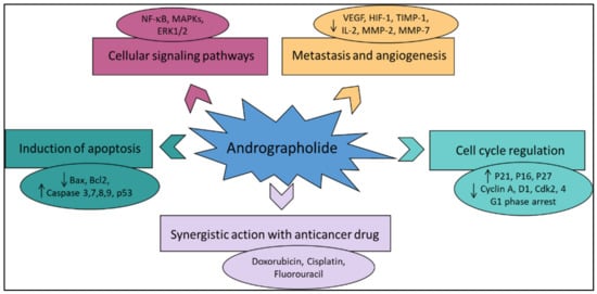
Figure 24.
The principal mechanisms of action of andrographolide as an anti-cancer agent.
5.1. Induction of Apoptosis and Growth Inhibitory Activity
Andrographolide has played a notable role in the recent advancement of pharmacophore development, especially anti-cancer drug development. In anti-cancer-drug development processes, apoptosis induction in carcinoma cells is known to be an important focus [53][57]. The process of apoptosis in the cell helps in the maintenance of tissue homeostasis by careful exclusion of undesirable cells [54][55][58,59]. Andrographolide is credited with potentially inducing apoptosis in several cancer cells and can enhance interleukin-2 secretion by cytotoxic T-lymphocytes for inhibition of tumor growth in mice [56][60]. A recent report on the identified compounds of A. paniculata states that andrographolide endorses the apoptosis process in human cancer cells through the induction of mitochondrial cytochrome c, accompanied by enhanced expression of Bax and reduced Bcl-2 in human leukemia HL-60 cells [57][61] and caspase 8 and caspase 3 activation in human prostate cancer cells PC-3 [58][62]. It also reported that this compound could inhibit the activity of NF-κB, one of the most important transcription factors accountable for cell proliferation and apoptosis [59][60][63,64] (Table 1). Andrographolide was proposed for potential application in cancer therapy for its apoptosis-induction activity [61][65]. Andrographolide was found to be accountable for phosphorylation of p53, as well as the transcriptional upregulation of death receptor 4 (DR4) induced by p53. This stimulation process caused the stimulation of the apoptosis via the tumor-necrosis-factor-related apoptosis-inducing ligand (TRAIL). Andrographolide also increased the TRAIL-induced apoptosis process through the DR4 in the TRAIL-resistant cells [61][65]. Treatment of T-47D mammary cells by andrographolide caused epidermal growth-factor receptor (EGFR) and transferring receptor (TfR) internalization owing to the downregulation of cell-surface receptors and dilapidation of the EGFRs and TfRs [23][27]. Andrographolide also caused apoptotic cell death by reducing the mRNA and protein levels of IL6 (needed for prostate cancer proliferation) [62][66]. Andrographolide is explicitly studied for its anti-cancer activity and is reported to possess capability to induce cell-cycle arrest in human colorectal carcinoma LoVo cells [63][67] and to inhibit cell proliferation of cell [64][68]. Induction of apoptosis in human ovarian teratocarcinoma (PA-1) cells was recently described by Bhat et al. [31][35]. An increased number of cells with activated caspase 3 and a low level of Bcl-2 after treatment with andrographolide was reported in comparison to the untreated cells. Shi et al. [63][67] described the pharmacophore activity of andrographolide and stated that it arrests the cell at the G1/S phase of the cell cycle via the CKI–cyclin–Cdk network. Andrographolide also showed G0/G1 phase arrest in MCF-7 cells [65][69]. In addition, 10–30 μM of andrographolide showed pro-apoptotic and growth inhibitory activity in rheumatoid arthritis by G0/G1 phase arrest of the cell cycle via p21 and p27 inhibition, reduced ratio of Bcl2/Bax, and decreased level of CDK-4 protein [66][70]. The arrest of the G2/M phase of the cell cycle was also caused by the action of andrographolide in glioblastoma U251 and U87 [67][71], human leukemia (K562) [17] and breast cancer cells [68][69][72,73]. Similarly, 3,19-(3-chloro-4fluorobenzylidene) and 3,19-(2-bromobenzylidene), derivatives of andrographolide exhibited superior cytotoxic and growth-inhibition activity in HCT- 116 and MCF-7 cell lines. Both derivatives showed potent inhibitory activity by arrest in the G1/S phase of the cell cycle and apoptosis in MCF-7 and HCT-116 cells [70][74]. Recent research has shown that andrographolide inhibits cell-cycle progression at the G2/M checkpoint in LNCaP, C4-2b, and PC3 cells and at the G1/S checkpoint in DU-145 cells. Cyclin B1 was also upregulated by andrographolide in LNCaP and PC3 cells [71][75]. Wang et al. [72][76] have shown that the osteosarcoma cell proliferation was inhibited by andrographolide by the process of arresting of the cell cycle at the G2/M phase and by enhancing the caspase-mediated apoptosis process. In vitro andrographolide inhibited the growth of osteosarcoma cells by causing G2/M phase cell-cycle arrest and inducing apoptosis via the reactive oxygen species (ROS)/c-Jun N-terminal kinase (JNK) signaling pathway. In vivo, andrographolide exhibited significant anti-tumor activity with minimal toxicity.5.2. Inhibition of Tumor Angiogenesis
The term tumor angiogenesis refers to the formation of new blood vessels within a tumor, which provide the growth center with a constant supply of oxygen and nutrients. Andrographolide decreased tumor-specific angiogenesis by lowering the manufacture of the pro- and anti-angiogenic factors such as interleukin-2, vascular endothelial growth factor, nitric oxide, and tumor necrosis factor TNF-α, in the C57BL/6 mice infected with the B16F-10 melanoma cells (Table 1) [71][75]. In addition, it was able of inhibit the angiogenesis-critical matrix metalloproteinase 2 (MMP-2) and metalloproteinase 9 (MMP-9) activities in colon cancer cells [73][77]. When it comes to A549 cells and non-small-cell lung cancer (NSCLC), HIF-1 is responsible for cancer growth. Andrographolide inhibited HIF-1, reduced vascular endothelial growth factor (VEGF), and boosted hydroxyl-HIF-1 and prolyl hydroxylase expression [74][78]. These results emphasize the promise of andrographolide as a potential chemotherapeutic or anti-angiogenesis drug for the treatment of NSCLC. Serum levels of tissue inhibitors of metalloproteinase 1 (TIMP-1), VEGF, and pro-inflammatory cytokines such as TNF-α, IL-1b, and IL-6, and granulocyte monocyte colony stimulating factor (GM-CSF) were decreased by andrographolide after being induced by the B16F-10 melanoma cell line in the C57BL/6 mice [56][60]. The andrographolide derivative 17-hydro-9-dehydro-andrographolide inhibited vascular endothelial cell proliferation and angiogenesis in rats at 1–10 mM [75][79]. At 50 mg/kg, andrographolide inhibited the expression of PCNA, vascular endothelial growth factor, and cyclin D1 in hamster buccal cells [34][38]. In another study, it is stated that biochemical analysis had identified andrographolide as a significant docking molecule that can bind to the ATP-binding pocket of vascular endothelial growth-factor receptor (VEGFR2) and thus inhibit its kinase activity by potentially interacting with the kinase domain of VEGFR2 [76][80]. VEGFR2 is the major receptor of VEGF. It is expressed in vascular endothelial cells and plays a very significant role in angiogenesis. In fact, by binding and activating VEGFR2, VEGF mediates endothelial invasion, migration, cell proliferation, and survival, and increases vascular permeability and neo-vascularization. In addition, in another study the author discussed the inhibition of angiogenesis by andrographolide by the process of inhibition of the Mir-21-5p/TIMP3 signaling pathway [77][81]. The results showed that andrographolide was able to inhibit the growth of the vascular tissues in the membranes of chick embryo chorioallantois and yolk sac, along with the suppression of the tumor angiogenesis [77][81]. Furthermore, they also stated that the proliferation, migration, and tube formation of the vascular endothelial cells was also inhibited by andrographolide action under in vitro action. The outcome of the process was principally facilitated through the inhibition of the expression of miR-21-5p and added targeting of the TIMP3; this proved that andrographolide was directly involved in the inhibition of angiogenesis [77][81]. Another article discussed the overall mechanism of action of andrographolide by the inhibition of the PI3K/AKT, NF-κB, v-Src, and STAT3 activities followed by the downregulation of the mediators of progression of the cell cycle, metastasis, and angiogenesis [78][82]. In a study by Li et al. [79][83], the author stated that a new andrographolide derivative (AGS-30) was able to display anti-angiogenic properties through the inhibition of the endothelial-cell proliferation, incursion, and relocation, as well as tube formation. The author also stated that the AGS-30 was able to inhibit cell proliferation and the phosphorylation of cell-survival-related proteins followed by the reduction of the VEGF expression in the HT-29 colon cancer cells [79][83]. Moreover, AGS-30 also suppressed the tumor growth and angiogenesis process in the HT-29 colon cancer cell xenografts in nude mice [79][83].5.3. Anti-Proliferative Activity
The anti-proliferative effects of andrographolide have been investigated against several cancer cell lines (Table 1). Udomwan et al. [80][84] investigated the cytotoxic activity of andrographolide by using the 3-(4,5-dimethylthiazole-2-yl)-2,5-biphenyl tetrazolium bromide (MTT) assay against three cervical cancer cell lines, namely CaSki, SiHa, and C33A. The most sensitive cells after treatment with andrographolide (at 20, 40, 80, and 160 µM concentrations) for 48 h were SiHa cells. Cell viability of SiHa cells was reduced to 50% at a concentration of 85.59 µM followed by the value of 87.52 µM for CaSki cells, and 96.05 µM for C33A cells. Successively, Tohkayomatee et al. [81][85] assessed the andrographolide effects on the cell viability against MCF-7 and MDAMB-231 breast cancer cell lines by MTT assay. At concentrations in the range 7.5–120 µM, the diterpene considerably reduced, in a concentration- and time-dependent manner, the cell viability of both cell lines with IC50 values after 48 h of exposure of 32.90 and 37.56 µM against MCF-7 and MDAMB-231, respectively. Treatment with andrographolide at the concentration of 50 µM reduced the viability of the THP-1 (human monocytic leukemia) cell line and NCI-H929 (human IgAkappa-producing multiple myeloma) cell line to 39.2 and 13.0%, respectively, with respect to the untreated cells in a concentration-dependent manner [82][86]. The IC50 values for treating were 31 and 8 µM, for THP-1 and H929 cells, respectively. Andrographolide was demonstrated to be active also against human malignant melanoma A375 and C8161 cell lines [83][87]. The cell viability was assessed by MTT assay. The IC50 values after 48 h of exposure were 12.07 and 10.92 µM for A375 and C8161, respectively, suggesting the promising anti-proliferative activity of andrographolide against malignant melanoma cells in a concentration- and time-dependent manner. Previously, the anti-proliferative activity of andrographolide on HT-29 colon cancer cells was studied by using MTT assay, colony formation assay, trypan blue exclusion assay, and morphological analysis [51][55]. The diterpene reduced cell viability of HT-29 cells in a concentration- and time-dependent manner. An interesting IC50 value of 3.7 µg/mL was found against human ovarian teratocarcinoma (PA-1) cells [31][35]. Devendra et al. [39][43], demonstrated that a series of 3,19-O-acetal derivatives of andrographolide exhibited significant anti-cancer properties and the results specified that the protection of the 3,19-hydroxyl groups of andrographolide with the appropriate ethylidene/benzylidene moiety prompted a substantial cytotoxicity effect with either the acetylation or dehydration of the 14-hydroxyl of the lead compound cyclic acetal derivative, that could have triggered its cytotoxic effect on all the cell lines [39][43].5.4. Induction of Autophagy
Autophagy is a process associated with several diseases, including the cancer that destroys and processes the damaged macromolecules and organelles through lysosomal pathways to maintain the homeostasis of cells [84][88]. Autophagy may play contrasting roles in different types of cancers and in their different stages of development, including promoting survival and inducing death [85][89]. For this reason, research into the molecular mechanisms of autophagy-related signal-transduction pathways is of interest [86][87][88][89][90,91,92,93]. In this context, Liu et al. [90][94] demonstrated the effects on autophagy of andrographolide in human osteosarcoma cells by suppressing the phosphatidylinositol-3-kinase (PI3K)/Akt and the mammalian target of rapamycin (mTOR) signaling pathways and enhancing the c-Jun N-terminal kinase (JNK) pathway. Autophagy induced by andrographolide inhibits the invasion and metastasis of osteosarcoma cells suggesting that the diterpene may represent a promising targeted agent in the prevention and treatment of osteosarcoma. Yuwen et al. [38][42] demonstrated that cisplatin induced autophagy that attenuated the sensitivity of both A549 and Lewis lung cancer cells to cisplatin, whereas the clinical drug andrographolide suppressed autophagy and enhanced cisplatin-mediated apoptosis in these cells [38][42].5.5. Oxidative Stress and Antioxidant Properties of Andrographolide in Anti-Cancer Treatment
Andrographolide exhibited promising antioxidant properties acting through different mechanisms of action including the neutralization of free radicals, the activation of antioxidant enzymes, the inhibition of pro-oxidant enzymes, and the protection of mitochondrial integrity [91][95]. Some studies have shown the reduction of ROS in cellular models by andrographolide [92][93][96,97]. Treatment with andrographolide (10 and 30 M) decreased the production of ROS in RAW264.7 macrophages motivated by the ovalbumin or lipopolysaccharide (LPS) [91][95]. Previously, andrographolide has been reported to inhibit intracellular ROS production in N-formylmethionyl-leucyl-phenylalanine-induced neutrophils [92][96]. Zhan et al. [94][98] showed a dose-dependent increase in catalase (CAT) and super oxide dismutase (SOD) activities after topical application of andrographolide to mouse skin that was exposed to UV radiation, as compared to the untreated mice. Andrographolide is also considered to be a potent inhibitor of the enzyme xanthine oxidase (XOD), that catalyzes the terminal steps of purine degradation. XOD has been proposed as a source of oxygen radicals in epithelial, connective, and endothelial tissue cells. In fact, the enzyme is described as playing an important role in cellular oxidative status. An in silico study revealed strong binding interactions between the andrographolide and the XOD enzyme [95][99] and a recent work showed the ability of the diterpene (30 mg/kg/day) to reduce liver XOD activity [96][100]. NADPH oxidase (NOX) is a membrane enzyme complex that catalyzes the oxidation reaction of NADPH by oxygen, inducing ROS production in cells. Andrographolide has been reported to reduce the expression of NOX2 through limiting the activation of phosphoinositide 3-kinase/protein kinase B (PI3K/AKT)-dependent nuclear factor-kappa B (NF-B) [97][101]. In another work, andrographolide (10 and 20 mg/kg/day) significantly decreased NOX2 and NOX4 expression in myocardial tissues [98][102]. There are reports that andrographolide can improve mitochondrial dysfunction in some in vitro and in vivo models. Geng et al. [99][103] demonstrated that andrographolide sulfonate treatment could reduce oxidative stress and protect the mitochondria in a transgenic mouse model (amyloid precursor protein/presenilin 1). In another model, it was found that andrographolide supplementation could reduce the production of nitric oxide, carbonyl protein, and malondialdehyde, and enhance mitochondrial complex activities in the electron transport chain [100][104]. On the other hand, increased activity of CAT, SOD, glutathione peroxidase, glutathione reductase, and glutathione-S-transferase, and reduced concentrations of glutathione and glutathione disulfite were observed [100][104].Table 1.
In vitro and in vivo study of andrographolide against cancer cell lines.
| Cancer Cell Line | Study Type | Main Effects | Ref. |
|---|---|---|---|
| Ovarian teratocarcinoma | In vitro | PA-1 cells—MTT test—IC50 3.7 µg/mL; induction of apoptosis | [31][35] |
| Lung cancer | In vitro | Suppression of autophagy and enhanced cisplatin-mediated apoptosis | [38][42] |
| Colon cancer | In vitro | HT-29 cells—induction of programed cell death and cell-cycle arrest through the increase of intracellular ROS level | [51][55] |
| Leukemia | In vitro | HL-60 cells—cell-cycle arrest and mitochondrial-mediated apoptosis | [57][61] |
| Prostate cancer | In vitro | PC-3 cells—caspase 8 and caspase 3 activation | [58][62] |
| Neuroblastoma | In vitro | Induction of p53- and caspase-independent cell death | [59][63] |
| Liver cancer | In vitro | HepG2 and Hep3B cells—sensitization of cancer cells to TRAIL-induced apoptosis via p53-mediated death receptor 4 upregulation | [61][65] |
| Cervical cancer | In vitro | HeLa cells—sensitization of cancer cells to TRAIL-induced apoptosis via p53-mediated death receptor 4 upregulation | [61][65] |
| Colorectal cancer | In vitro | HCT116 cells—sensitization of cancer cells to TRAIL-induced apoptosis via p53-mediated death receptor 4 upregulation | [61][65] |
| Prostate cancer | In vitro | LNCaP, DU145, and PC-3 cells—apoptotic cell death by reducing the mRNA and protein levels of IL6 | [62][66] |
| Colorectal carcinoma | In vitro | LoVo cells—induction of cell-cycle arrest and inhibition of cell proliferation | [63][67[64],68] |
| Brest cancer | In vitro | MCF-7 cells—G0/G1 phase arrest | [65][69] |
| Glioblastoma | U251 and U87 cells—arrest of the G2/M phase cell cycle phase | [67][71] | |
| Leukemia | K562 cells—arrest of the G2/M cell cycle phase | [17] | |
| Breast cancer | Arrest of the G2/M phase | [68][69][72,73] | |
| Prostate cancer | In vitro | LNCaP, C4-2b, and PC3 cells—inhibition of cell-cycle progression at the G2/M phase; decrease of tumor-specific angiogenesis | [71][75] |
| Osteosarcoma | In vitro | Inhibition of cell proliferation by arresting of the cell cycle at the G2/M phase | [72][76] |
| Osteosarcoma | In vivo | Female Balb/c-nu mice with HOS-Luc cells injected subcutaneously—induction of apoptosis via the ROS/JNK pathway | [72][76] |
| Colon cancer | In vitro | HT29 cell line—anti-invasive activity against colon cancer cells via inhibition of matrix metalloproteinase 2 (MMP2) | [73][77] |
| Cervical cancer | In vitro | CaSki cell line—MTT test—reduction of cell viability of 50% at 87.52 µM | [80][84] |
| Cervical cancer | In vitro | SiHa cell line—MTT test—reduction of cell viability of 50% at 85.59 µM | [80][84] |
| Cervical cancer | In vitro | C33A cell line—MTT test—reduction of cell viability of 50% at 96.05 µM | [80][84] |
| Breast cancer | In vitro | MCF-7 cell line—MTT test—IC50 32.90 µM | [81][85] |
| Breast cancer | In vitro | MDAMB-231 cell line—MTT test—IC50 37.56 µM | [81][85] |
| Monocytic leukemia | In vitro | THP-1 cell line—MTT test—IC50 31 µM | [82][86] |
| IgAkappa-producing multiple myeloma | In vitro | H929 cell line—MTT test—IC50 8 µM | [82][86] |
| Malignant melanoma | In vitro | A375 cell line—MTT test—IC50 12.07 µM | [83][87] |
| Malignant melanoma | In vitro | C8161 cell line—MTT test—IC50 10.92 µM | [83][87] |
| Osteosarcoma | In vitro | Suppression of phosphatidylinositol-3-kinase (PI3K)/Akt and the mammalian target of rapamycin (mTOR) signaling pathways; enhancement of the c-Jun N-terminal kinase (JNK) pathway | [90][94] |
| Mice | In vivo | Inhibition of PI3K/AKT-dependent NOX2 and iNOS expression | [97][101] |
| APP/PS1 transgenic mice | In vivo | Reduction of oxidative stress and protection of mitochondria | [99][103] |
| Male Wistar rats | In vivo | Reduction of the production of nitric oxide, carbonyl protein, and malondialdehyde, and enhancement of mitochondrial complex activities in the electron transport chain. Increased activity of CAT, SOD, glutathione peroxidase, glutathione reductase, and glutathione-S-transferase | [100][104] |