ADP-Ribosylation during Infection Establishment by Trypanosomatidae Parasites: Comparison
Please note this is a comparison between Version 1 by Josh Dowling and Version 2 by Lindsay Dong.

ADP-ribosylation is a reversible post-translational protein modification, which is evolutionarily conserved in prokaryotic and eukaryotic organisms. It governs critical cellular functions, including, but not limited to cellular proliferation, differentiation, RNA translation, and genomic repair. The addition of one or multiple ADP-ribose moieties can be catalysed by poly(ADP-ribose) polymerase (PARP) enzymes, while in eukaryotic organisms, ADP-ribosylation can be reversed through the action of specific enzymes capable of ADP-ribose signalling regulation. In several lower eukaryotic organisms, including Trypanosomatidae parasites, ADP-ribosylation is thought to be important for infection establishment. Trypanosomatidae encompasses several human disease-causing pathogens, including Trypanosoma cruzi, T. brucei, and the Leishmania genus. These parasites are the etiological agents of Chagas disease, African trypanosomiasis (sleeping sickness), and leishmaniasis, respectively.

  • ADP-ribosylation
  • PARP
  • PARG
  • Trypanosoma
  • Leishmania

1. ADP-Ribosylation in Infection

ADP-ribosylation is a fundamental post-translational protein modification in which single or several ADP-ribose units are covalently attached to proteins. The modification is commonly catalysed by members of the poly(ADP-ribose) polymerase (PARP) enzyme family. PARP enzymes attach ADP-ribose moieties to the aspartate, glutamate, lysine, arginine, cysteine, threonine, or serine residues, resulting in the creation of branched and linear polymers [1]. In addition to the actions of the PARP enzymes, ADP-ribosylation can also occur via the action of mono(ADP-ribosyl)transferases, which catalyse the attachment of ADP-ribose to arginine side chains via the activity of an essential and highly conserved R-S-EXE motif [2]. This motif is localised within a specialised loop used in target recognition for mono(ADP-ribosyl) transferases as well as for several other ADP-ribosyltransferases (ARTs), including human PARP-1.

1.1. ADP-Ribosylation in Viral Pathogens

PARP enzymes have long-documented actions in infection progression and protection against pathogens in humans [3]. The role of PARP-mediated protection, particularly against viral infection, has seen extensive study. Several human PARPs, including PARP 1, 5, 7, 9, 10, 12, and 13 are known antagonists of both DNA and RNA viruses [4][5][6][4,5,6]. Viral replication is disrupted by several PARP enzymes, predominantly targeting dysregulation in viral genomic translation and transcription, leading to inhibition or prevention of the completion of the viral life cycle. PARP13, in particular, demonstrates potent antiviral activity against many DNA and RNA viruses, including alphaviruses, influenza, filoviruses (including Ebola and Marburg viruses), herpes virus, HIV-1, coxsackie virus B3, hepatitis B, and Japanese encephalitis virus [7][8][9][10][7,8,9,10]. PARP13 utilizes multiple mechanisms in viral inhibition, binding viral RNA through its four zinc-finger motifs, to allow the inhibition of transcription and translation of the viral genome which disrupts the viral life cycle. PARP13 is, then, able to degrade the 5′ end of HIV-1 RNA via the recruitment of several degrading host factors, including poly(A)-specific ribonuclease (PARN) and the KHNYN endonuclease [11].
Viral replication is targeted by the host expressing PARP enzymes to defend against infection. For example, PARP1 and PARP5 serve as antagonists to critical binding sites used by viral proteins for genomic replication by Kaposi sarcoma-associated herpesvirus (KSHV) and Epstein–Barr virus (EBV), respectively [12]. PARP1 is also able to prevent transcriptional elongation of HIV-1 RNA through the binding of PARP1 to a TAR binding site, which is responsible for the binding of RNA elongation factor p-TEFb. PARP1 also binds with TAR more efficiently than p-TEFb, demonstrating its ability to inhibit viral replication through epigenetic modification [13]. Genome translation is essential for viral replication and a target for inhibition by PARPs. PARP13 has been shown to decrease the production of Nef, a protein that is present in HIV-1 and is critical for successful viral replication. PARP13 is also able to degrade viral RNA via exosome activation [14]. This is further evidence of the broad antiviral mechanisms exhibited by PARP13. Other PARPs demonstrate antiviral activity through the targeted degradation of essential viral proteins. PARP9 can form a protein-degrading complex with a ubiquitin ligase capable of Picronoviridae protein degradation [15]. PARP10 is capable of transfer to nuclei during avian influenza (AIV) viral infection to degrade the protein AIV NS1, which is important for AIV replication [16]. PARP12 can degrade Zika virus (ZIKV) proteins NS1 and NS3 via mon(ADP-ribosyl)ation, catalysed by PARP12 [17].

1.2. ADP-Ribosylation in Bacterial Pathogens

Studies examining the roles of PARPs during bacterial pathogen infection are not as extensive in comparison to those on PARPs in viral infections. Only a small number of bacterial species possess functional PARylation systems. Although the majority contain domains capable of PAR binding, in addition to PAR-degrading enzymes [18]. The majority of antibacterial studies on PARP enzymes have primarily focused on the use of PARP1. Studies have been performed on several notable human bacterial pathogens, including Helicobacter, Salmonella, Escherichia coli, Pseudomonas aeruginosa, Streptococcus pneumonia, Streptococcus pyogenes, and Chlamydophila. Experiments utilising these bacteria surmise a similar consensus, whereby shifts in PARP activity increase the difficulty of mounting an effective response in preventing damage from bacterial infection [19][20][21][22][23][19,20,21,22,23]. Several species of bacteria have also been found to possess PARG enzymes, including Thermomonospora curvata and Herpetosiphon aurantiacus [24][27]. In humans, PARG enzymes are a mechanism through which PAR can be removed from the cell via catabolism of poly(ADP-ribose), through hydrolysis of the ribose-ribose bonds. This prevents damage caused by excessive PAR accumulation in the cytoplasm [25][28]. PAR accumulation can lead to a PARP-mediated cell death pathway known as parthanatos, through which excessive PAR can lead to apoptosis via several mechanisms, including depletion of NAD and the PAR-mediated activation of an apoptosis-inducing factor (AIF) [26][29]. PAR can bind to AIF, which is followed by AIF translocation to the nucleus, resulting in extensive DNA fragmentation and chromatin damage [27][30]. PARG is the primary means through which excessive PAR is removed in human cells. Other human PAR hydrolases do exist, necessitated by PARG’s inability to remove the most proximal ADP-ribose moieties [28][31], including ARH3, which is present during the removal of PAR from the mitochondria [29][32]. There is much evidence to suggest the extensive roles of human PARP enzymes in immune protection during bacterial and viral infection. However, key questions remain for both. PARP activity is seemingly broader across enzymes in terms of antiviral activity (10 out of 17 human PARP enzymes have identified antiviral activity). Potentially as a result of primarily cytoplasmic and nuclear localisation, which allows the PARP enzymes to interrupt viral replication cycles at several distinct stages [30][34]. Therefore, it seems that the expression of multiple cytoplasmic PARP enzymes, developed alongside the evolution of vertebrates, are seemingly as equally as important as the nuclear-localised PARPs in maintaining cellular health through the maintenance of essential processes and antimicrobial activity.

2. Trypanosomatidae

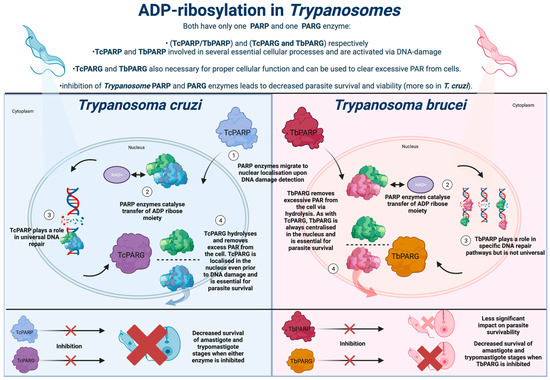
Trypanosomatidae is an order of singular flagellate kinetoplastid parasites, the most relevant to human health being Trypanosoma and Leishmania. The parasites T. cruzi, T. brucei, and Leishmania are all responsible for infections categorised by the World Health Organisation (WHO) as neglected tropical diseases (NTDs) [31][37]. As such, these infections are responsible for profound impacts as they primarily arise in vulnerable people inhabiting developing countries. ADP-ribosylation and the associated enzymes, including PARP and PARG, play an essential role in the ability of Trypanosomatidae to establish a successful infection in the human host. Given the lack of safe and effective medications available to combat these parasites, and the parasites’ reliance on the functioning ADP-ribosylation to maintain parasitic viability, the disruption of their ADP-ribosylation systems may present a novel and effective therapeutic option. T. cruzi is the causative agent of Chagas disease, which is endemic to Central and South America. An initial infection during the acute stage results in flu-like systems. These symptoms mask the diagnosis of T. cruzi infection [32][38]. During chronic stages, an infection can lay dormant for decades, characterised by potentially minimal symptoms and low parasitemia, which causes issues in detecting the infection. The chronic infection eventually results in organ enlargement, with parasites primarily targeting the heart, leading to numerous cardiac complications and potential death. T. brucei is the etiological agent of African trypanosomiasis. Vectoral transmission is the most common route of infection, in which parasites enter the human host via bites inflicted by the primary tsetse fly host. Similar to Chagas disease, the initial stage of infection presents aspecific symptoms, followed by parasites eventually migrating to the brain, leading to neurological complications, and ultimately death, without proper treatment [33][41]. Available therapies for the neurological stage of infection are also limited, with Melarsoprol the only available drug, although this causes death in 5% of the patients who ingest it since Melarsoprol resistance is present in some strains [34][42]. The anti-parasitic Fexinidazole has shown activity against both the CNS and peripheral stages of African trypanosomiasis, although studies remain in clinical trials and effective drug alternatives are required to limit resistance [35][43]. Leishmaniasis is an umbrella term for three distinct diseases: visceral, cutaneous, and mucocutaneous leishmaniasis. Infections are predominantly found in Asia, the Middle East, and Northern Africa to differing degrees, The diseases are caused by several different Leishmania species and are transmitted commonly by bite wounds inflicted by an infected female phlebotomine sand fly. Visceral leishmaniasis is the most severe, as it causes a systemic infection that almost invariably is fatal without rapid treatment. The cutaneous infection leads to superficial skin lesions, whilst mucocutaneous infection leads to significant damage of the buccal and nasal cavities via the formation of damaging mucocutaneous lesions, which may disappear and reoccur repeatedly [36][44]. Given the severe nature of the infection, the majority of attention in the development of novel therapies for leishmaniasis has been focused on visceral leishmaniasis [37][38][45,46]. Consequently, given the scarcity of funding available to research the development of novel therapeutics to combat these NTDs and the lack of efficacy for existing treatments, the identification of novel ways to combat these infections is paramount. T. cruzi, T. brucei, and Leishmania all utilise ADP-ribosylation in several distinct ways to facilitate successful infection in the human host. Given the avenues with potential to be explored, whereby ADP-ribosylation manipulation can be harnessed to combat viral and bacterial infection, it is feasible that inhibiting or disrupting ADP-ribosylation in these parasites could lead to diminished parasite survival and proliferation. In contrast to the extensive PARP network found in humans, the PARP system present in Trypanosomatidae is much simpler. T. cruzi and T. brucei both possess a singular PARP enzyme, designated TcPARP and TbPARP, respectively. Both parasites also utilise a sole poly(ADP-ribose) glycohydrolase (PARG) enzyme, which is used to reverse the action of PARP via the hydrolysis of ribose–ribose bonds present in PARP, which helps prevent extensive DNA damage by excessive PARP accumulation [39][49]. The primary ADP-ribosylation mechanisms within these parasites use polyADP-ribosylation, although there is also evidence of them using monoADP-ribosylation systems. Both T. cruzi and T. brucei possess proteins that are homologous to human MacroD1 and MacroD2, which are domains that hydrolyse and cleave ADP-ribose attachments to proteins in monoADP-ribosylation within human systems [40][50].

3. ADP-Ribosylation in Trypanosoma cruzi

3.1. Use of ADP-Ribosylation and Associated PARP and PARG Enzymes in Trypanosoma cruzi

Trypanosoma cruzi expresses a sole PARP enzyme throughout its lifecycle, known as TcPARP (Figure 1). An initial study of TcPARP revealed several structural and molecular similarities to its human homologue, hPARP-1 [41][52]. Similar to hPARP-1, TcPARP is activated via DNA strand nicks, upon exposure of the parasite DNA to damaging agents such as H2O2. Initial studies on TcPARP revealed a highly evolutionarily conserved C-terminal catalytic domain that is homologous to T. brucei PARP, human PARP-1, and PARP-4. Furthermore, the catalytic triad of histidine, tyrosine, and glutamic acid is utilised for PAR elongation in hPARP-1, the closest human homologue to TcPARP is conserved within the parasite [42][53].
Figure 1. ADP-ribosylation in Trypanosomes. In contrast to pathways in eukaryotes, ADP-ribosylation in Trypanosomatidae is more linear, with the parasites only expressing one PARP enzyme. If this pathway can be understood and exploited, it may reveal new avenues for combatting Trypanosomatidae infection.
TcPARP activity is dependent upon the detection of DNA damage and is key in successful DNA repair and signalling. It serves roles in the ability of the parasite to differentiate into different life-cycle stages and establish infection in a human host. Consequently, the use of PARP inhibitors in the treatment of T. cruzi has been studied and has seen some success. Olaparib, a common PARP inhibitor, exerts strong inhibitory effects on hPARP-1 and TcPARP activities [41][43][52,57]. Olaparib has shown the ability to reduce amastigote (non-motile form lacking flagella) formation in a range of cell lines [41][52].

3.2. The Potential of ADP-Ribosylation Targeted Therapy for Trypanosoma cruzi

Intriguingly, there may be an interplay between hPARP-1 and TcPARP during infection establishment by T. cruzi. hPARP-1 silencing experiments in A549 cells demonstrated a significantly decreased amastigote number as well as a reduction in trypomastigotes in the culture media [41][52]. T. cruzi is dependent upon the ability of healthy trypomastigotes to establish a lasting infection and the subsequent differentiation and multiplication of amastigotes [44][60]. Therefore, successful inhibition and prevention of T. cruzi require the disruption of differentiation and growth. T. cruzi trypomastigote penetration of cardiac tissue in vitro leads to ROS generation, through activation of hPARP-1. ROS accumulation subsequently triggers the expression of NF-kB, which is a transcription factor that facilitates the transcription of multiple cytokines acting in concert to allow successful penetration of the parasite into cell lines in vitro [45][46][61,62]. The absence of cytokines TNF-α and IL-1β resulted in decreased levels of infection, demonstrating that successful infection by T. cruzi requires some activation of NF-kB [47][63].
However, chronic exposure to PARP inhibitors can be profoundly detrimental. Should PARP inhibitors be assessed regarding Chagas therapy, protection from DNA damage is a fundamental consideration. PARG enzymes can reverse the effects of PARP enzymes, via clearance of excessive PAR accumulation to circumvent any harm to cells [48][64]. The PARG enzyme present in T. cruzi, denoted TcPARG, has been demonstrated as essential for epimastigote growth and the infection cycle in vitro. TcPARG shares 46.5% sequence identity with human PARG and possesses a preserved domain, including the tyrosine residues utilised to bind PARG inhibitors [49][65] Importantly, as with hPARP-1, T. cruzi seemingly uses host PARG during infection establishment. 

4. ADP-Ribosylation in Trypanosoma brucei

4.1. PARP Enzymes in Trypanosoma brucei and Potential Therapies

As in T. cruzi, T. brucei possesses one PARG and one PARP enzyme, denoted as TbPARG and TbPARP, respectively (Figure 1). TbPARP is highly conserved and similar to TcPARP and hPARP-1 in terms of its sequence, structure, and function [50][51][68,69]. A basic sequence of amino acids is likely responsible for DNA damage detection by TbPARP and the subsequent activity of the enzyme. Notably, TbPARP lacks a reliance on metal ions for TbPARP activity, both of which are characteristics that are shared with TcPARP. Metal ions (including Mn2+, Ni2+, and Zn2+) can have an inhibitory effect on the activity of both TbPARP and TcPARP, which is likely due to the metal ions binding to sulfhydryl groups, thought to be necessary for disulphide bond formation and reduction reactions [50][68]. This is in contrast to the impacts that metal ions have on human PARP enzymes, with Ca2+ and Mn2+ able to increase the levels of PAR synthesis, with human PARP enzymes relying on an alternative mechanism for oxygen reduction and disulphide bond formation [52][70]. Studies on TbPARP structure have also elucidated N-terminus disorder, which is a characteristic shared by both hPARP2 and hPARP3 [53][24]. TbPARP, along with TcPARP, has also been shown to have a WGR binding domain composed of arginine, glycine, and tryptophan, which is present in several eukaryotic organisms, and has a demonstrated role in DNA-dependent PARP activity for PARP enzymes lacking the characteristic zinc-finger binding domain of hPARP-1 [54][71].

4.2. PARG Enzymes in Trypanosoma brucei and Potential Therapies Targeting PARG Enzymes

The role of PARG-mediated suppression of PARP enzymes in T. brucei is unclear. However, a relationship between PARG and PARP enzymes exists within T. brucei. Therefore, although it is likely that a PARP/PARG-mediated treatment is closer for Chagas disease, a similar achievement could be attained for African trypanosomiasis. TbPARG shares 60% sequence similarity with human PARG, including the adenosine diphosphate hydroxymethyl pyrrolidinediol (ADP-HPD) binding site, with ADP-HPD commonly involved in PARG inhibition, meaning PARG inhibitors used in mammalian systems may offer relevance in T. brucei [49][65]. In T. bruce, the depletion of PARG has been shown to result in the increased nuclear accumulation of PAR in Trypanosomes, even in the absence of oxidative stress [55][75]. The duality of PARP and PARG enzymes likely exerts a similar effect in T. brucei as in T. cruzi, and their regulation strikes a fine balance between cellular protection and cell death. PARP enzymes exert protective effects against DNA damage in these parasites, but excessive PAR accumulation can lead to disrupted DNA repair, and NAD+ and ATP depletion resulting in apoptosis. PARP and PARG enzymes remain credible targets for intervention in T. brucei as the parasite relies on both enzymes to establish infection. Nevertheless, the current state of understanding is poor in T. brucei in comparison to T. cruzi.

5. ADP-Ribosylation in Leishmania

5.1. LdARL-3A Ribosylation Factor as a Therapeutic Target in Leishmania

ADP-ribosylation in Leishmania is less studied and poorly understood compared to the Trypanosomes, though work has taken place to better understand the importance of ADP-ribosylation in relation to Leishmania viability and infection. LdARL-3A is an ADP-ribosylation factor identified in Leishmania donovani, which is expressed specifically during the promastigote stage, during the insect stage of the lifecycle. ARL (ADP ribosylation-like) enzymes are essential for numerous cellular processes, including trafficking, endocytosis, and other cell signalling pathways. LdARL-3A seemingly plays a role in flagellum formation and viability, as overexpression of a constitutively active LdARL-3A mutant led to a correlated decrease in flagellum length, with stronger levels of overexpression leading to larger decreases in flagellar length [56][77]. Therefore, if the function of LdARL-3A can be inhibited, the motility of the parasite in the insect host may be reduced before the infection of the human host.

5.2. LiSIR2RP1 as a Therapeutic Target in Leishmania

Leishmania infantum possesses a gene denoted LiSIR2RP1 that encodes the SIR2 protein, which is a deacetylase to several cellular substrates, including histone lysine residues [57][79]. This deacetylation activity is dependent upon the use of NAD+. Disruption of LiSIR2RP1 has been shown to decrease the viability of Leishmania infantum amastigotes both in vivo and in vitro, with the close association of LiSIR2RP1 with the cytoskeletal structure of both L. infantum promastigote and amastigotes. LiSIR2RP1 can exert deacetylase activity upon tubulin, which is critical for parasite structural viability as well as the parasite’s ability to interact with host cells [58][80].

5.3. Targeting NAD+ Salvage Pathways as a Therapeutic Option in Leishmania

Leishmania species are NAD+ auxotrophs and require NAD+ sequestered from host cells. Leishmania lacks both intrinsic de novo pathways that can be used for innate biosynthesis of NAD, which use either L-tryptophan or aspartic acid as a precursor (which are in eukaryotic and prokaryotic de novo pathways, respectively). Leishmania instead uses NR (nicotinamide riboside) as a precursor for NAD+ production in salvage pathways, wherein NAD+ is sequestered from the host [59][81]. The NAD nucelotidase (NadN) enzyme first described in Haemophilus influenzae was found to also be present in Leishmania [60][82]. NadN is a periplasmic enzyme thought to be involved in NAD+ synthesis via NAD+ hydrolysis into NR, adenosine, and phosphate, and finally, NR uptake across the inner membrane of the parasite and NadR catalysed resynthesis of NR into NAD+ [61][83].
Video Production Service