Membrane filtration processes are best known for their application in the water, oil, and gas sectors, but also in food production they play an eminent role. Filtration processes are known to suffer from a decrease in efficiency in time due to e.g., particle deposition, also known as fouling and pore blocking. Although these processes are not very well understood at a small scale, smart engineering approaches have been used to keep membrane processes running. Microfluidic devices have been increasingly applied to study membrane filtration processes and accommodate observation and understanding of the filtration process at different scales, from nanometer to millimeter and more. In combination with microscopes and high-speed imaging, microfluidic devices allow real time observation of filtration processes.
- microfluidics
- membrane
- pore design
1. Introduction
Membrane filtration is used for a wide variety of applications due to the availability of an abundance of membranes targeted at different separations [1]. The versatility of membranes and membrane modules allows for their application in many different fields, ranging from water purification [2][3] and blood dialysis [4][5] to fermentation broth purification [6] and milk fractionation [7]. For some applications such as water purification, membranes and membrane modules are produced and used on a large scale [8][9]. For other applications, such as dehydration of bioethanol from fermentation broths, scale up is still a challenge due to the high complexity of the process leading to the need to use hybrid systems such as a pervaporation-distillation hybrids [10]. In all membrane processes there are challenges with which researchers and engineers must deal. One of the most researched and reported challenge is the process of flux decline due to concentration polarization and membrane fouling [11][12]. More practical issues are the mechanical and chemical stability, and costs related to production, running and cleaning the membrane modules, which is also directly linked to energy consumption and general environmental impact [13]. These parameters are expected to become increasingly relevant in the coming years and lead to the development of even more sustainable processes.
Membrane separation is mostly considered a mild processing technique since it uses low processing temperatures and low applied pressures (microfiltration and ultrafiltration). For these reasons it is expected to contribute to current world challenges, mainly those related to high energy consumption. For example, the higher demand for food products [14] can benefit from mildly resourced raw materials, leading to more efficient use by purification and isolation of substances of interest and commercial value [6][15][16]. Obviously, this can only be achieved by tuning membranes and membrane processes to the desired application. This may seem very straightforward, but at the same time, doing this repeatedly for every feed stream of interest is not efficient. Especially when keeping in mind that many membranes and membrane processes revolve around similar effects such as fluid mechanics, the understanding of the underlying mechanisms of membrane failure and solute behavior during filtration would greatly speed up process design, and lead to substantial improvements in membrane filtration.
The effects that are ruling flux decrease generally occur at a very small size, short time scales, and in modules that are not that accessible for detailed observations. Computer simulations have been instrumental in achieving better insights, and developing reliable membrane processes; however, validation is always difficult, and for these, microfluidic devices may hold the key. Microfluidic devices have proven to be valuable and powerful tools for fundamental research in many fields such as fluid mechanics [17][18], soft matter [19][20], and biology [21][22] and are currently also starting their way into membrane research. Some of their advantages are their relative low cost, flexibility of design, and the possibility of coupling it with microscopy/high speed imaging techniques [23]. A disadvantage of this approach is the current resolution limitation on the techniques for microfluidic device fabrication. This results in the availability of devices with channels that are much wider than the pores of "real" microfiltration and ultrafiltration membranes; however, these devices still allow for very insightful and interesting experiments and results that can certainly contribute to the advancement of the membrane separation field. We expect these limitations to be overcome in the future as will be discussed later in the outlook session.
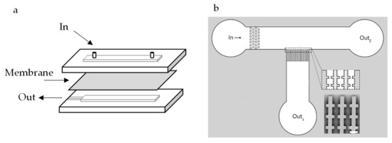
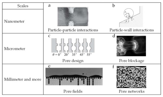
In the membrane separation field, several configurations of microfluidic devices have been reported. Microfluidic devices with embedded membranes (see Figure 1a) have been described for particle sorting [24], membrane fouling [25][26], and to study flow in membrane microreactors [27]. In these studies, microfluidic devices are used to support small pieces of membranes and allow for in situ observation of the filtration process. Alternatively, microfluidic devices can be designed as model membrane systems [28][29][30] (see Figure 1b), containing channels that mimic, for example, membrane pores under highly ideal conditions. In this way, microfluidic devices can be used to study and understand phenomena occurring at different scales [31], ranging from nanometers to even millimeters and more (see Figure 2 for illustrative examples).

Figure 1. (a) Microfluidic configuration with an embedded membrane. (b) Microfluidic configuration with an array of parallel channels with the objective to mimic a membrane in an ideal situation. The image has been cropped and modified from the original work [32] and reproduced under the Creative Commons Attribution 4.0 International License [33].

Figure 2. Examples of how microfluidic tools are applied to investigate mechanisms occurring at various scales. At nanometer scale particle–particle (a) and particle–wall (b) interactions can be studied [34]. Pore design (c) [34] and pore blockage (d) [35] are topics that can be investigated at micrometer scale, and finally at millimeter scale pore fields (e) [32] and pore networks (f) [36] have been used to study the collective behavior of particles in porous media. Images a-e have been cropped from their original work and reproduced under the Creative Commons Attribution 4.0 International License [33]. Image f has been reproduced with permission from the author.
At the nanometer scale, the study of colloidal particle–particle interaction during flow and particle surface interactions are some examples of topics that have been studied [32]. Material surface modification and functionalization is also part of these investigations, and geared toward minimizing the interactions occurring at nanometer scale [4] (Figure 2 top line). At micrometer scale, idealized membrane mimicking microfluidic devices have been proposed to investigate, for instance, pore geometry in relation to blocking and process optimization, using both hard and deformable particles [28][29][30] (Figure 2, middle line). Finally, at millimeter scale, microfluidic devices have been useful for investigating phenomena related to collective particle behavior, such as surface deposition/cake formation and the (collective) movement of particles in flow [26][37][38] (Figure 2, bottom line). It is good to mention that insights obtained with microfluidics in other fields for example, to study flow [39][40][41] and to separate individual cells [42][43] are very relevant for membrane separation.
A number of reviews have been written on membranes and microfluidics [44][45][46][47]. Some of them describe the applications of microfluidic chips with integrated membranes as used for cell and protein research and gas detection [44][45]. In their review, published in 2006, De Jong et al. [45] discussed the integration of membrane functionality into microfluidic chips. They stated that bridging the fields of microfluidics and membranes can be beneficial for both fields of study. Indeed, in the last years, the use of microfluidic devices to study membrane filtration phenomena has flourished, although the focus has been mostly on microchip fabrication techniques as reviewed in [46][47], and not much effort has gone into bringing the findings that were obtained toward improvement of membrane processes. Therefore, we chose this as the topic of our review.
2. Microfluidic Devices as Tools
2.1. Structure
As indicated previously, microfluidic devices used to investigate membrane filtration can be separated in two broad categories: microfluidic devices with embedded membranes [4][26][27][48] and microfluidic devices that mimic membrane structures [28][29][30]. As the name already states, devices with embedded membranes are microfluidic devices designed to accommodate a small piece of a membrane in their structure. The advantage of this configuration is that a real membrane is being used, so if measurements of flux and selectivity are important for the study, this might be a good option. However, in situ observation possibilities in these devices can be limited, as is also the case in larger scale observation techniques that monitor cake layer buildup, droplet deposition or sorption of macromolecules (with fluorescent probes) for instance [12][49]. Membrane mimicking devices have designs that reproduce, in an ideal way, the structure of a membrane. Most of these devices contain an array of parallel channels or structures, that would represent the pores in a real membrane. The advantage of this approach is the possibility of observation of particle behavior at individual level. Additionally, the observation of cake layer structure and pore blocking mechanisms in these systems is facilitated through sideways 2D observation. Due to the flexibility of design of microfluidic devices, the structures mimicking a membrane can be fabricated practically at will. Parallel straight through channels [50], parallel channels with constrictions [51], round pillars [52][53] and non-aligned squares [29] are some examples of structures used to simulate membrane filtration with microfluidic devices. Most membrane mimicking microfluidic devices, including the ones cited before, are made of Polydimethylsiloxane (PDMS) via soft lithography. However, new fabrication techniques are being developed such as the fabrication of polyethylene glycol (PEG)-based hydrogel membranes via photo-patterning. These membranes can have their permeability easily controlled with pore sizes closer to those present in "real" membranes, are pressure-resistant and have been reported to be used for the study of microfiltration and ultrafiltration processes [54][55].
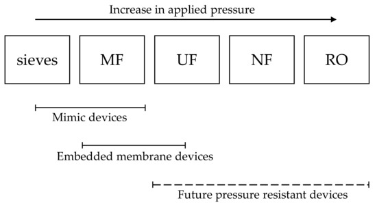
Although microfluidics can be a highly ideal system to study membrane filtration, the results and insights obtained can be of great interest for real membrane processes, mainly microfiltration. Microfiltration is the membrane process that can be most easily investigated since the current available technology allows the production of devices that have pore/channels sizes that are the closest to the ones present in microfiltration membranes. Additionally, microfiltration processes target mostly micrometer sized particles, and these are easy to model and observe with simple optical microscopy techniques. Ultrafiltration processes can still be investigated with microfluidic devices but producing mimicking microfluidic devices can be challenge due to the resolution of the current available techniques that does not allow the production of channels in the smaller range. Microfluidic devices with embedded membranes can be easily used instead where a piece of ultrafiltration membrane is attached to a microfluidic system. Visualization of the process when dealing with ultrafiltration processes can also be more challenging but still possible. Direct observation of accumulation of proteins for example, can be achieved by using fluorescence microscopy of tagged proteins. Other filtration processes such as nanofiltration and reverse osmosis are still not eligible for investigation studies with microfluidic devices mainly due to the high pressure required in these processes and also due to the limitations on microscopy techniques that currently do not allow in situ and real time observation of small specimens such as salts and small molecules (Figure 3).

Figure 3. Scheme showing which type of microfluidic devices are currently suitable for the study of different porous media—sieves, microfiltration (MF), ultrafiltration (UF), nanofiltration (NF), and reverse osmosis (RO).
2.2. Foulants
2.2. Foulants
The to-be-tested suspension/foulant will influence the outcomes of the experiments greatly, as would also be the case in any membrane separation experiment. While some studies use real suspensions such as blood in their experiments [56][57], most prefer to use model suspensions such as latex beads suspended in water [28][46][58]. Model suspensions contain in general only one solvent and type of particle, and their choice depends on the objective of the experiment. Water is the most used solvent, while the selected suspended particles can be more diverse. If the suspension is used to gain general insights, basically any particle will do, but if the system should somehow reflect the characteristics of a suspension of interest, various particle properties need to be considered: soft or hard, how large, monodisperse or polydisperse, etc.
The choice between soft or hard model particles depends on either the system that is being modelled or the specific characteristics that the study is aiming to assess. Hard particles are suitable, e.g., for studies aiming at modelling filtration behavior in general since the particle size remains constant during the process, thereby limiting the number of variables to be considered (such as compaction and deformation). Soft particles are mostly chosen as models for cells and protein aggregates, and often microgels are used due to their ease of production, and tunable properties. Soft particles are able to modify size and shape during filtration, therefore making their filtration behavior much more complex [19][59], e.g., leading to added flow resistance when pressurized in a filtration cake layer [60].
The size of the particles will also influence the outcome of the experiments and are ideally as close as possible to the final application. For example, the migration of particles in flow highly depends on their size, and thus also the build-up of concentration polarization and cake layer [61][62]. If colloidal interactions during the filtration process are of interest, smaller particles should be preferred; however, in various cases large particles can also show the behavior of interest making observation much easier. The current technological constraints regarding microfluidic device fabrication should also be considered when selecting particle size. At the moment, the fabrication of devices mimicking membranes with channels smaller than 5 µm can still be a challenge and for that reason, when observation of individual particle behavior is the main objective of a study, the use larger particles will be more adequate while smaller particles can still be used for collective behavior observations in larger channels. The minimum particle size to be used can also be limited by the resolution of the optical methods used to observe the particles during the experiments.
From the previous point, it also follows that particle size distribution is a parameter to consider since it can largely influence the observations. The use of both monodisperse and polydisperse particle size distributions for experiments in microfluidic devices have been reported in the literature [37][38][63]. For experiments focusing on process modelling, monodispersed distributions are preferred since they will not bring extra complexity to the system. However, the use of polydisperse model particles brings model systems closer to feed streams as used in industrial applications, and that will contain particles with a wide range of sizes.
References
- Iulianelli, A.; Drioli, E. Membrane engineering: Latest advancements in gas separation and pre-treatment processes, petrochemical industry and refinery, and future perspectives in emerging applications. Fuel Process. Technol. 2020, 206, 106464, doi:10.1016/j.fuproc.2020.106464.
- Bassyouni, M.; Abdel-Aziz, M.; Zoromba, M.S.; Abdel-Hamid, S.; Drioli, E. A review of polymeric nanocomposite membranes for water purification. J. Ind. Eng. Chem. 2019, 73, 19–46, doi:10.1016/j.jiec.2019.01.045.
- Jun, B.-M.; Al-Hamadani, Y.A.; Son, A.; Park, C.M.; Jang, M.; Jang, A.; Kim, N.C.; Yoon, Y. Applications of metal-organic framework based membranes in water purification: A review. Sep. Purif. Technol. 2020, 247, 116947, doi:10.1016/j.seppur.2020.116947.
- Ausri, I.R.; Feygin, E.M.; Cheng, C.Q.; Wang, Y.; Lin, Z.Y. (William); Tang, X. (Shirley) A highly efficient and antifouling microfluidic platform for portable hemodialysis devices. MRS Commun. 2018, 8, 474–479, doi:10.1557/mrc.2018.43.
- Snisarenko, D.; Pavlenko, D.; Stamatialis, D.; Aimar, P.; Causseranda, C.; Bacchin, P. Insight into the transport mechanism of solute removed in dialysis by a membrane with double functionality. Chem. Eng. Res. Des. 2017, 126, 97–108, doi:10.1016/j.cherd.2017.08.017.
- Nazir, A.; Khan, K.; Maan, A.; Zia, R.; Giorno, L.; Schroën, K. Membrane separation technology for the recovery of nutraceuticals from food industrial streams. Trends Food Sci. Technol. 2019, 86, 426–438, doi:10.1016/j.tifs.2019.02.049.
- Brans, G.; Schroën, C.; Van Der Sman, R.; Boom, R. Membrane fractionation of milk: state of the art and challenges. J. Membr. Sci. 2004, 243, 263–272, doi:10.1016/j.memsci.2004.06.029.
- Huang, Y.; MacKenzie, A.; Meteer, L.; Hofmann, R. Evaluation of phosphorus removal from a lake by two drinking water treatment plants. Environ. Technol. 2018, 41, 863–869, doi:10.1080/09593330.2018.1512656.
- Robinson, S.; Bérubé, P. Membrane ageing in full-scale water treatment plants. Water Res. 2019, 169, 115212, doi:10.1016/j.watres.2019.115212.
- Khalid, A.; Aslam, M.; Qyyum, M.A.; Faisal, A.; Khan, A.L.; Ahmed, F.; Lee, M.; Kim, J.; Jang, N.; Chang, I.S.; et al. Membrane separation processes for dehydration of bioethanol from fermentation broths: Recent developments, challenges, and prospects. Renew. Sustain. Energy Rev. 2019, 105, 427–443, doi:10.1016/j.rser.2019.02.002.
- Guo, W.; Ngo, H.-H.; Li, J. A mini-review on membrane fouling. Bioresour. Technol. 2012, 122, 27–34, doi:10.1016/j.biortech.2012.04.089.
- Chen, J.C.; Li, Q.; Elimelech, M. In situ monitoring techniques for concentration polarization and fouling phenomena in membrane filtration. Adv. Colloid Interface Sci. 2004, 107, 83–108, doi:10.1016/j.cis.2003.10.018.
- Keil, F.J. Process intensification. Rev. Chem. Eng. 2018, 34, 135–200, doi:10.1515/revce-2017-0085.
- Schroën, K.; De Ruiter, J.; Berton-Carabin, C.C. Microtechnological Tools to Achieve Sustainable Food Processes, Products, and Ingredients. Food Eng. Rev. 2020, 12, 101–120, doi:10.1007/s12393-020-09212-5.
- Fu, Y.; Liu, W.; Soladoye, O.P. Towards potato protein utilisation: Insights into separation, functionality and bioactivity of patatin. Int. J. Food Sci. Technol. 2019, 55, 2314–2322, doi:10.1111/ijfs.14343.
- Kaur, N.; Sharma, P.; Jaimni, S.; Kehinde, B.A.; Kaur, S. Recent developments in purification techniques and industrial applications for whey valorization: A review. Chem. Eng. Commun. 2019, 207, 123–138, doi:10.1080/00986445.2019.1573169.
- Kashani, M.N.; Kriel, F.H.; Binder, C.; Priest, C. Analysis of co-flowing immiscible liquid streams and their interfaces in a high-throughput solvent extraction chip. Microfluid. Nanofluidics 2020, 24, 1–10, doi:10.1007/s10404-020-2320-0.
- Browne, C.A.; Shih, A.; Datta, S.S. Pore-Scale Flow Characterization of Polymer Solutions in Microfluidic Porous Media. Small 2019, 16, e1903944, doi:10.1002/smll.201903944.
- Linkhorst, J.; Rabe, J.; Hirschwald, L.T.; Kuehne, A.J.C.; Wessling, M. Direct Observation of Deformation in Microgel Filtration. Sci. Rep. 2019, 9, 1–7, doi:10.1038/s41598-019-55516-w.
- De Aguiar, I.B.; Van De Laar, T.; Meireles, M.; Bouchoux, A.; Sprakel, J.; Schroën, K. Deswelling and deformation of microgels in concentrated packings. Sci. Rep. 2017, 7, 1–11, doi:10.1038/s41598-017-10788-y.
- Pappas, D. Microfluidics and cancer analysis: cell separation, cell/tissue culture, cell mechanics, and integrated analysis systems. Anal. 2016, 141, 525–535, doi:10.1039/c5an01778e.
- Shen, S.; Ma, C.; Zhao, L.; Wang, Y.; Wang, J.-C.; Xu, J.; Li, T.; Pang, L.; Shen, S. High-throughput rare cell separation from blood samples using steric hindrance and inertial microfluidics. Lab. a Chip 2014, 14, 2525–2538, doi:10.1039/c3lc51384j.
- Dressaire, E.; Sauret, A. Clogging of microfluidic systems. Soft Matter 2017, 13, 37–48, doi:10.1039/c6sm01879c.
- Wei, H.; Chueh, B.-H.; Wu, H.; Hall, E.W.; Li, C.-W.; Schirhagl, R.; Lin, J.-M.; Zare, R.N. Particle sorting using a porous membrane in a microfluidic device. Lab. a Chip 2011, 11, 238–245, doi:10.1039/c0lc00121j.
- Warkiani, M.E.; Wicaksana, F.; Fane, A.G.; Gong, H.-Q. Investigation of membrane fouling at the microscale using isopore filters. Microfluid. Nanofluidics 2014, 19, 307–315, doi:10.1007/s10404-014-1538-0.
- Ngene, I.S.; Lammertink, R.G.; Wessling, M.; Van Der Meer, W.G.J. A microfluidic membrane chip for in situ fouling characterization. J. Membr. Sci. 2010, 346, 202–207, doi:10.1016/j.memsci.2009.09.035.
- Alfadhel, K.A.; Kothare, M.V. Microfluidic modeling and simulation of flow in membrane microreactors. Chem. Eng. Sci. 2005, 60, 2911–2926, doi:10.1016/j.ces.2005.01.013.
- Linkhorst, J.; Beckmann, T.; Go, D.; Kuehne, A.J.C.; Wessling, M. Microfluidic colloid filtration. Sci. Rep. 2016, 6, 22376, doi:10.1038/srep22376.
- Bacchin, P.; Derekx, Q.; Veyret, D.; Glucina, K.; Moulin, P. Clogging of microporous channels networks: role of connectivity and tortuosity. Microfluid. Nanofluidics 2013, 17, 85–96, doi:10.1007/s10404-013-1288-4.
- De Aguiar, I.B.; Meireles, M.; Bouchoux, A.; Schroën, K. Microfluidic model systems used to emulate processes occurring during soft particle filtration. Sci. Rep. 2019, 9, 3063, doi:10.1038/s41598-019-39820-z.
- Hu, G.; Li, D. Multiscale phenomena in microfluidics and nanofluidics. Chem. Eng. Sci. 2007, 62, 3443–3454, doi:10.1016/j.ces.2006.11.058.
- Van Zwieten, R.; Van De Laar, T.; Sprakel, J.; Schroën, K. From cooperative to uncorrelated clogging in cross-flow microfluidic membranes. Sci. Rep. 2018, 8, 1–10, doi:10.1038/s41598-018-24088-6.
- Creative Commons Attribution 4.0 International License. Available online: http://creativecommons.org/licenses/by/4.0/ (accessed on 22 September 2020 ).
- Van De Laar, T.; Klooster, S.T.; Schroën, K.; Sprakel, J. Transition-state theory predicts clogging at the microscale. Sci. Rep. 2016, 6, 28450, doi:10.1038/srep28450.
- De Aguiar, I.B.; Meireles, M.; Bouchoux, A.; Schroën, K. Conformational changes influence clogging behavior of micrometer-sized microgels in idealized multiple constrictions. Sci. Rep. 2019, 9, 1–9, doi:10.1038/s41598-019-45791-y.
- Van De Laar, T. Sticky, Squishy & Stuck: A soft matter approach to membrane failure. Ph.D. Thesis, Wageningen University and Research, Wageningen, The Netherlands, May 2018.
- Ngene, I.S.; Lammertink, R.G.H.; Wessling, M.; Van Der Meer, W.G. Visual characterization of fouling with bidisperse solution. J. Membr. Sci. 2011, 368, 110–115, doi:10.1016/j.memsci.2010.11.026.
- Mustin, B.; Stoeber, B. Deposition of particles from polydisperse suspensions in microfluidic systems. Microfluid. Nanofluidics 2010, 9, 905–913, doi:10.1007/s10404-010-0613-4.
- Kim, H.-S.; Michielsen, S.; Denhartog, E. New wicking measurement system to mimic human sweating phenomena with continuous microfluidic flow. J. Mater. Sci. 2020, 55, 7816–7832, doi:10.1007/s10853-020-04543-4.
- Zhang, S.; Cagney, N.; Lacassagne, T.; Balabani, S.; Naveira-Cotta, C.P.; Tiwari, M.K. Mixing in flows past confined microfluidic cylinders: Effects of pin and fluid interface offsetting. Chem. Eng. J. 2020, 397, 125358, doi:10.1016/j.cej.2020.125358.
- Dudek, M.; Fernandes, D.; Herø, E.H.; Øye, G. Microfluidic method for determining drop-drop coalescence and contact times in flow. Colloids Surfaces A: Physicochem. Eng. Asp. 2020, 586, 124265, doi:10.1016/j.colsurfa.2019.124265.
- Hanson, C.; Sieverts, M.; Tew, K.; Dykes, A.; Salisbury, M.; Vargis, E. In The use of microfluidics and dielectrophoresis for separation, concentration, and identification of bacteria. In Proceedings of the SPIE BiOS, San Fransico, CA, USA, 21 March 2016.
- Jimenez, M.; Bridle, H. Angry pathogens, how to get rid of them: introducing microfluidics for waterborne pathogen separation to children. Lab. a Chip 2015, 15, 947–957, doi:10.1039/c4lc00944d.
- Chen, X.; Shen, J. Review of membranes in microfluidics. J. Chem. Technol. Biotechnol. 2016, 92, 271–282, doi:10.1002/jctb.5105.
- De Jong, J.; Lammertink, R.G.H.; Wessling, M. Membranes and microfluidics: a review. Lab. a Chip 2006, 6, 1125–1139, doi:10.1039/b603275c.
- Debnath, N.; Sadrzadeh, M. Microfluidic Mimic for Colloid Membrane Filtration: A Review. J. Indian Inst. Sci. 2018, 98, 137–157, doi:10.1007/s41745-018-0071-7.
- Gerami, A.; AlZahid, Y.; Mostaghimi, P.; Kashaninejad, N.; Kazemifar, F.; Amirian, T.; Mosavat, N.; Warkiani, M.E.; Armstrong, R.T. Microfluidics for Porous Systems: Fabrication, Microscopy and Applications. Transp. Porous Media 2018, 130, 277–304, doi:10.1007/s11242-018-1202-3.
- Jiao, Y.; Zhao, C.; Kang, Y.; Yang, C. Microfluidics-based fundamental characterization of external concentration polarization in forward osmosis. Microfluid. Nanofluidics 2019, 23, 36, doi:10.1007/s10404-019-2202-5.
- Chen, V.; Li, H.; Fane, A. Non-invasive observation of synthetic membrane processes—a review of methods. J. Membr. Sci. 2004, 241, 23–44, doi:10.1016/j.memsci.2004.04.029.
- Agbangla, G.C.; Climent, E.; Bacchin, P. Experimental investigation of pore clogging by microparticles: Evidence for a critical flux density of particle yielding arches and deposits. Sep. Purif. Technol. 2012, 101, 42–48, doi:10.1016/j.seppur.2012.09.011.
- Bacchin, P.; Marty, A.; Duru, P.; Meireles, M.; Aimar, P. Colloidal surface interactions and membrane fouling: Investigations at pore scale. Adv. Colloid Interface Sci. 2011, 164, 2–11, doi:10.1016/j.cis.2010.10.005.
- Maitri, R.V.; De, S.; Koesen, S.P.; Wyss, H.M.; Van Der Schaaf, J.; Kuipers, J.A.M.; Padding, J.T.; Peters, E.A.J.F. (Frank) Effect of microchannel structure and fluid properties on non-inertial particle migration. Soft Matter 2019, 15, 2648–2656, doi:10.1039/c8sm02348d.
- Debnath, N.; Kumar, A.; Thundat, T.; Sadrzadeh, M. Investigating fouling at the pore-scale using a microfluidic membrane mimic filtration system. Sci. Rep. 2019, 9, 1–10, doi:10.1038/s41598-019-47096-6.
- Decock, J.; Schlenk, M.; Salmon, J.-B. In situphoto-patterning of pressure-resistant hydrogel membranes with controlled permeabilities in PEGDA microfluidic channels. Lab a Chip 2018, 18, 1075–1083, doi:10.1039/c7lc01342f.
- Nguyen, H.-T.; Massino, M.; Keita, C.; Salmon, J.-B. Microfluidic dialysis using photo-patterned hydrogel membranes in PDMS chips. Lab. a Chip 2020, 20, 2383–2393, doi:10.1039/d0lc00279h.
- Chen, X.; Cui, D.; Liu, C.; Li, H. Microfluidic chip for blood cell separation and collection based on crossflow filtration. Sensors Actuators B: Chem. 2008, 130, 216–221, doi:10.1016/j.snb.2007.07.126.
- Kim, C.H.; Park, J.; Kim, S.J.; Ko, D.-H.; Lee, S.H.; Lee, S.J.; Park, J.-K.; Lee, M.-K. On-site extraction and purification of bacterial nucleic acids from blood samples using an unpowered microfluidic device. Sensors Actuators B: Chem. 2020, 320, 128346, doi:10.1016/j.snb.2020.128346.
- Wiese, M.; Malkomes, C.; Krause, B.; Wessling, M. Flow and filtration imaging of single use sterile membrane filters. J. Membr. Sci. 2018, 552, 274–285, doi:10.1016/j.memsci.2018.02.002.
- Choi, G.; Nouri, R.; Zarzar, L.; Guan, W. Microfluidic deformability-activated sorting of single particles. Microsystems Nanoeng. 2020, 6, 1–9, doi:10.1038/s41378-019-0107-9.
- De Aguiar, I.B.; Schroën, K.; Meireles, M.; Bouchoux, A. Compressive resistance of granular-scale microgels: From loose to dense packing. Colloids Surfaces A: Physicochem. Eng. Asp. 2018, 553, 406–416, doi:10.1016/j.colsurfa.2018.05.064.
- Schroën, K.; Van Dinther, A.; Stockmann, R. Particle migration in laminar shear fields: A new basis for large scale separation technology? Sep. Purif. Technol. 2017, 174, 372–388, doi:10.1016/j.seppur.2016.10.057.
- Davis, R.H. Modeling of Fouling of Crossflow Microfiltration Membranes. Sep. Purif. Methods 1992, 21, 75–126, doi:10.1080/03602549208021420.
- Cejas, C.M.; Maini, L.; Monti, F.; Tabeling, P. Deposition kinetics of bi- and tridisperse colloidal suspensions in microchannels under the van der Waals regime. Soft Matter 2019, 15, 7438–7447, doi:10.1039/c9sm01098j.
