Fundamental Knowledge of the Biochemical Properties of 1O2: Comparison
Please note this is a comparison between Version 2 by Jason Zhu and Version 1 by Junichi Fujii.

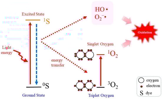
Energy transfer to ground state triplet molecular oxygen results in the generation of singlet molecular oxygen (1O2), which has potent oxidizing ability. Irradiation of light, notably ultraviolet A, to a photosensitizing molecule results in the generation of 1O2, which is thought to play a role in causing skin damage and aging. It should also be noted that 1O2 is a dominant tumoricidal component that is generated during the photodynamic therapy (PDT). While type II photodynamic action generates not only 1O2 but also other reactive species, endoperoxides release pure 1O2 upon mild exposure to heat and, hence, are considered to be beneficial compounds for research purposes. Concerning target molecules, 1O2 preferentially reacts with unsaturated fatty acids to produce lipid peroxidation. Enzymes that contain a reactive cysteine group at the catalytic center are vulnerable to 1O2 exposure. Guanine base in nucleic acids is also susceptible to oxidative modification, and cells carrying DNA with oxidized guanine units may experience mutations. 

  • photodynamic therapy
  • ultraviolet
  • endoperoxides

1. Introduction

Interactions of oxygen molecules with electrons leaked from enzymatic and non-enzymatic processes produces reactive oxygen species (ROS), such as superoxide (O2•) and hydrogen peroxides (H2O2) [1,2,3][1][2][3]. Hydroxyl radicals (HO•) are the most reactive type of ROS and are likely produced by the reaction of H2O2 and ferrous iron, via the so called Fenton reaction [4], although there is some debate as to which ROS are the primary products in this reaction [5,6][5][6]. Singlet molecular oxygen (1O2), is a high-energy oxygen species but possesses unique properties that are different from other ROS [7,8,9][7][8][9]. While most ROS are produced by electron transfer reactions, 1O2 is generated by the transfer of energy to the ground state, triplet molecular oxygen (3O2), the most abundant oxygen molecule in atmosphere. The type II photodynamic reaction promoted by the presence of photosensitizing molecules is widely employed to generate 1O2 in biological systems [10,11][10][11]. However, the problem with the photodynamic action is that other ROS are also generated as byproducts [12,13][12][13]. This methodological issue appears to have hindered progress in research on the biological effects of 1O2, despite its importance.
1O2 possesses high energy and is considered to be a major cause for skin damage induced by ultraviolet (UV) irradiation [14,15][14][15]. Meanwhile, due to its strong cytotoxicity, 1O2 is the molecule that is responsible for killing tumor cells during photodynamic therapy (PDT) [10,11][10][11]. 1O2 as well as HO• preferentially reacts with conjugated double bonds, and hence polyunsaturated fatty acids (PUFA), which are dominantly present in the form of phospholipids in the cell membrane, are likely targets [9]. It is known that, upon mitotic stimuli, a small amount of ROS, notably H2O2, is produced, and this species modulates phosphorylation-mediated signaling pathways [16,17][16][17]. While signal modulation by H2O2 involves the transient oxidation of cysteine (Cys), reactions with 1O2 tend to result in irreversible oxidation. In most cases, exposure to 1O2 impairs cellular function but also occasionally stimulates tumorigenic cell growth [18,19][18][19]. Concerning cell death, results reported in many studies indicate that the apoptotic pathway is activated by 1O2 [20,21,22][20][21][22]. However, recent studies suggest that ferroptosis, an iron-dependent necrotic cell death [23[23][24],24], is also involved in 1O2-promoted cell death [25,26][25][26].

2. Properties of 1O2 as a Potent Oxidant

Oxidative stress is induced by either the production of large amounts of ROS or an insufficient amount of antioxidants which include enzymes that eliminate ROS or small antioxidant compounds, such as glutathione (GSH), carotenoids and tocopherols [2]. Electrons that are leaked from enzymatic and non-enzymatic reactions initiate the generation of ROS, as represented by O2•, H2O2 and HO•, and hence, the radical electron plays pivotal roles in the development of oxidative stress in many situations [1,3][1][3]. However, 1O2 is generated when the oxygen molecule in the ground triplet state 3O2 is excited by receiving energy without the transfer of an electron. The 1O2-generating system involves enzymatic reactions, such as myeloperoxidase, lipoxygenase and cyclooxygenase as well as chemical reactions, such as O2•-mediated GSH oxidation and the interaction of peroxides with hypochlorite or peroxynitrite [13,28,29,30][13][27][28][29]. Photoaging and PDT are the subjects that have been most extensively investigated in terms of 1O2–mediated reactions that are associated with human physiology and the pathogenesis of related diseases. In spite of the high oxidizing power, reactions of 1O2 are thought to exert only limited effects compared to those of HO• in biological systems.
HO• is considered to be the most reactive ROS and appears to be responsible for a variety of pathological conditions. However, the half-life of HO• is quite short (10 nsec), so it only reacts with molecules that are in close proximity to the site where it is generated. On the other hand, the half-life of 1O2 is approximately 4 µsec in aqueous solution, which allows it to diffuse 150–220 nm [31,32][30][31]. Thus, 1O2 may react at various locations beyond where it is generated and, therefore, can affect surrounding molecules and organelles more widely compared to HO•. Nevertheless, this distance is insufficient for extracellularly produced 1O2 to move to the interior of a cell. Therefore, 1O2 that is generated inside the cell has the ability to damage various cellular components including DNA and organelles.

3. Chemical Probes for Detecting 1O2

Analyses employing a cell biological approach are essential for answering basic questions as to which part of the cell produces 1O2 in photoaging and during PDT and how cellular responses proceed in such situations. For that purpose, the use of a fluorescent chemical probe is the most convenient approach. 1O2 sensor green (SOSG) is a prototype that is popularly used in studies for detecting 1O2, although it has some disadvantages such as lack in membrane permeability [33][32]. Other compounds have been designed to overcome the disadvantage of SOSG. For example, Aarhus Sensor Green, which is a tetrafluoro-substituted fluorescein derivative that is covalently linked to a 9,10-diphenyl anthracene moiety [34][33] and the classic indocyanine green probe may also be applicable for this objective in certain experiments [35][34]. To increase the cellular delivery of SOSG, biocompatible nanosensors, with SOSG encapsulated within their hydrophobic core, have been developed, and these modifications appear to improve its delivery [36,37,38][35][36][37].
The cell membrane permeable far-red fluorescence probe Si-DMA, which is composed of silicon-containing rhodamine and anthracene moieties as a chromophore, has also been developed [39][38]. Upon reaction with 1O2, Si-DMA is converted into an endoperoxide at the anthracene moiety that emits strongly. The use of Si-DMA reportedly enables the visualization of 1O2 generated in a single mitochondrial tubule during PDT. After the treatment of cells with the endoperoxide, dose-dependent increases in fluorescence of Si-DMA were observed [40][39]. Thus, these results suggest that chemical probes may be applicable for studies concerning the cellular effects of 1O2. This compound is now commercially available. Another compound, a rhodamine 6G-aminomethylanthracene-linked donor–acceptor molecule (RA), was reported to exhibit unique properties [41][40]. RA acts as a fluorogenic 1O2 sensor molecule and also acts as a photosensitizer to generate 1O2 upon exposure to green light. Other fluorescent reagents, such as one based on an aminocoumarin-methylanthracene-based electron donor–acceptor molecule [42][41], are also being developed. Since information on the use of these newly developed probes is currently limited, it is necessary to carefully choose which compounds are suitable for the intended research.

4. Photodynamic Reaction as a 1O2 -Generating System

PDT mainly contributes to 1O2 generation in biological systems through type II mechanisms that involve energy transfer from triplet excited molecules to triplet oxygen. Photosensitizers may also act according to competitive type I photosensitization mechanism that mostly involves charge transfer between suitable targets and a photosensitizer in its triplet excited state [43,44][42][43]. In order to protect cells against the deteriorating action of UV light, the effects of 1O2 on skin tissues have been extensively investigated. In the meantime, 1O2 is considered to be the main molecule that promotes cytotoxic processes during PDT, and hence, multiple studies are currently underway with the aim of understanding the mechanism responsible for 1O2-mediated cell death and developing efficient photosensitizers [8,10][8][10]. UV radiation causes skin photoaging and oxidatively generated damage to dermal cells and is especially troublesome in cases of sunburn which occurs by exposure to excessive UV for long periods of time [14,45][14][44]. UVB (280–315 nm) comprises approximately 5% of the solar UV and causes the direct photodamage to many molecules including DNA and proteins in skin tissues through its high energy photochemical reactions. Genetic damage caused by the oxidative modification of DNA and other molecules emerges in a short time after exposure to UVB. In the case of UVA (315–400 nm) that accounts for approximately 95% of the solar UV, cellular damage occurs through the activation of chromophores that act as photosensitizers to generate 1O2 and other ROS, and hence, the oxidative reaction proceeds indirectly via the ROS that are generated. It is rather difficult to determine if changes in cells that have been exposed to UVA are the consequence of the generation of either 1O2 or other ROS because they are produced simultaneously by the photodynamic reactions and result in essentially the same end products [46][45].
In order to elucidate the reactions caused by 1O2, reliable methods for generating 1O are required [12]. The most common method for this purpose is irradiation of the photosensitizer with UV or visible light because it is simple and easy to control its production [27,47][46][47]. Figure 1 represents the “Jablonski diagram” that depicts conceptual images of the generation of 1O2 by the irradiation of a photosensitizing molecule (S) with light followed by transferring energy to 3O2 [48]. When a photosensitizer is exposed to light, most likely the UVA in natural light, photon energy converts the photosensitizer in the ground state (0S) to that in the excited state (1S). On returning to the ground state 0S, a part of the released photoenergy can be transferred to 3O2, which results in the electron spin state being altered and the generation of 1O2. Under this situation, photodynamic action generates not only 1O2 but also other ROS such as O2• and HO• [8].
Figure 1. Photodynamic 1O2 generation. Photoirradiation of a photosensitizing molecule in the ground state (0S) leads the production of the excited form (1S). On returning to the ground state, energy is transferred to 3O2, which becomes excited to 1O2. In the meantime, however, other ROS such as O2• and HO• may also be produced.
To observe cellular responses to 1O2, cell-permeable and non-cytotoxic compounds need to be used as the photosensitizer. For example, Rose Bengal and methylene blue meet the conditions and, hence, are popularly used for the purpose of examining biological action of 1O2 [47]. Since PDT is a useful therapeutic for eliminating tumors, many attempts have been made to improve the treatment by developing convenient photosensitizers [11,27,49][11][46][49].

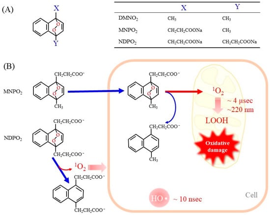
5. Endoperoxides as Donor Compounds for Generating Pure 1O2

Endoperoxides that release 1O2 without other ROS have been developed to evaluate its unique reaction [13,50][13][50]. Naphthalene endoperoxide-based 1O2 donor compounds were first developed, and the structure–function relationships of the compounds have been described in detail in a review article [13]. Consequently, several naphthalene endoperoxides have been established and utilized for the in vitro evaluation of the effects of 1O2, as representative structures in Figure 2A. Upon mild heating at 37 °C, the endoperoxides spontaneously release pure 1O2, which then directly reacts with surrounding compounds and organelles. Heat-labile endoperoxides are considered clean sources of 1O2 for highly specific oxidation of cellular biomolecules and have been applied for 1O2-mediated oxidation of the DNA guanine base in cells [51,52][51][52].
Figure 2. Representative endoperoxides and the release of 1O2. (A) The general structure of naphthalene endoperoxides is shown on the left. The table on the right shows the chemical groups attached to the naphthalene ring. DMNO2, 1,4-dimethylnaphthalene endoperoxide; MNPO2, 1-methylnaphthalene-4-propanonate endoperoxide; NDPO2, 1,4-naphthalenedipropanoate endoperoxide. (B) Endoperoxides represented by MNPE and NDPE generate 1O2 spontaneously at physiologic temperature 37 °C. MNPE, which is relatively hydrophobic, can enter the cell, but NDPE cannot cross the cell membrane. The short life of 1O2 (~4 µsec) makes it diffuse only 150–220 nm in aqueous solution. As a result, 1O2 released from NDPE is present outside the cell only, while 1O2 from MNPE can act inside the cell. For reference, hydroxy radicals are also shown.
Here discuss the advantages and disadvantages of 1O2 donor compounds in comparison with the photodynamic action. The benefits include the following: (1) Endoperoxides produce pure 1O2. (2) The concentrations of the released 1O2 are easily controlled. (3) Heating at physiological temperature, generally under cell culture conditions, can promote the release of 1O2 from endoperoxides. (4) It may be possible to design endoperoxides that are localized to a specific organelle by appropriate chemical modification of the compounds. Limitations include the following: (1) A high concentration of endoperoxides is required to generate sufficient levels of 1O2. (2) It is essential to consider effects of the raw material after the release of 1O2 because they are sometimes toxic to cells. (3) Endoperoxides may not be evenly distributed inside cells due to their chemical nature. (4) The amount of 1O2 released is initially maximal but then gradually decreases with increasing endoperoxide consumption.
When endoperoxides are applied to a cell culture system, it is necessary to use compounds that are able to pass through the cell membrane. In fact, the side chains of naphthalene endoperoxides determine whether they enter cells or remain outside of cells (Figure 2B) [53]. 1-Methylnaphthalene-4-propanonate endoperoxide (MNPO2) is cell membrane-permeable and generates 1O2 within cells. However, 1,4-naphthalenedipropanoate endoperoxide (NDPO2) cannot enter cells. Accordingly, while MNPO2 induces cell damage, NDPO2 at the same concentration has no effects, although both compounds trigger the release of cyt c from isolated mitochondria to a similar extent [53].
While the generation of 1O2 by a light-irradiated photosensitizer is frequently used, the use of 1O2 donor compounds has been limited because they are complex molecules that are difficult to synthesize. Some naphthalene endoperoxides are now commercially available. New compounds other than naphthalene-based endoperoxides are also being developed. For the efficient delivery of a 1O2 donor to cancer cells, a porphyrin-based covalent organic framework that contains a naphthalene endoperoxide has also been developed [54]. Trials to develop new types of endoperoxides, which are based on 2-pyridone and anthracene, are also underway [50]. Two 1O2-producing systems, photodynamic reactions and naphthalene endoperoxides, have provided rather consistent results so far [13], implying that the contribution of other byproducts may be negligible.

6. Natural or Synthetic 1O2-Scavenging Compounds

The body is protected from oxidatively generated damage by a variety of natural and synthetic compounds that scavenge 1O2. The quantitative evaluation of the 1O2-scavenging ability of a compound provides useful information not only for basic research but also for developing functional foods and medicines concerning antioxidation [55]. Many nutritional compounds, such as tocopherols, carotenoids and flavonoids, possess antioxidant capacity and protect susceptible molecules from 1O2. The oxygen radical absorption capacity (ORAC) assay is a representative method for the detection of 1O2-scavenging ability of food ingredients [56]. Thereafter, a simple method called a singlet oxygen absorption capacity (SOAC) assay has been established for the evaluation of 1O2-scavenging ability [56,57][56][57]. These methods are useful in exploring popularly used 1O2-scavenging compounds, especially in the field of food chemistry.
While carotenoids react with 1O2 more rapidly than α-tocopherol, in a nearly diffusion-limited manner (~1010 M−1s−1) [58[58][59],59], lycopene, which is found in fruits and vegetables such as tomato, is one of the strongest natural carotenoids [60]. By employing the SOAC assay method, carotenoids have been found to quench 1O2 approximately 30–100 times faster than α-tocopherol [57]. After transferring excitation energy to carotenoids, 1O2 returns to the ground state. The excited carotenoids spontaneously release thermal energy and then return to the ground state. Hence, carotenoids are spontaneously recycled and have the advantage of quenching 1O2 without affecting other molecules. This chemical property of 1O2 is a major difference from other radicals that are generated by electron transfer reactions that require another radical to quench.
Based on in vitro data on the action of anti-oxidants, the biological benefit of carotenoids has also been examined by some studies on humans. The administration of a representative carotenoid β-carotene to humans failed to alleviate sunburn reactions under the conditions used [61]. However, a later study reported that carotenoids, β-carotene and lycopene effectively protect erythema formation induced using a solar light simulator [62,63][62][63]. Lycopene has also attracted attention as a nutrient with anticancer effects [64]. For another example, lutein is a xanthophyll carotenoid that is found in foods such as dark green leafy vegetables and exhibits strong antioxidant activity via its ability to scavenge ROS including 1O2 and lipid peroxyl radicals [65]. Lutein appears to exert anti-inflammatory actions against some diseases, including neurodegenerative disorders, eye diseases, cardiovascular diseases and skin diseases.
Many synthetic antioxidant compounds that scavenge ROS including 1O2 have been developed as medicines. Edaravone (3-methyl-1-phenyl-2-pyrazolin-5-one) is a compound that eliminates a variety of radical species and was the first approved compound for use as a medicine for the treatment of acute brain infarctions. Edaravone can scavenge 1O2 that is generated by activated human neutrophils [66] and by photoactivated Rose Bengal [67]. The plasma lipid peroxidation caused by 1O2, however, cannot be suppressed by edaravone and other clinical drugs with antioxidant ability, which include roglitazone, probucol, carvedilol, pentoxifylline and ebselen, although they exhibit suppressive effects on lipid peroxidation caused by free radicals, peroxynitrite, hypochlorite, and lipoxygenase reactions [68]. Because blood plasma contains high concentrations of proteins and many other compounds that could potentially interfere with the scavenging reaction by these chemicals, such biological compounds may have influenced the results.

References

  1. Fridovich, I. Superoxide radical and superoxide dismutases. Annu. Rev. Biochem. 1995, 64, 97–112.
  2. Sies, H.; Berndt, C.; Jones, D.P. Oxidative Stress. Annu. Rev. Biochem. 2017, 86, 715–748.
  3. Fujii, J.; Homma, T.; Osaki, T. Superoxide Radicals in the Execution of Cell Death. Antioxidants 2022, 11, 501.
  4. Tang, Z.; Zhao, P.; Wang, H.; Liu, Y.; Bu, W. Biomedicine Meets Fenton Chemistry. Chem Rev. 2021, 121, 1981–2019.
  5. Enami, S.; Sakamoto, Y.; Colussi, A.J. Fenton chemistry at aqueous interfaces. Proc. Natl. Acad. Sci. USA 2014, 111, 623–628.
  6. Koppenol, W.H.; Hider, R.H. Iron and redox cycling. Do’s and don’ts. Free Radic. Biol. Med. 2019, 133, 3–10.
  7. Petrou, A.L.; Terzidaki, A. A meta-analysis and review examining a possible role for oxidative stress and singlet oxygen in diverse diseases. Biochem. J. 2017, 474, 2713–2731.
  8. Di Mascio, P.; Martinez, G.R.; Miyamoto, S.; Ronsein, G.E.; Medeiros, M.H.G.; Cadet, J. Singlet Molecular Oxygen Reactions with Nucleic Acids, Lipids, and Proteins. Chem. Rev. 2019, 119, 2043–2086.
  9. Murotomi, K.; Umeno, A.; Shichiri, M.; Tanito, M.; Yoshida, Y. Significance of Singlet Oxygen Molecule in Pathologies. Int. J. Mol. Sci. 2023, 24, 2739.
  10. Ahmad, N.; Mukhtar, H. Mechanism of photodynamic therapy-induced cell death. Methods Enzymol. 2000, 319, 342–358.
  11. Chilakamarthi, U.; Giribabu, L. Photodynamic Therapy: Past, Present and Future. Chem. Rec. 2017, 17, 775–802.
  12. Garcia-Diaz, M.; Huang, Y.Y.; Hamblin, M.R. Use of fluorescent probes for ROS to tease apart Type I and Type II photochemical pathways in photodynamic therapy. Methods 2016, 109, 158–166.
  13. Pierlot, C.; Aubry, J.M.; Briviba, K.; Sies, H.; Di Mascio, P. Naphthalene endoperoxides as generators of singlet oxygen in biological media. Methods Enzymol. 2000, 319, 3–20.
  14. Krutmann, J. Ultraviolet A radiation-induced biological effects in human skin: Relevance for photoaging and photodermatosis. J. Dermatol. Sci. 2000, 23 (Suppl. S1), S22–S26.
  15. Tyrrell, R.M. Solar ultraviolet A radiation: An oxidizing skin carcinogen that activates heme oxygenase-1. Antioxid. Redox Signal. 2004, 6, 835–840.
  16. Rhee, S.G. Cell signaling. H2O2, a necessary evil for cell signaling. Science 2006, 312, 1882–1883.
  17. Finkel, T. Signal transduction by reactive oxygen species. J. Cell Biol. 2011, 194, 7–15.
  18. Zhuang, S.; Kochevar, I.E. Singlet oxygen-induced activation of Akt/protein kinase B is independent of growth factor receptors. Photochem. Photobiol. 2003, 78, 361–371.
  19. Le Panse, R.; Dubertret, L.; Coulomb, B. p38 mitogen-activated protein kinase activation by ultraviolet A radiation in human dermal fibroblasts. Photochem. Photobiol. 2003, 78, 168–174.
  20. Morita, A.; Werfel, T.; Stege, H.; Ahrens, C.; Karmann, K.; Grewe, M.; Grether-Beck, S.; Ruzicka, T.; Kapp, A.; Klotz, L.O.; et al. Evidence that singlet oxygen-induced human T helper cell apoptosis is the basic mechanism of ultraviolet-A radiation phototherapy. J. Exp. Med. 1997, 186, 1763–1768.
  21. Klotz, L.O.; Kröncke, K.D.; Sies, H. Singlet oxygen-induced signaling effects in mammalian cells. Photochem. Photobiol. Sci. 2003, 2, 88–94.
  22. Novikova, I.N.; Potapova, E.V.; Dremin, V.V.; Dunaev, A.V.; Abramov, A.Y. Laser-induced singlet oxygen selectively triggers oscillatory mitochondrial permeability transition and apoptosis in melanoma cell lines. Life Sci. 2022, 304, 120720.
  23. Dixon, S.J.; Lemberg, K.M.; Lamprecht, M.R.; Skouta, R.; Zaitsev, E.M.; Gleason, C.E.; Patel, D.N.; Bauer, A.J.; Cantley, A.M.; Yang, W.S.; et al. Ferroptosis: An iron-dependent form of nonapoptotic cell death. Cell 2012, 149, 1060–1072.
  24. Stockwell, B.R. Ferroptosis turns 10: Emerging mechanisms, physiological functions, and therapeutic applications. Cell 2022, 185, 2401–2421.
  25. Homma, T.; Kobayashi, S.; Fujii, J. Induction of ferroptosis by singlet oxygen generated from naphthalene endoperoxide. Biochem. Biophys. Res. Commun. 2019, 518, 519–525.
  26. Meng, X.; Deng, J.; Liu, F.; Guo, T.; Liu, M.; Dai, P.; Fan, A.; Wang, Z.; Zhao, Y. Triggered All-Active Metal Organic Framework: Ferroptosis Machinery Contributes to the Apoptotic Photodynamic Antitumor Therapy. Nano Lett. 2019, 19, 7866–7876.
  27. Wefers, H.; Sies, H. Oxidation of glutathione by the superoxide radical to the disulfide and the sulfonate yielding singlet oxygen. Eur. J. Biochem. 1983, 137, 29–36.
  28. Di Mascio, P.; Briviba, K.; Sasaki, S.T.; Catalani, L.H.; Medeiros, M.H.; Bechara, E.J.; Sies, H. The reaction of peroxynitrite with tert-butyl hydroperoxide produces singlet molecular oxygen. Biol. Chem. 1997, 378, 1071–1074.
  29. Miyamoto, S.; Martinez, G.R.; Medeiros, M.H.; Di Mascio, P. Singlet molecular oxygen generated by biological hydroperoxides. J. Photochem. Photobiol. B. 2014, 139, 24–33.
  30. Redmond, R.W.; Kochevar, I.E. Spatially resolved cellular responses to singlet oxygen. Photochem. Photobiol. 2006, 82, 1178–1186.
  31. Jiménez-Banzo, A.; Sagristà, M.L.; Mora, M.; Nonell, S. Kinetics of singlet oxygen photosensitization in human skin fibroblasts. Free Radic. Biol. Med. 2008, 44, 1926–1934.
  32. Driever, S.M.; Fryer, M.J.; Mullineaux, P.M.; Baker, N.R. Imaging of reactive oxygen species in vivo. Methods Mol. Biol. 2009, 479, 109–116.
  33. Pedersen, S.K.; Holmehave, J.; Blaikie, F.H.; Gollmer, A.; Breitenbach, T.; Jensen, H.H.; Ogilby, P.R. Aarhus sensor green: A fluorescent probe for singlet oxygen. J. Org. Chem. 2014, 79, 3079–3087.
  34. Tang, C.Y.; Wu, F.Y.; Yang, M.K.; Guo, Y.M.; Lu, G.H.; Yang, Y.H. A Classic Near-Infrared Probe Indocyanine Green for Detecting Singlet Oxygen. Int. J. Mol. Sci. 2016, 17, 219.
  35. Ruiz-González, R.; Bresolí-Obach, R.; Gulías, Ò.; Agut, M.; Savoie, H.; Boyle, R.W.; Nonell, S.; Giuntini, F. NanoSOSG: A Nanostructured Fluorescent Probe for the Detection of Intracellular Singlet Oxygen. Angew Chem. Int. Ed. Engl. 2017, 56, 2885–2888.
  36. Chen, B.; Yang, Y.; Wang, Y.; Yan, Y.; Wang, Z.; Yin, Q.; Zhang, Q.; Wang, Y. ACS Precise Monitoring of Singlet Oxygen in Specific Endocytic Organelles by Super-pH-Resolved Nanosensors. Appl. Mater. Interfaces 2021, 13, 18533–18544.
  37. Nath, P.; Hamadna, S.S.; Karamchand, L.; Foster, J.; Kopelman, R.; Amar, J.G.; Ray, A. Intracellular detection of singlet oxygen using fluorescent nanosensors. Analyst 2021, 146, 3933–3941.
  38. Kim, S.; Tachikawa, T.; Fujitsuka, M.; Majima, T. Far-red fluorescence probe for monitoring singlet oxygen during photodynamic therapy. J. Am. Chem. Soc. 2014, 136, 11707–11715.
  39. Murotomi, K.; Umeno, A.; Sugino, S.; Yoshida, Y. Quantitative kinetics of intracellular singlet oxygen generation using a fluorescence probe. Sci. Rep. 2020, 10, 10616.
  40. Zhao, H.; Takano, Y.; Sasikumar, D.; Miyatake, Y.; Biju, V. Excitation-Wavelength-Dependent Functionalities of Temporally Controlled Sensing and Generation of Singlet Oxygen by a Photoexcited State Engineered Rhodamine 6G-Anthracene Conjugate. Chemistry 2022, 28, e202202014.
  41. Sasikumar, D.; Takano, Y.; Zhao, H.; Kohara, R.; Hamada, M.; Kobori, Y.; Biju, V. Caging and photo-triggered uncaging of singlet oxygen by excited state engineering of electron donor-acceptor-linked molecular sensors. Sci. Rep. 2022, 12, 11371.
  42. Baptista, M.S.; Cadet, J.; Di Mascio, P.; Ghogare, A.A.; Greer, A.; Hamblin, M.R.; Lorente, C.; Nunez, S.C.; Ribeiro, M.S.; Thomas, A.H.; et al. Type I and Type II Photosensitized Oxidation Reactions: Guidelines and Mechanistic Pathways. Photochem. Photobiol. 2017, 93, 912–919.
  43. Baptistal, M.S.; Cadet, J.; Greer, A.; Thomas, A.H. Photosensitization Reactions of Biomolecules: Definition, Targets and Mechanisms. Photochem. Photobiol. 2021, 97, 1456–1483.
  44. Girotti, A.W.; Kriska, T. Role of lipid hydroperoxides in photo-oxidative stress signaling. Antioxid. Redox Signal. 2004, 6, 301–310.
  45. Reis, A.; Spickett, C.M. Chemistry of phospholipid oxidation. Biochim. Biophys. Acta 2012, 1818, 2374–2387.
  46. Maharjan, P.S.; Bhattarai, H.K. Singlet Oxygen, Photodynamic Therapy, and Mechanisms of Cancer Cell Death. J. Oncol. 2022, 2022, 7211485.
  47. Kochevar, I.E.; Redmond, R.W. Photosensitized production of singlet oxygen. Methods Enzymol. 2000, 319, 20–28.
  48. Jablonski, A. Efficiency of anti-stokes fluorescence in dyes. Nature 1933, 131, 839–840.
  49. Aziz, B.; Aziz, I.; Khurshid, A.; Raoufi, E.; Esfahani, F.N.; Jalilian, Z.; Mozafari, M.R.; Taghavi, E.; Ikram, M. An Overview of Potential Natural Photosensitizers in Cancer Photodynamic Therapy. Biomedicines 2023, 11, 224.
  50. Wei, L.; Zhang, Z.; Kumar, A.; Banerjee, S.; Huang, H. Endoperoxides Compounds for Highly Efficient Cancer Treatment under Hypoxia. Chemistry 2022, 28, e202202233.
  51. Ravanat, J.L.; Di Mascio, P.; Martinez, G.R.; Medeiros, M.H.; Cadet, J. Singlet oxygen induces oxidation of cellular DNA. J. Biol. Chem. 2000, 275, 40601–40604.
  52. Ravanat, J.L.; Douki, T.; Duez, P.; Gremaud, E.; Herbert, K.; Hofer, T.; Lasserre, L.; Saint-Pierre, C.; Favier, A.; Cadet, J. Cellular background level of 8-oxo-7,8-dihydro-2’-deoxyguanosine: An isotope based method to evaluate artefactual oxidation of DNA during its extraction and subsequent work-up. Carcinogenesis 2002, 23, 1911–1918.
  53. Otsu, K.; Sato, K.; Ikeda, Y.; Imai, H.; Nakagawa, Y.; Ohba, Y.; Fujii, J. An abortive apoptotic pathway induced by singlet oxygen is due to the suppression of caspase activation. Biochem. J. 2005, 389, 197–206.
  54. Lu, F.; Pan, L.; Wu, T.; Pan, W.; Gao, W.; Li, N.; Tang, B. An endoperoxide-containing covalent organic framework as a singlet oxygen reservoir for cancer therapy. Chem. Commun. 2022, 58, 11013–11016.
  55. Gülçin, İ. Antioxidant activity of food constituents: An overview. Arch. Toxicol. 2012, 86, 345–391.
  56. Ou, B.; Hampsch-Woodill, M.; Prior, R.L. Development and validation of an improved oxygen radical absorbance capacity assay using fluorescein as the fluorescent probe. J. Agric. Food Chem. 2001, 49, 4619–4626.
  57. Ouchi, A.; Aizawa, K.; Iwasaki, Y.; Inakuma, T.; Terao, J.; Nagaoka, S.; Mukai, K. Kinetic study of the quenching reaction of singlet oxygen by carotenoids and food extracts in solution. Development of a singlet oxygen absorption capacity (SOAC) assay method. J. Agric. Food Chem. 2010, 58, 9967–9978.
  58. Foote, C.S.; Chang, Y.C.; Denny, R.W. Chemistry of singlet oxygen. X. Carotenoid quenching parallels biological protection. J. Am. Chem. Soc. 1970, 92, 5216–5218.
  59. Edge, R.; Truscott, T.G. Singlet Oxygen and Free Radical Reactions of Retinoids and Carotenoids-A Review. Antioxidants 2018, 7, 5.
  60. Di Mascio, P.; Kaiser, S.; Sies, H. Lycopene as the most efficient biological carotenoid singlet oxygen quencher. Arch. Biochem. Biophys. 1989, 274, 532–538.
  61. Garmyn, M.; Ribaya-Mercado, J.D.; Russel, R.M.; Bhawan, J.; Gilchrest, B.A. Effect of beta-carotene supplementation on the human sunburn reaction. Exp. Dermatol. 1995, 4, 104–111.
  62. Stahl, W.; Sies, H. Carotenoids and protection against solar UV radiation. Skin Pharmacol. Appl. Skin Physiol. 2002, 15, 291–296.
  63. Sies, H.; Stahl, W. Carotenoids and UV protection. Photochem. Photobiol. Sci. 2004, 3, 749–752.
  64. Ozkan, G.; Günal-Köroğlu, D.; Karadag, A.; Capanoglu, E.; Cardoso, S.M.; Al-Omari, B.; Calina, D.; Sharifi-Rad, J.; Cho, W.C. A mechanistic updated overview on lycopene as potential anticancer agent. Biomed. Pharmacother. 2023, 161, 114428.
  65. Ahn, Y.J.; Kim, H. Lutein as a Modulator of Oxidative Stress-Mediated Inflammatory Diseases. Antioxidants 2021, 10, 1448.
  66. Sommani, P.; Arai, T.; Yamashita, K.; Miyoshi, T.; Mori, H.; Sasada, M.; Makino, K. Effects of edaravone on singlet oxygen released from activated human neutrophils. J. Pharmacol. Sci. 2007, 103, 117–120.
  67. Nishinaka, Y.; Mori, H.; Endo, N.; Miyoshi, T.; Yamashita, K.; Adachi, S.; Arai, T. Edaravone directly reacts with singlet oxygen and protects cells from attack. Life Sci. 2010, 86, 808–813.
  68. Morita, M.; Naito, Y.; Yoshikawa, T.; Niki, E. Inhibition of plasma lipid oxidation induced by peroxyl radicals, peroxynitrite, hypochlorite, 15-lipoxygenase, and singlet oxygen by clinical drugs. Bioorg. Med. Chem. Lett. 2016, 26, 5411–5417.
More
Video Production Service