Endoplasmic Reticulum Stress and Unfolded Protein Response Signaling: Comparison
Please note this is a comparison between Version 1 by Hakim Manghwar and Version 3 by Hakim Manghwar.

The endoplasmic reticulum (ER) is an important subcellular organelle, primarily recognized as a checkpoint for protein folding. It plays an essential role in ensuring the proper folding and maturation of newly secreted and transmembrane proteins. Different processes are activated when around one-third of newly synthesized proteins enter the ER in the eukaryote cells, such as glycosylation, folding, and/or the assembling of these proteins into protein complexes. However, protein folding in the ER is an error-prone process whereby various stresses easily interfere, leading to the accumulation of unfolded/misfolded proteins and causing ER stress. The unfolded protein response (UPR) is a process that involves sensing ER stress. Many strategies have been developed to reduce ER stress, such as UPR, ER-associated degradation (ERAD), and autophagy. 

  • plants
  • ER
  • ER stress
  • UPR
  • IRE1
  • bZIP17
  • bZIP28
  • bZIP60

1. Introduction

Plants are becoming exposed to numerous environmental changes during their lifecycle and use complex integrated mechanisms to sense and adapt to these conditions for their growth and development [1][2][1,2]. Plants have evolved a number of strategies to respond to various types of stresses at diverse levels, from gene expression alterations to changes in morphology [3][4][5][3,4,5]. The sensing and transduction of environmental signals have been extensively studied in stressed plants, revealing potential strategies for improving agricultural productivity and plant tolerance against different stresses [6]. The environmental sensors ultimately induce changes in metabolic pathways, protein synthesis, and gene expression to enhance plant tolerance against various stresses [6]

2. Endoplasmic Reticulum (ER)

ER is a large, complex, and highly dynamic cytoplasmic membrane system of eukaryotic cells and is considered to be a central network of interconnected tubules and flattened cisternae that extend across the cytoplasm [7][8][7,8]. The ER network occupies a significant portion of the cytoplasm with its membrane, accounting for ~50% of total cellular membranes [9]. ER plays a crucial role in protein synthesis, peptide chain folding and processing, post-translational modifications, lipid biosynthesis, Ca2+ storage and homeostasis, and the regulation of glucose concentration [10][11][10,11] (Figure 1). This organelle provides an oxidative environment to facilitate the formation of a disulfide bond and is loaded with molecular chaperones [12]. In addition, the ER is involved in regulating the stress responses in animal and plant cells [13].
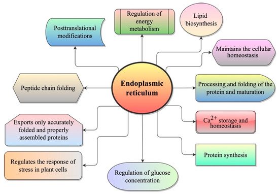
Figure 1.
 Different functions of the ER in plants.

3. ER Stress

ER plays a vital role in maintaining cellular homeostasis in different cellular processes through its functions, such as initial modification and folding of transmembrane and secretory proteins. Endogenous factors and environmental conditions can increase the demand for protein folding machinery. Many factors induce ER stress in plants, such as pathogens, environmental stresses, salinity, and drought, resulting in a higher load on secretory proteins in the ER [14][15][14,15]. These stresses accumulate misfolded or unfolded proteins that induce ER homeostasis imbalance, which is called ER stress [16][17][18][16,17,18]. Protein synthesis and modification lead to errors in almost one-third of the nascent proteins in the ER [19][20][21][19,20,21]. ER stress often leads to growth retardation in Arabidopsis (Arabidopsis thaliana) [22]. To overcome ER stress, the stress sensors localized in the ER activate distinct downstream organelle-nucleus signaling pathways to invoke a cytoprotective response, which is known as UPR [23]. UPR has recently been recognized as an important intracellular signaling pathway for linking ER proteostasis with gene regulation in the nucleus to reduce ER stress [6]. Although the molecular mechanism of ER stress in plants is not as well understood as it is in animals [24]. The expansion and abundance of genes related to ER stress, revealed by the genome sequencing of various plant species, suggest that plants use more ER stress responses than animals in order to adapt to the environment [24].

4. Chemical Inducers for the Accumulation of the Unfolded Protein

4.1. Tunicamycin (TM) Stress

4.1. Tunicamycin (TM) Stress

TM and Dithiothreitol (DTT) have been found to induce UPR by interfering with protein folding in the ER [25][26][25,26]. TM triggers ER-mediated stress in various eukaryotic species, such as plants, yeast, and humans [27][28][29][30][27,28,29,30]. TM inhibits the N-linked glycosylation (N-glycans formation) by interfering with the GlcNac phosphotransferase enzyme, which is responsible for the initial glycosylation steps [31]. The N-glycans are essential for the proper folding and stability of proteins, as well as their transportation to the Golgi apparatus for the packaging of vesicles and secretion. Besides, N-glycans are involved in the post-translational alteration of microbe-associated molecular patterns (MAMPs) receptors and immune response in plants [30][32][33][30,32,33].

4.2. Dithiothreitol (DTT) Stress

4.2. Dithiothreitol (DTT) Stress

DTT is a powerful reducing agent that induces acute ER stress by disrupting the redox conditions required to form disulfide bridges in proteins [34][35][34,35]. DTT is a robust reducing agent commonly used to promote reductive pressure. It can cross membranes and inhibit the formation of a disulfide bond. DTT treatment induces reductive stress, leading to the accumulation of misfolded proteins in the ER [36][37][36,37]. However, as a strong inducer of ER stress, DTT has not been considered as an ideal UPR triggering agent for in vivo studies because it can inhibit disulfide bond formation in the ER and cytosol during protein formation, making it unspecific to ER stress [38]. Besides, TM and DTT have differential effects on ER stress kinetics and can influence the expression of UPR target genes [30][39][30,39]. Arabidopsis basic leucine zipper 28 (bZIP28), an ER membrane-associated transcription factor (TF) (ER membrane-associated basic leucine zipper), was triggered by an ER stress response induced by exposure to DTT and TM or adverse environmental conditions [37][40][37,40]. In addition, TM or DTT may chemically induce basic leucine zipper TF 60 (bZIP60) splicing [41][42][41,42].

5. UPR Signaling in Plant Development

UPR has been found to play a crucial role in both reproductive and vegetative development [14]. Additionally, UPR plays an unexpected role in hormone biology, which may explain the effect of UPR on vegetative growth and development [14]. Normal plant growth and development require UPR. Normal growth and development require the mobilization of basic leucine zipper 17 (bZIP17) into the nucleus [43]. The triple mutant inositol-requiring enzyme 1 (IRE1a IRE1b) bZIP17 grew abnormally under normal growth conditions and was also defective in the stress signaling pathways. The functions of bZIP17 in A. thaliana development were observed in another study using genomic and genetic approaches. In contrast to bZIP28 and bZIP60, the bZIP17 is not a primary UPR activator, but works in conjunction with bZIP28 to regulate development-related genes, particularly stress maintenance and root elongation [44]. In A. thaliana, ER stress induces the expression of NAC103NAC103 overexpression has pleiotropic effects on plant growth, plays a vital role in inducing the expression of some UPR downstream genes under normal growth conditions [45]

6. UPR Signaling in Different Stresses

The UPR can be activated by various stresses in plants that induce the accumulation of unfolded proteins in the ER lumen [46][47][50,51] (Figure 2). UPR has been involved in the immunity and development of plants and provides defense against different stresses [14][42][44][14,42,44], such as heat [48][52], drought [49][53], salinity [50][51][54,55], osmotic pressure, high light intensity and heavy metals. These stresses disturb protein folding [15][52][15,56]. In addition, UPR is activated under protein synthesis overload conditions when the need for protein folding simply does not meet demands [53][54][57,58]. However, cells commit to programmed cell death (PCD) during the failure of UPR in chronic or unresolved ER stress conditions [23][55][56][57][23,59,60,61]. Biotic agents have more complex effects on the UPR. Various biotic agents have been reported to induce the UPR or require the UPR to infect plants. A study observed that when Nicotiana benthamiana was inoculated with the host-pathogen and non-host pathogens, Pseudomonas syringae and Pseudomonas cichorii, respectively, the host pathogen P. syringae did not induce the expression of bZIP60. At the same time, the expression of bZIP60P was induced by the non-host pathogen, P. cichorii. However, the plants became more susceptible to P. syringae when virus-induced gene silencing (VIGS) was used to silence the bZIP60 in N. benthamiana [58][62]. UPR is induced and required by various plant viruses for successful infection. 
Figure 2. Activation of the UPR. A variety of stresses (biotic and abiotic) and plant development processes (vegetative and reproductive) trigger the UPR by excessive accumulation of unfolded proteins in the ER or cause an imbalance in the supply of amino acids, which leads to the activation of one or more UPR arms. This figure was created by using BioRender software.

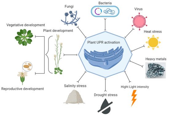
7. Strategies to Reduce ER Stress

Many strategies have been used to reduce ER stress, including the UPR, ER-associated degradation (ERAD), and autophagy.

7.1. Unfolded Protein Response (UPR)

7.1. Unfolded Protein Response (UPR)

UPR is an intracellular signaling mechanism activated by ER stress and has been designed to restore the ER function and to ignite the PCD processes when ER stress remains unresolved [59][100]. ER stress leads to the accumulation and aggregation of the unfolded proteins in the ER lumen. Moreover, UPR originates at the ER, where it overcomes the ER stress, restores the ER homeostasis, and leads to the ER chaperones and foldases synthesis to attenuate the ER stress [60][61][95,101]. These ER chaperones and foldases are BiP, protein disulfide isomerase (PDI), glucose-regulated protein (GRP94), peptidyl-prolyl isomerases (PPI) or immunophilins, calnexin and calreticulin [62][102]. Both signaling pathways eventually result in the upregulation of genes to either correctly fold or degrade misfolded proteins and regulate translation and transcription for restoring the ER homeostasis [28][63][28,103]. UPR may relieve the transient ER stress, whereas persistent ER stress can result in PCD [64][65][104,105].

7.2. Mechanism of UPR Signaling Pathway in Plants

7.2. Mechanism of UPR Signaling Pathway in Plants

7.2.1. Regulated IRE-1 Dependent Splicing (RIDS)

A wide range of stresses affect protein folding, causing ER stress that is communicated to the nucleus via the UPR, a cellular homeostatic response to ER stress [66][67][106,107]. As a result, genes involved in the folding, import, export, and quality control of proteins are up-regulated. In plants, signal transducers mediate the signaling that forms two arms of the UPR signaling pathway [14][38][43][14,38,43]. One arm includes membrane-associated TFs, for instance, bZIP17 and bZIP28, and the other arm includes IRE1, which is an RNA splicing factor. These two arms shape the stress transcriptome, the upregulation and the downregulation of the expression of genes to combat the stress effects [63][103]. On the other hand, cells undergo PCD if the adaptation mechanisms are inadequate to manage the unfolded protein load. In plants, the PCD regulatory mechanism and the key factors that regulate various outputs of ER stress receptors remain unclear. The bZIP17/28 are retained in the ER under normal conditions, associating with UPR regulator BiP. However, when unfolded proteins accumulate under stress conditions, BiP is sequestered and released from bZIP17/28 [68][69][108,109].

7.2.2. ER-Associated Degradation (ERAD)

ERAD is part of the ER protein quality-control system (ERQC), which is considered essential for the conformation fidelity of most of the membrane and secretory proteins in eukaryotes. The ERAD process is associated with the ubiquitin/proteasome system (UPS), which relieves ER stress. The UPS function involves the use of a ubiquitin-activating enzyme (E1), the ubiquitin-conjugating enzyme (E2), the ubiquitin ligase (E3), and the 26S proteasome [70][116]. ERAD is accomplished through multistep reactions involving the sequential recruitment of E1, E2, and E3 enzymes. E2 and E3 enzymes are responsible for the specificity of the substrate [71][72][92,117]

7.2.3. Autophagy

Autophagy is a self-eating cellular process that has been conserved throughout evolution. Autophagy functions as a degradation process in recycling cellular cytoplasmic contents and removing damaged proteins or organelles under adverse growth conditions [61][101]. There are three main types of autophagy on the basis of their mechanism; chaperone-mediated autophagy [73][119], macroautophagy [74][120], and microautophagy [75][121]. Chaperone-mediated autophagy is highly selective, while macroautophagy and microautophagy may be selective or non-selective [76][77][122,123]. In plants, the following two types of autophagy are known: macroautophagy and microautophagy [78][124]. During macroautophagy, the majority of cytosolic constituents are sequestered into a double-membrane structure, known as an autophagosome. The autophagosome is a specific organelle that mediates macroautophagy. For degradation, autophagy delivers cytoplasmic materials or organelles to the vacuole/lysosome by forming autophagosome [79][80][99,125]. The autophagosome’s outer membrane fuse with the vacuolar membrane upon delivery to the vacuole, delivering the inner membrane structure and its cargo, i.e., the autophagic body into the vacuolar lumen for degradation by vacuolar hydrolases [81][82][126,127]