In this work, we show a deep revision of the viral vector vaccines that have been developed to counteract bluetongue virus (BTV), an arthropod-borne disease that whips domestic and wild ruminants. We analyzed the main advantages and disadvantages of every of them, as well as the immunological features and efficacy that these candidates provided in both murine models and natural hosts.
- bluetongue virus (BTV)
- recombinant vaccines
- vira
Note: The following contents are extract from your paper. The entry will be online only after author check and submit it.
1. Introduction
Bluetongue virus (BTV) is a virus classified under the genus Orbivirus, within the family Reoviridae, and is transmitted via biting midges of the genus Culicoides. BTV is the causative agent of bluetongue (BT), a noncontagious arthropod-borne viral disease that affects both wild and domestic ruminants [1]. Certain breeds of sheep, especially fine-wool European breeds, and some species of wild ruminants, such as white-tailed deer, are the most commonly affected hosts, as they can show significant mortality rates [2,3][2][3], whereas cattle, goats, and the majority of wild ruminant species are usually asymptomatic. Nonetheless, cattle can be clinical upon infection (specially by BTV-8) [4] and, along with goats, can act as reservoirs for virus transmission from infected animals to other susceptible ruminants.
BTV virion is a non-enveloped icosahedral particle composed of three concentric protein capsid layers that surround a segmented genome [5,6][5][6]. Ten linear double-stranded RNA genome segments (S1 to S10) encode for seven structural (VP1–VP7) and five nonstructural proteins (NS1–NS5) [7,8][7][8]. The outer capsid layer contains two major proteins, VP2 and VP5, which are involved in cell attachment and membrane penetration, while the core is made up of the surface VP7 shell and the underlying VP3 layer [9]. Inside the core, there are transcriptase complexes formed by three minor enzymatic proteins, VP1, VP4, and VP6 [10,11][10][11]. The segmented nature of the BTV dsRNA genome enables the reassortment of genome segments when different serotypes or strains infect the host cell simultaneously [12[12][13],13], playing an important role in generating viral diversity. To date, 29 distinct serotypes of BTV, some of which are considered putative (serotypes 27–29) [14–16][14][15][16], have been identified all over the world [14[14][16],16], except in Antarctica.
BTV causes severe economic losses that are associated with its considerable impact on animal health, both direct such as weight loss, reduced fertility rate, reduced meat and milk production efficiency, and death, and indirect like lost revenue and trade restrictions [17,18][17][18]. To minimize these losses, vaccines have emerged as the most effective prophylactic measure to control BT disease and to potentially interrupt the cycle from the infected animal to the hematophagous vector. The focus of most current BTV vaccine research is on neutralizing antibody-based approaches; however, these are serotype specific. In fact, the specificity of interactions between BTV outer capsid proteins and neutralizing antibodies (Nabs) determines the identity of the BTV serotypes [19,20][19][20]. Cytotoxic T lymphocytes (CTLs) also play an important role in protective immunity against BTV; particularly, cell-mediated immune responses against nonstructural proteins are likely to be crucial in protecting against heterologous BTV serotypes [21–24][21][22][23][24]. However, antibody and CTL-based protection largely depends upon the nature of the vaccine platform applied. Typically, inactivated and subunit vaccines stimulate mainly antibody-based mechanisms, but they are poor stimulators of CTLs. On the other hand, live-attenuated and vectored vaccines may be potent inducers of both antibodies and CTLs [25]. Although inactivated vaccines are safer and can limit BTV dissemination, they cannot address the need for cross-protection among the different serotypes and do not allow for the distinction between infected and vaccinated animals (DIVA strategy). Live-attenuated vaccines (LAVs) have been widely used to control BTV in the past [26]. However, they are associated with teratogenicity, reversion to virulence, viremia that allows transmission to the insect vector, and risk of reassortment events with virulent wild-type viruses, giving rise to new virulent strains [27]. Recently, new strategies such as LAV based on reverse genetics [28,29][28][29] and viral vector vaccines have been designed to avoid these drawbacks.
2. Viral Vectors for Vaccine Applications
Viral vectors are regarded as potential tools for gene therapy and vaccine development. Their utility is predominantly based on the ability of viruses to infect cells, and the main advantages offered by viral vectors for vaccine development can be summarized as follows: (a) highly efficient gene transduction, (b) highly specific delivery of genes to target cells, (c) transient antigen expression, and (d) induction of robust immune responses, maintaining strong humoral immune responses and enhancing cellular immunity [30]. A successful presentation and delivery of antigens are crucial for inducing immunity and lifelong protection. Recombinant viral vectors have a potential for prophylactic use because they enable intracellular antigen expression and induce robust CTL response, leading to the removal of virus-infected cells. They are, therefore, ideal shuttles for delivering foreign proteins and also induce immune response by mimicking natural infection [30].
In addition, some attributes, such as the achievement of stable insertion of coding sequences into the genome, the aforementioned induction of a protective immune response, a proven safety record, and the potential for large-scale production, are required in order to qualify as a vaccine vector.
Multiple viruses have been used as vaccine viral vectors, ranging from very complex large DNA viruses such as poxviruses, down to simple RNA viruses such as parainfluenza viruses [31–33] [31][32][33][40][41][42][43][44][45][46][47][48][49][50][51][52][53][54][55], where there are few restrictions imposed by gene packaging limits. Viral vector vaccines have been applied extensively in veterinary medicine. An outstanding example of this is Raboral V-RG (Merial), the first oral live vaccinia virus vector vaccine expressing the glycoprotein (GP) of Evelyn-Rokitnicki-Abelseth rabies virus [34,35][34][35].
3. Poxviruses
3.1. Vaccinia Virus and Modified Vaccinia Virus Ankara
Vaccinia viruses (VVs) have been engineered to express foreign genes, turning them into powerful vectors for recombinant protein expression. These were originated from highly efficacious vaccines for the eradication of smallpox [36], serving as a highly appealing delivery system for heterologous viral antigens [37]. The first approach to develop recombinant VV against BTV was described by Lobato et al., using the Western Reserve (WR) VV strain to construct recombinant VV expressing VP2 or VP5 of BTV-1, or coexpressing both BTV antigens [38] (Table 1). Notably, sheep immunized with the recombinant VV coexpressing VP2 and VP5 were able to develop high titers of Nabs, but lower in comparison with those sheep that received the LAV. Moreover, these two groups were not viremic, and animals did not display pyrexia following a challenge. Despite the non-negligible results of this work, the virulence of VV strains, particularly the WR strain, and the observation of a lower immunogenic profile compared with other highly attenuated VV strains entailed an insurmountable obstacle for their use as vaccine vectors [39].
Table 1. Overview of viral vector vaccine candidates against BTV.
|
Viral Vector |
Type of Virus |
Antigen Included |
Type of Induced Immune Response |
Animal Model |
Multiserotype Protection |
Safety |
Ref. |
|
VV Western Reserve strain |
dsDNA |
VP2/VP5/VP2 + VP5 |
Humoral |
Merino sheep |
In vitro cross-neutralization |
Virulent strain |
[38] |
|
Modified vaccinia virus Ankara (MVA) |
dsDNA |
VP2, VP5, VP7, and NS1 |
Humoral and cellular |
IFNAR(−/−) mice Churra sheep |
Yes (Not tested in sheep) |
Safe |
[22,23,40–44] |
|
Fowlpox virus (FPV) |
dsDNA |
VP2 + VP5 |
Humoral |
BALB/c mice Sheep (breed not specified) |
No |
Safe |
[45] |
|
Canarypox virus (CPV) |
dsDNA |
VP2 + VP5 |
Humoral |
Dorset sheep |
No |
Safe |
[46] |
|
Myxomavirus (MYXV) |
dsDNA |
VP2/VP2 + VP5 |
Humoral |
Lacaune lambs |
No |
Safe |
[47] |
|
Capripoxvirus (CaPV) |
dsDNA |
VP7 |
Cellular |
Dorset sheep |
Partial |
Safe |
[48] |
|
VP2 + VP7 + NS1 + NS3 |
Humoral and Cellular |
Saanen goats and Préalpes sheep |
Presumable (not tested) |
Safe |
[49] |
||
|
Bovine herpesvirus 4 (BoHV-4) |
dsDNA |
VP2 |
Humoral |
IFNAR(−/−) mice |
No |
Safe |
[50] |
|
Equine herpesvirus 1 (EHV-1) |
dsDNA |
VP2 + VP5 |
Humoral |
IFNAR(−/−) mice |
No |
Safe |
[51] |
|
Canine adenovirus type 2 (CAV-2) |
dsDNA |
VP7 |
Cellular |
Préalpes sheep |
No |
Safe |
[52] |
|
Adenovirus type 5 (Ad5) |
dsDNA |
VP7, VP2, and NS3 (different combinations) |
Humoral and cellular |
IFNAR(−/−) mice Colmenareña sheep |
Presumable (not tested) |
Safe |
[53] |
|
Chimpanzee adenovirus 1 (ChAdOx1) |
dsDNA |
NS1/NS1-Nt |
Cellular |
IFNAR(−/−) mice Churra sheep |
Yes |
Safe |
[43] |
|
Vesicular stomatitis virus (VSV) |
ssRNA(−) |
VP2/VP5/VP2 + VP5 |
Humoral |
Swiss White Alp sheep |
No |
Safe |
[54] |
|
Rift Valley fever virus (RVFV) |
ssRNA(−) |
VP2/NS1-Nt |
Humoral and cellular |
BALB/c mice Churra sheep |
Presumable (not tested) |
Safe |
[55] |
dsDNA: double-stranded DNA; ssRNA(−): negative-sense single-stranded RNA
To overcome these issues, researchers focused their efforts on the development of safer and immunogenic vectors. One of these highly attenuated strains, the modified vaccinia virus Ankara (MVA) strain, has been found to be immunogenic and useful for the application of protection against a vast number of infectious diseases [56]. Historical research focused on MVA and its use as vaccine against smallpox has allowed the scientific community to establish an extraordinary safety profile of this vector. This strain can be used under biosafety level 1 (BSL1) conditions because of its nature and its deficiency to productively grow in mammalian hosts. In addition, this replication-deficient viral vector has intrinsic capacities to induce both humoral and cellular immune responses. Historically, MVA was developed by serial tissue culture passage in primary chicken cells of vaccinia virus strain Ankara, and clinically used to avoid the undesirable side effects of conventional smallpox vaccination [57]. Adapted to grow in avian cells, MVA lost the ability to replicate in mammalian hosts and lacks many immunomodulatory genes that orthopoxviruses use to regulate the host cell environment [58–60]. Its ancestor virus is the vaccinia virus strain Ankara, which was originally propagated on the skin of calves and donkeys for smallpox vaccine production at the Turkish Vaccine Institute in Ankara. In 1953, the vaccinia virus strain Ankara was brought to Munich and added to the strain collection of the Institute for Infectious Diseases and Tropical Medicine at the University of Munich, where Herrlich and Mayr grew the virus on the chorioallantois membranes of embryonated chicken eggs and, therefore, named it as chorioallantois vaccinia virus Ankara (CVA) [61]. After serial passages in chicken (516th), it was renamed as modified vaccinia virus Ankara and given to the Bavarian State Institute for Vaccines to test its suitability for smallpox vaccine production [57,62].
Comparison with the genome maps of CVA ancestor viruses revealed that the MVA genome harbors six major deletions and mutations, resulting in the loss of ~30Kb of genetic material, which have altered virus–host interactions, as the absence of the A-type inclusion body protein or truncations in the HA promoter sequence [63].
The most common method used to produce recombinant MVAs involves the insertion of foreign genes into the thymidine kinase (TK) gene of the VV via homologous recombination [64]. This is accomplished through the construction of a recombination plasmid containing the gene of interest flanked by the VV TK gene. Afterwards, this plasmid is used to transfect previously MVA-infected cells, in which recombinant MVAs carrying the foreign gene are obtained by marker selection (Figure 1) [65].
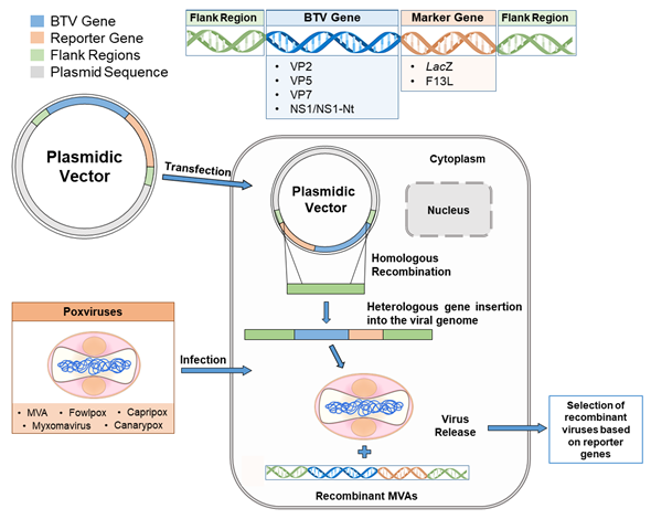
Figure 1. Diagrammatic representation of one example of cloning strategy for the generation of recombinant poxviral vector. A homologous recombination process takes place in the cytoplasm of eukaryotic cells simultaneously infected with the virus vector and transfected with a plasmid vector encoding the BTV gene of interest (blue) along with a marker gene (orange). Flank regions (green) allow for recombination and heterologous gene insertion into the viral genome. The BTV genes cloned and the marker genes used in several works are listed above. Finally, after a selection process based on the marker gene, the recombinant poxvirus expressing BTV genes is recovered.
The use of MVA as an expression vector for foreign genes was first described by Sutter and Moss, in which the expression of foreign reporter proteins such as LacZ was tested [66]. Since then, multiple recombinant MVAs have been generated against a plethora of human diseases [67–72]. The MVA vector has also been widely used against veterinary viral diseases [73–76]. For BTV (Table 1), the recombinant MVAs were first used for vaccination studies along with DNA vaccines, expressing the outer capsid proteins VP2 and VP5 from BTV-4. This regimen conferred a partial protection with reduced levels of viremia in the IFNAR(-/-) mouse model, using recombinant MVAs as boosters [40]. In the same work, the authors demonstrated the key role of the viral antigen VP7, observing a sterile protective effect in mice immunized with MVAs expressing these three antigens, suggesting the important role of Nabs induction (due to VP2) and the IFN-γ-secreting T cell activation (triggered by VP2 and VP7). Similar analysis was performed against BTV-8, a serotype with enhanced tropism for cattle that suddenly emerged in Northern-Central Europe in 2006 [77]. MVA–MVA or DNA–MVA prime-boost immunizations were performed, expressing VP2 alone, VP7 alone, or as a cocktail of MVAs expressing VP2, VP5, and VP7. The authors showed the capacity of VP2 in conferring protection against a lethal challenge of BTV-8 in the IFNAR(-/-) mouse model, whereas this level of protection was not achieved with VP7 alone [41].
As we already described, a myriad of different BTV serotypes have been reported. The approaches previously described have demonstrated good results regarding protection against homologous infections. However, an ideal vaccine against BTV would have to confer protection against multiple serotypes. To this end, researchers began to focus on those viral antigens that are more conserved among different serotypes and are able to induce a robust immune response in the host, when used as a vaccine candidate. Unlike VP2, which is the most genetically diverse antigen, the nonstructural protein NS1 is the most conserved protein among serotypes, and it has been described as a strong inducer of cellular immune response in both the mouse model and sheep [78]. Following this rationale, NS1 antigen was introduced in the vaccine composition along with VP2 and VP7, following a prime-boost immunization with DNA (prime) and MVAs (boost). The protective capacity of this strategy was evaluated in mice against the homologous challenge with BTV-4, showing sterile protection [22]. Subsequently, this strategy was also probed against heterologous infections with BTV-1 and BTV-8, showing 84% and 100% protection in immunized mice, respectively [22]. A Nab response was achieved (VP2 mediated), as well as a strong induction of the CD8+ T cell population, which was observed after stimulation of this cell subset with the three antigens used in the vaccine composition [22]. The broad protection observed against multiple BTV serotypes suggested that the protective role of NS1 and the cellular immune responses could be critical to achieve multiserotype protection.
Thereafter, the multiserotype protective role of NS1 was confirmed, observing that only this nonstructural protein vectorized in MVA in a homologous prime-boost immunization is necessary for conferring sterile protection against different BTV serotypes, like BTV-1, 4, 8, and 16, as well as the reassortant BTV-4 Morocco strain (BTV-4/MOR09) [23]. This study showed that mono- and multiserotype protection against BTV can be achieved in the complete absence of Nabs by enhancing cytotoxic CD8+ cellular immune responses. This work also showed that the protective capacity of NS1 resides in the N-terminal region (NS1-Nt), being dependent of a specific T cell epitope located in the amino acid position 152 (GQIVNPTFI) (peptide 152). The absence of this peptide in the NS1 amino acid sequence totally abrogates its protective ability [23].
MVAs have also been found to be protective in combination with other vaccine platforms, such as antigen presenting protein microspheres (µNS) carrying VP2, VP7, and NS1. In this case, this heterologous immunization strategy based on BTV-4 antigens was able to protect IFNAR(-/-) mice against serotypes 1 and 4 [42]. Moreover, MVAs have been combined with other viral vectors, like chimpanzee adenovirus Oxford 1 (ChadOx1), which will be discussed later [43]. In this study, the protective immune effect of MVA-NS1 was also evaluated in mice in a single-dose vaccination experiment, observing a delay in mortality and partial protection against a lethal challenge of BTV.
Another interesting approach was the generation of MVAs developed to combat viral infectious diseases that overlap in distribution or host and present a potential risk of expansion in nonendemic but close-to-endemic areas. This is the case of engineered recombinant MVAs against BTV and Rift Valley fever virus (RVFV). RVFV is a zoonosis that affects livestock, mainly sheep, and it is endemic in Africa and some regions in the Middle East. The appearance of outbreaks of RVFV in nonendemic areas like Europe is a potential threat, as there are different species of competent mosquito vectors, such as Culex and Aedes, already established in the area. Dual MVAs were generated in this study, cloning the GnGc gene of RVFV and the segments that encode VP2, NS1, and NS1-Nt from BTV in the F13L and TK loci, respectively, and under the control of VV early/late promoters. After a BTV challenge, all the immunized groups of IFNAR(-/-) mice showed protection, especially those immunized with NS1 and NS1-Nt, where 100% sterile protection was observed [44].
Finally, prompted by the high vaccination efficacy observed in the mouse model, the effectiveness of some of these promising candidates have been tested in the natural host. The dual MVA-GnGc-NS1 previously mentioned was tested against BTV-4 in sheep, using two doses of 108 PFU per animal and observing very similar results in terms of rectal temperature and viremia. Additionally, vaccinated sheep were aviremic for an RVFV challenge (except one animal at day 3 postinfection), maintaining stable biochemical parameters (aspartate transaminase, gamma-glutamyltransferase, lactate dehydrogenase, and albumin), and had mild histological lesions compared with the nonvaccinated group, which indicated the bivalent character of the designed vaccine [44]. A similar trend was observed when MVA-NS1 was used as a booster of ChAdOx1-NS1 in a heterologous prime-boost immunization, as immunized sheep showed reduced levels of viremia and lower temperatures than the control group [43].
Although these results pave the way for the development of multiserotype vaccines against BTV in ruminants, further questions will need to be addressed, such as the exploration of other BTV viral antigens able to activate broad immune responses, the assessment of the long-term efficacy elicited by these candidates, and their capacity to reduce viremia that sufficiently avoids potential transmission by midge bites.
3.2. Other Poxviruses
Besides MVA, a variable set of other viruses belonging to the family Poxviridae, including capripoxviruses, avian poxviruses, and myxomaviruses, has been proposed as alternative vaccine platforms against BTV, but to a lesser extent (Table 1).
The genus Capripoxvirus (CaPV) comprises three closely related species (up to 97% nucleotide homology [79]) that are restricted to ruminant hosts: sheeppox virus (SPPV), goatpox virus (GTPV), and lumpy skin disease virus (LSDV). Attenuated capripoxviruses have been positively evaluated as vaccine vectors in ruminants [80–83], proving its safety and immunogenicity, and are considered ideal viral vectors because of their thermostability, large genome size, and ruminant host restriction, and because they are nonpathogenic to human hosts [84,85]. Interestingly, inoculation of these recombinant viral vectors induces a vector-specific immunity, which could eventually enhance the valence of the attenuated CaPV vaccine or even offer the possibility of constructing bivalent vaccines against both the viral vector used (CaPV) and the targeted viral agent [83,85]. Nonetheless, this pre-existing immunity may constrain their potential as vaccine vectors in ruminants, as it has been shown after the immunization of cattle with a recombinant CaPV encoding heterologous antigens from rinderpest virus [86]. Concerning BTV, a serotype cross-reactive, cell-mediated immunity was elicited in sheep by the recombinant live-attenuated strain KS-1 of LSDV expressing VP7 of BTV-1, observing partial protection against a heterotypic challenge with BTV-3 after a homologous prime-boost immunization regime [48]. BTV-specific ex vivo lymphocyte proliferation was also observed in goats after subcutaneous injection of a single dose of a recombinant capripoxvirus (KS-1 strain) individually expressing VP2, VP7, NS1, and NS3 of BTV-2 [49]. Nonstructural proteins of BTV are the predominant sources of antigens recognized by BTV-specific CD8+ CTLs [87], which have been described as critical for the development of a long-lasting immunity in animals infected with BTV [88,89]. However, mild protection was observed after a homotypic challenge in both sheep and goat, as animals displayed mild clinical signs but detectable levels of viremia after a challenge despite the inclusion of nonstructural proteins in vaccine design [49].
To date, two avipoxviruses have been exploited as viral vectors against BTV: fowlpox (FPV) and canarypox viruses (CPV). Recombinant FPV and CPV vaccines expressing foreign antigens have been proved safe and effective in mammalian hosts [90–100]. In addition to having a large cargo capacity of both viral vectors, these exhibit an ideal safety profile due to their natural host range restriction to avian species and abortive replication in mammalian and insect cells, which makes them a safer but effective alternative to other live virus vectors [101–104]. For BTV, a recombinant FPV coexpressing genes encoding the VP2 and VP5 outer capsid proteins of BTV-1 administered in combination with a DNA vaccine prime elicited humoral and BTV-specific T-cell responses in BALB/c mice and significant and sustained levels of serum Nabs in sheep [45]. However, the protective capability against BTV was not analyzed further. Regarding CPV, serotype-specific protection was observed in sheep subjected to a homologous prime-boost vaccination regime with a recombinant CPV coexpressing VP2 and VP5 proteins of BTV-17, as BTV particles were not isolated from the blood of vaccinated sheep after challenge [46].
Myxomavirus (MYXV), a leporide-specific poxvirus, is also a potential nonreplicative vector for ruminant immunization. Like FPV and CPV, MYXV abortively infects ruminant cells, allowing the expression of substantial amounts of foreign genetic material [105,106]. Furthermore, this viral vector has shown vaccine efficacy and safety in several mammalian hosts, including sheep [106–108]. With regard to BTV, VP2, individually or in combination with VP5, was vectorized in a recombinant MYXV. After a homologous prime-boost immunization regime with the recombinant MYXV expressing VP2 alone, immunized sheep displayed higher levels of viremia and more severe clinical signs than sheep vaccinated with inactivated BTV-8, but animals were better protected from a homotypic BTV-8 viral challenge than the non-immunized control group [47]. It has been described that VP5 enhances the protective immune response elicited by VP2 alone [109,110]. Conversely, the protection conferred by the recombinant MYXV simultaneously expressing VP2 and VP5 was similar to that of the negative control group, which could rely on a diminished expression of VP2 by the recombinant vector and/or nuclear localization of VP5 observed, which could mismatch its likely conformational influence on VP2 (mainly located in the cytoplasm) as pointed out by the authors, thus impairing the induction of a potent humoral immune response. The immunogenicity of another recombinant MYXV (derived from the attenuated vaccine strain of MYXV SG33) expressing the VP7 protein of BTV-2 has also been assessed, observing an induction of a VP7-specific CD4+ cell subset [52].
In summary, these poxviral vectors are a potential alternative to inactivated vaccines or LAVs as they are immunogenic and naturally attenuated in ruminants. Moreover, these reviewed strategies could be further used in combination with MVA to evade vector-specific immune responses, thus boosting the immunogenicity of recombinant MVAs. Nonetheless, further research is needed to improve the efficacy of this poxviral vector in ruminant species, which will be important not only for BTV but also for other relevant virulent veterinary diseases.
References
- Erasmus, B.J. Bluetongue in sheep and goats. Aust Vet. J. 1975, 51, 165–170.
- Maclachlan, N.J.; Drew, C.P.; Darpel, K.E.; Worwa, G. The pathology and pathogenesis of blue-tongue. J. Comp. Pathol. 2009, 141, 1–16.
- Drolet, B.S.; Reister, L.M.; Rigg, T.D.; Nol, P.; Podell, B.K.; Mecham, J.O.; VerCauteren, K.C.; van Rijn, P.A.; Wilson, W.C.; Bowen, R.A. Experimental infection of white-tailed deer (Odocoileus virgini-anus) with Northern European bluetongue virus serotype 8. Vet. Microbiol 2013, 166, 347–355.
- Elbers, A.R.W.; van der Spek, A.N.; van Rijn, P.A. Epidemiologic characteristics of bluetongue virus serotype 8 laboratory-confirmed outbreaks in The Netherlands in 2007 and a comparison with the situation in 2006. Prev. Vet. Med. 2009, 92, 1–8.
- Grimes, J.M.; Burroughs, J.N.; Gouet, P.; Diprose, J.M.; Malby, R.; Ziéntara, S.; Mertens, P.P.; Stuart, D.I. The atomic structure of the bluetongue virus core. Nature 1998, 395, 470–478.
- Roy, P. Functional mapping of bluetongue virus proteins and their interactions with host proteins during virus replication. Cell Biochem. Biophys. 2008, 50, 143–157.
- Ratinier, M.; Caporale, M.; Golder, M.; Franzoni, G.; Allan, K.; Nunes, S.F.; Armezzani, A.; Bay-oumy, A.; Rixon, F.; Shaw, A.; et al. Identification and characterization of a novel non-structural protein of bluetongue virus. PLoS Pathog. 2011, 7, e1002477.
- Stewart, M.; Hardy, A.; Barry, G.; Pinto, R.M.; Caporale, M.; Melzi, E.; Hughes, J.; Taggart, A.; Jan-owicz, A.; Varela, M.; et al. Characterization of a second open reading frame in genome segment 10 of bluetongue virus. J. Gen. Virol. 2015, 96, 3280–3293.
- Mertens, P.P.C.; Diprose, J. The bluetongue virus core: A nano-scale transcription machine. Virus Res. 2004, 101, 29–43.
- Verwoerd, D.W. Purification and characterization of bluetongue virus. Virology 1969, 38, 203–212.
- Roy, P. Orbivirus structure and assembly. Virology 1996, 216, 1–11, doi:10.1006/viro.1996.0028.
- Oberst, R.D.; Stott, J.L.; Blanchard-Channell, M.; Osburn, B.I. Genetic reassortment of bluetongue vi-rus serotype 11 strains in the bovine. Vet. Microbiol. 1987, 15, 11–18.
- Samal, S.K.; el-Hussein, A.; Holbrook, F.R.; Beaty, B.J.; Ramig, R.F. Mixed infection of Culicoides var-iipennis with bluetongue virus serotypes 10 and 17: Evidence for high frequency reassortment in the vector. J. Gen. Virol. 1987, 68, 2319–2329.
- Schulz, C.; Bréard, E.; Sailleau, C.; Jenckel, M.; Viarouge, C.; Vitour, D.; Palmarini, M.; Gallois, M.; Höper, D.; Hoffmann, B.; et al. Bluetongue virus serotype 27: Detection and characterization of two novel variants in Corsica, France. J. Gen. Virol. 2016, 97, 2073–2083.
- Bumbarov, V.; Golender, N.; Jenckel, M.; Wernike, K.; Beer, M.; Khinich, E.; Zalesky, O.; Erster, O. Characterization of bluetongue virus serotype 28. Transbound. Emerg. Dis 2020, 67, 171–182.
- Yang, H.; Gu, W.; Li, Z.; Zhang, L.; Liao, D.; Song, J.; Baoxin, S.; Hasimu, J.; Li, Z.; Yang, Z.; et al. Novel Putative Bluetongue Virus Serotype 29 Isolated from Inapparently Infected Goat in Xinjiang of China. Transbound. Emerg. Dis. 2020, doi:10.1111/tbed.13927, accepted for publication.
- Gethmann, J.; Probst, C.; Conraths, F.J. Economic Impact of a Bluetongue Serotype 8 Epidemic in Germany. Front. Vet. Sci. 2020, 7, 65.
- Rushton, J.; Lyons, N. Economic impact of Bluetongue: A review of the effects on production. Vet. Ital. 2015, 51, 401–406.
- Roy, P. Bluetongue virus proteins. J. Gen. Virol. 1992, 73, 3051–3064.
- Maan, S.; Maan, N.S.; Samuel, A.R.; Rao, S.; Attoui, H.; Mertens, P.P.C. Analysis and phylogenetic comparisons of full-length VP2 genes of the 24 bluetongue virus serotypes. J. Gen. Virol. 2007, 88, 621–630.
- Jeggo, M.H.; Wardley, R.C.; Brownlie, J. A study of the role of cell-mediated immunity in bluetongue virus infection in sheep, using cellular adoptive transfer techniques. Immunology 1984, 52, 403–410.
- Calvo-Pinilla, E.; Navasa, N.; Anguita, J.; Ortego, J. Multiserotype Protection Elicited by a Combina-torial Prime-Boost Vaccination Strategy against Bluetongue Virus. PLoS ONE 2012, 7, e34735.
- Marín-López, A.; Calvo-Pinilla, E.; Barriales, D.; Lorenzo, G.; Brun, A.; Anguita, J.; Ortego, J. CD8 T Cell Responses to an Immunodominant Epitope within the Nonstructural Protein NS1 Provide Wide Immunoprotection against Bluetongue Virus in IFNAR −/− Mice. J. Virol. 2018, 92, e00938-18.
- Anderson, J.; Hägglund, S.; Bréard, E.; Riou, M.; Zohari, S.; Comtet, L.; Olofson, A.-S.; Gélineau, R.; Martin, G.; Elvander, M.; et al. Strong protection induced by an experimental DIVA subunit vaccine against bluetongue virus serotype 8 in cattle. Vaccine 2014, 32, 6614–6621.
- Plotkin, S.A. Complex correlates of protection after vaccination. Clin. Infect. Dis. 2013, 56, 1458–1465.
- Savini, G.; MacLachlan, N.J.; Sánchez-Vizcaino, J.-M.; Zientara, S. Vaccines against bluetongue in Europe. Comp. Immunol. Microbiol. Infect. Dis. 2008, 31, 101–120.
- Batten, C.A.; Maan, S.; Shaw, A.E.; Maan, N.S.; Mertens, P.P.C. A European field strain of blue-tongue virus derived from two parental vaccine strains by genome segment reassortment. Virus Res. 2008, 137, 56–63.
- Matsuo, E.; Celma, C.C.P.; Boyce, M.; Viarouge, C.; Sailleau, C.; Dubois, E.; Bréard, E.; Thiéry, R.; Zientara, S.; Roy, P. Generation of replication-defective virus-based vaccines that confer full protec-tion in sheep against virulent bluetongue virus challenge. J. Virol. 2011, 85, 10213–10221.
- Feenstra, F.; Pap, J.S.; van Rijn, P.A. Application of Bluetongue Disabled Infectious Single Animal (DISA) vaccine for different serotypes by VP2 exchange or incorporation of chimeric VP2. Vaccine 2015, 33, 812–818.
- Ura, T.; Okuda, K.; Shimada, M. Developments in Viral Vector-Based Vaccines. Vaccines 2014, 2, 624–641.
- Ertl, H.C. Viral vectors as vaccine carriers. Curr. Opin. Virol. 2016, 21, 1–8.
- Moss, B.; Smith, G.L.; Mackett, M. Use of vaccinia virus as an infectious molecular cloning and ex-pression vector. Gene Amplif. Anal. 1983, 3, 201–213.
- Russell, C.J.; Hurwitz, J.L. Sendai Virus as a Backbone for Vaccines against RSV and other Human Paramyxoviruses. Expert Rev. Vaccines 2016, 15, 189–200.
- Lauer, K.B.; Borrow, R.; Blanchard, T.J. Multivalent and Multipathogen Viral Vector Vaccines. Clin. Vaccine Immunol. 2017, 24, doi:10.1128/CVI.00298-16.
- Pastoret, P.P.; Brochier, B.; Languet, B.; Thomas, I.; Paquot, A.; Bauduin, B.; Kieny, M.P.; Lecocq, J.P.; De Bruyn, J.; Costy, F. First field trial of fox vaccination against rabies using a vaccinia-rabies recom-binant virus. Vet. Rec. 1988, 123, 481–483.
- Ladnyĭ, I.D. Global program of smallpox eradication. 1. Smallpox in the world before acceptance of the program of its eradication by the World Health Organization. Zh. Mikrobiol. Epidemiol. Immunobi-ol. 1977, 3, 98–105.
- Sutter, G.; Staib, C. Vaccinia vectors as candidate vaccines: The development of modified vaccinia virus Ankara for antigen delivery. Curr. Drug Targets Infect. Disord. 2003, 3, 263–271.
- Lobato, Z.I.P.; Coupar, B.E.H.; Gray, C.P.; Lunt, R.; Andrew, M.E. Antibody responses and protec-tive immunity to recombinant vaccinia virus-expressed bluetongue virus antigens. Vet. Immunol. Im-munopathol. 1997, 59, 293–309.
- Freitas, L.F.D. de; Oliveira, R.P.; Miranda, M.C.G.; Rocha, R.P.; Barbosa-Stancioli, E.F.; Faria, A.M.C.; Fonseca, F.G. da The Virulence of Different Vaccinia Virus Strains Is Directly Proportional to Their Ability To Downmodulate Specific Cell-Mediated Immune Compartments In Vivo. J. Virol. 2019, 93, doi:10.1128/JVI.02191-18
- Calvo-Pinilla, E.; Rodríguez-Calvo, T.; Sevilla, N.; Ortego, J. Heterologous prime boost vaccination with DNA and recombinant modified vaccinia virus Ankara protects IFNAR (−/−) mice against le-thal bluetongue infection. Vaccine 2009, 28, 437–445.
- Jabbar, T.K.; Calvo-Pinilla, E.; Mateos, F.; Gubbins, S.; Bin-Tarif, A.; Bachanek-Bankowska, K.; Alpar, O.; Ortego, J.; Takamatsu, H.-H.; Mertens, P.P.C.; et al. Protection of IFNAR (−/−) Mice against Blue-tongue Virus Serotype 8, by Heterologous (DNA/rMVA) and Homologous (rMVA/rMVA) Vaccina-tion, Expressing Outer-Capsid Protein VP2. PLoS ONE 2013, 8, e60574.
- Marín-López, A.; Calvo-Pinilla, E.; Barriales, D.; Lorenzo, G.; Benavente, J.; Brun, A.; Mar-tínez-Costas, J.M.; Ortego, J. Microspheres-prime/rMVA-boost vaccination enhances humoral and cellular immune response in IFNAR (−/−) mice conferring protection against serotypes 1 and 4 of bluetongue virus. Antivir. Res. 2017, 142, 55–62.
- Utrilla-Trigo, S.; Jiménez-Cabello, L.; Alonso-Ravelo, R.; Calvo-Pinilla, E.; Marín-López, A.; Moreno, S.; Lorenzo, G.; Benavides, J.; Gilbert, S.; Nogales, A.; et al. Heterologous Combination of ChAdOx1 and MVA Vectors Expressing Protein NS1 as Vaccination Strategy to Induce Durable and Cross-Protective CD8+ T Cell Immunity to Bluetongue Virus. Vaccines 2020, 8, 346.
- Calvo-Pinilla, E.; Marín-López, A.; Moreno, S.; Lorenzo, G.; Utrilla-Trigo, S.; Jiménez-Cabello, L.; Benavides, J.; Nogales, A.; Blasco, R.; Brun, A.; et al. A protective bivalent vaccine against Rift Valley fever and bluetongue. NPJ Vaccines 2020, 5, 1–12.
- Li, J.; Yang, T.; Xu, Q.; Sun, E.; Feng, Y.; Lv, S.; Zhang, Q.; Wang, H.; Wu, D. DNA vaccine prime and recombinant FPV vaccine boost: An important candidate immunization strategy to control blue-tongue virus type 1. Appl. Microbiol. Biotechnol. 2015, 99, 8643–8652.
- Boone, J.D.; Balasuriya, U.B.; Karaca, K.; Audonnet, J.-C.; Yao, J.; He, L.; Nordgren, R.; Monaco, F.; Savini, G.; Gardner, I.A.; et al. Recombinant canarypox virus vaccine co-expressing genes encoding the VP2 and VP5 outer capsid proteins of bluetongue virus induces high level protection in sheep. Vaccine 2007, 25, 672–678.
- Top, S.; Foucras, G.; Deplanche, M.; Rives, G.; Calvalido, J.; Comtet, L.; Bertagnoli, S.; Meyer, G. Myxomavirus as a vector for the immunisation of sheep: Protection study against challenge with bluetongue virus. Vaccine 2012, 30, 1609–1616.
- Wade-Evans, A.M.; Romero, C.H.; Mellor, P.; Takamatsu, H.; Anderson, J.; Thevasagayam, J.; Flem-ing, M.J.; Mertens, P.P.; Black, D.N. Expression of the major core structural protein (VP7) of blue-tongue virus, by a recombinant capripox virus, provides partial protection of sheep against a virulent heterotypic bluetongue virus challenge. Virology 1996, 220, 227–231.
- Perrin, A.; Albina, E.; Bréard, E.; Sailleau, C.; Promé, S.; Grillet, C.; Kwiatek, O.; Russo, P.; Thiéry, R.; Zientara, S.; et al. Recombinant capripoxviruses expressing proteins of bluetongue virus: Evaluation of immune responses and protection in small ruminants. Vaccine 2007, 25, 6774–6783.
- Franceschi, V.; Capocefalo, A.; Calvo-Pinilla, E.; Redaelli, M.; Mucignat-Caretta, C.; Mertens, P.; Or-tego, J.; Donofrio, G. Immunization of knock-out α/β interferon receptor mice against lethal blue-tongue infection with a BoHV-4-based vector expressing BTV-8 VP2 antigen. Vaccine 2011, 29, 3074–3082.
- Ma, G.; Eschbaumer, M.; Said, A.; Hoffmann, B.; Beer, M.; Osterrieder, N. An Equine Herpesvirus Type 1 (EHV-1) Expressing VP2 and VP5 of Serotype 8 Bluetongue Virus (BTV-8) Induces Protection in a Murine Infection Model. PLoS ONE 2012, 7, e34425.
- Bouet-Cararo, C.; Contreras, V.; Caruso, A.; Top, S.; Szelechowski, M.; Bergeron, C.; Viarouge, C.; Desprat, A.; Relmy, A.; Guibert, J.-M.; et al. Expression of VP7, a Bluetongue Virus Group Specific Antigen by Viral Vectors: Analysis of the Induced Immune Responses and Evaluation of Protective Potential in Sheep. PLoS ONE 2014, 9, e111605.
- Martín, V.; Pascual, E.; Avia, M.; Peña, L.; Valcárcel, F.; Sevilla, N. Protective Efficacy in Sheep of Adenovirus-Vectored Vaccines against Bluetongue Virus Is Associated with Specific T Cell Respons-es. PLoS ONE 2015, 10, e0143273.
- Kochinger, S.; Renevey, N.; Hofmann, M.A.; Zimmer, G. Vesicular stomatitis virus replicon express-ing the VP2 outer capsid protein of bluetongue virus serotype 8 induces complete protection of sheep against challenge infection. Vet. Res. 2014, 45, 64.
- Moreno, S.; Calvo-Pinilla, E.; Devignot, S.; Weber, F.; Ortego, J.; Brun, A. Recombinant Rift Valley fever viruses encoding bluetongue virus (BTV) antigens: Immunity and efficacy studies upon a BTV-4 challenge. PLoS Negl. Trop. Dis. 2020, 14, e0008942.