Submitted Successfully!
To reward your contribution, here is a gift for you: A free trial for our video production service.
Thank you for your contribution! You can also upload a video entry or images related to this topic.
Version Summary Created by Modification Content Size Created at Operation
1 + 2783 word(s) 2783 2021-07-07 06:19:01 |
2 format correction Meta information modification 2783 2021-07-13 07:41:44 |

Video Upload Options

Do you have a full video?

Confirm

Are you sure to Delete?
Cite
If you have any further questions, please contact Encyclopedia Editorial Office.
Rustgi, S. Gluten Detection Methods. Encyclopedia. Available online: https://encyclopedia.pub/entry/11995 (accessed on 25 April 2024).
Rustgi S. Gluten Detection Methods. Encyclopedia. Available at: https://encyclopedia.pub/entry/11995. Accessed April 25, 2024.
Rustgi, Sachin. "Gluten Detection Methods" Encyclopedia, https://encyclopedia.pub/entry/11995 (accessed April 25, 2024).
Rustgi, S. (2021, July 13). Gluten Detection Methods. In Encyclopedia. https://encyclopedia.pub/entry/11995
Rustgi, Sachin. "Gluten Detection Methods." Encyclopedia. Web. 13 July, 2021.
Gluten Detection Methods
Edit

Celiac disease, wheat sensitivity, and allergy represent three different reactions, which may occur in genetically predisposed individuals on the ingestion of wheat and derived products with various manifestations. Improvements in the disease diagnostics and understanding of disease etiology unveiled that these disorders are widespread around the globe affecting about 7% of the population. The only known treatment so far is a life-long gluten-free diet, which is almost impossible to follow because of the contamination of allegedly “gluten-free” products. Accidental contamination of inherently gluten-free products could take place at any level from field to shelf because of the ubiquity of these proteins/grains. Gluten contamination of allegedly “gluten-free” products is a constant threat to celiac patients and a major health concern. Several detection procedures have been proposed to determine the level of contamination in products for celiac patients. 

gluten contamination celiac disease gluten detection food labeling aptamers prolamins wheat

1. Introduction

Wheat feeds one-quarter of the annual worldwide demand for plant proteins (60 metric tons) and has been the source of nutrition since the dawn of human civilization [1][2][3][4]. It is not only a primary staple worldwide. But it is also responsible for numerous foodborne disorders, which remained one of the major causes of premature deaths in the most resource-deprived parts of the world since the prehistoric-times [3][5][6][7][8]. Given its vast influence on human health, domestication of bread wheat and its subsequent industrialization has been considered a “mistake of evolution” that created conditions for human diseases related to gluten exposure [7][9][10]. The word “gluten” refers to a complex mixture of proline and glutamine-rich seed-storage proteins that serves as fuel for multiple disorders [11][12][13][14][15][16]. Different overlapping or non-overlapping epitopes have been shown to elicit various reactions in different individuals in accordance to their genetic constitutions [14][17][18][19][20]. Gluten-intake in sensitive individuals can lead to gastrointestinal, neurological, and fatal symptoms such as non-Hodgkin lymphoma [21][22][23]. These symptoms can be classified grossly into celiac disease, wheat sensitivity, and allergy [11][23][24], with the celiac disease being the most prevalent gastrointestinal disorder [23]. In celiac disease, the response to gluten is mediated by the adaptive immune system and the induction of autoantibodies against the indigestible gluten peptides and tissue transglutaminase 2 (tTG2), an enzyme involved in tissue homeostasis [25]. The tTG2 is also responsible for chemical modification of gluten peptides, which enhance their recognition by the immune system. In this process, the faulty immune system in genetically predisposed individuals recognizes tTG2 as an enemy and triggers an autoimmune response against it [14][26]. Because of their mode of action, gluten peptides were compared with the non-replicating pathogen [27]. Since like pathogens, these peptides evade “host” defenses by escaping digestion through gastrointestinal enzymes, invade the intestinal epithelium, take a more aggressive form after the modification by tTG2, and trigger a cascade of reactions leading to the intestinal and extra-intestinal symptoms [14][27][28]. The first reaction initiated by gluten-peptides gets amplified to take a more severe form of an autoimmune disorder upon recognition of tTG2 by the immune system as antigen [29][30]. The second kind of reaction is gluten-allergy, which involves both the innate and adaptive immune systems [23][31]. It is a quick reaction against the external allergen within few minutes to hours after ingestion or inhalation and results in a variety of symptoms such as dermatitis, anaphylaxis, etc., [32]. The third kind of reaction known as wheat sensitivity involves the innate immune system and is associated with diverse symptoms ranging from fatigue, distress, depression, and migraines to gastrointestinal disorders [31][33].
The only known treatment for gluten-associated disorders is a life-long wheat exclusion diet [14][16]. Such a diet is difficult to follow because of the unintended contamination of “gluten-free” products, improper labeling, social constraints, and ubiquity of gluten proteins in raw or cooked food and pharmaceuticals [34][35][36]. Thus, accidental gluten encounters are likely [37][38]. Different celiac patients show sensitivity to different gluten proteins [9][39]. Besides, different individuals show different tolerance levels for gluten intake. In general, celiac patients were shown to tolerate up to 20 mg gluten in a kg of food consumed in a day [40][41]. Therefore, it is crucial to precisely monitor the gluten content of the food prepared for celiac patients and to maintain gluten-levels below the prescribed limits in their diets [42].
According to the Codex definition, any food product containing >20 mg/kg gluten cannot be considered or labeled as “gluten-free” [43]. Because of the gluten contamination, many inherently gluten-free products (derived from corn, rice, millet, oats, etc.,) cannot be consumed by celiac patients [37][38]. These products, if misbranded as “gluten-free” and used by the celiac patients, will result in recurrence of symptoms [34]. The gluten contamination can take place at any level from field to the shelf during harvesting, transportation, and/or processing [44][45][46]. In bakeries where gluten is present ubiquitously, it is almost impossible to decontaminate all equipment, thus unconsciously contaminate the “gluten-free” products [47]. For instance, in analysis of R5 antibody-based enzyme-linked immunosorbent assay (ELISA) of 22 commonly available, “gluten-free” commodities including grains, seeds, and flours, seven showed mean gluten levels of more than 20 mg/kg [44]. Antibody-based methods of gluten detection, specifically those relying on R5 and G12 antibodies, are also endorsed by the Prolamin Working Group of the Codex Alimentarius Commission to test gluten contamination in raw and processed food samples (cf. Table 1) [48]. There is sufficient evidence to support that even products derived from inherently gluten-free grains cannot be considered safe under the proposed FDA rules for gluten-free labeling [43][44][49][50][51][52][53][54][55]. As most of these surveys were performed on the raw material, it is very likely, that processed food or convenience products, which have more chances of getting contaminated, will show even higher gluten contamination levels. Besides, most of the existing detection methods employ the sandwich ELISA system, which suffers from the following inherent problems. i) Sandwich ELISA can only be applied to antigens larger than 5-kDa in size and with at least two sterically distant epitopes for their binding and detection by capture and detection antibodies. It thus is unsuitable for the detection of hydrolyzed protein products. ii) The detection limit for most of the assays is 10 mg/kg, with a high error rate close to the lower detection limits. iii) Biased detection of one family of proteins over others, leading to the overestimation of one and underestimation of the other protein family. iv) Protein contaminations in heat-processed food samples (especially glutenins) are difficult to detect in these assays.
Table 1. A list of commonly available gluten detection kits, associated antibodies, target proteins, detection procedures, and extraction systems.

Company

Neogen Corp.

R-Biopharm AG

R-Biopharm AG

Inmunología y Genética Aplicada SA

Romer Labs

Tepnel Biosystem

Morinaga Inc.

Product

Veratox

RIDA-

SCREEN

Ridascreen® Gliadin Competitive

INgezim Gluten

AgraQuant® Gluten G12

Gluten assay

Wheat protein

Antibody

2 mAb

R5 mAb

R5 mAb

R5 mAb

G12 mAb

Skerritt mAb

Wheat pAb

ELISA type

Sandwich

Sandwich

Competitive

Sandwich

Sandwich

Sandwich

Sandwich

Time

30 min

1.5 h

40 min

60 min

60 min

30 min

2.5 h

Target

gliadin

ω, α/β- & γ-gliadins and LMWg

ω, α/β- & γ-gliadins and LMWg

ω, α/β- & γ-gliadins and LMWg

α gliadins

ω gliadins and HMWg

Wheat proteins

Antigen


LOD (mg/kg)

n/a

3

1.36

3

2

1

0.3

LOQ (mg/kg)

10

5

5

10

4

10

3.12

2. Celiac Disease Prevalence

The availability of better tests such as serological analysis for antibodies against tissue transglutaminase (tTG), deamidated gliadin peptide (DGP), and anti-endomysial antibodies (EMA), as well as small bowel biopsy, have improved celiac disease (CD) diagnosis and distinction between CD and non-celiac wheat sensitivity (NCWS) [26][56][57][58]. Worldwide prevalence of celiac disease has been documented to range between 0.5 to 1.7% [59][60]. It is noteworthy that the prevalence of CD has increased over the decades, with an incidence rate of 0.6% in the 1990s to 0.8% between 2001 to 2016. Better diagnostics and health awareness might be responsible for this increased CD prevalence. In the northern hemisphere, the incidence in adults diagnosed by biopsy ranges from 0.96% in Canada, 1% in the United States to ~2% in Europe [61][62]. In the southern hemisphere, the once thought to be the “celiac-free” region of the world has reported having similar CD prevalence to European countries. The latest surveys have established an equal incidence of celiac disease in Asia and Africa [59][63][64]. Specifically, in India, a celiac disease frequency of 1.04% was reported [65][66][67]. In some provinces of China, the CD prevalence, similar to the global incidence of two individuals diagnosed every hundred, was reported [68][69]. In Northern Africa, the CD prevalence of about 0.3 to 5.6% was observed. In Australia and New Zealand, a CD incidence of 0.5% and 1.2 %, respectively, was reported. In South America, specifically Argentina and Brazil, a range of 0.2 to 0.6% was observed. Whereas, in Mexico, a higher rate close to 3% of CD occurrence was reported [59]. It is possible, over the years, with the advancement in technology and better diagnostics, several patients currently considered to suffer from NCWS will be diagnosed with celiac disease, leading to the changes in our understanding of CD prevalence. The prevalence of NCWS has been documented to range between 0.6 to 13% in the general population. However, because of the lack of diagnostic criteria, biomarkers, and misconceptions on self-diagnosis, the actual prevalence of NCWS cannot be established with a level of confidence today [70]. These epidemiological studies have suggested that gluten-associated disorders are common around the globe, and reflected toward a need for the “gluten-free” products. However, given the market trend and consumer safety in mind, it is imperative to ascertain that the products labeled as “gluten-free” should have and maintain a gluten-free status.

3. Hidden Gluten or Gluten Contamination

The products containing hidden gluten include sausages, fish fingers, cheese spreads, soups, sauces, mixed seasonings, mincemeats, some medications, and food supplements like vitamin preparations [34]. Some beverages like real ales, beers, and stout are also contaminated with gluten generally [36]. The primary causes of contamination are either use of common machinery during the harvest, transportation, and processing or the use of shared storage space [71]. Mixed, parallel or sequential cultivation of gluten-containing and gluten-free cereals could also lead to contamination of the inherently gluten-free grains [49]. Contamination is unavoidable if the same milling equipment is used for gluten-free and gluten-containing grains. Interestingly there is no legislature in place regarding the maximum levels of foreign grains in gluten-free cereals. However, in general, 2% of other grains are used as the maximum limit.
The ubiquitous nature of gluten constitutes a global problem. Contamination of food has been reported as early as 1987 in Sweden, United Kingdom, Canada, South Africa, Brazil, the United States, Italy, Australia, and Ireland [46]. The existence of gluten contamination was reported in industrial and non-industrial products, with or without a “gluten-free” label, with an overall prevalence of 23.2% [46]. Besides the methodological limitations, such as different detection methods, the experimental design, or non-consensus on the gluten reference material, there is an agreement on the importance of strict regulations to check gluten contamination. Studies conducted over time have shown a decrease in the number of gluten-contaminated samples worldwide [72][73]. After analysis of a fifteen-year period, more significant decrease was observed in cereals and additives, which were down from 37.7% and 33.0% of gluten contaminated samples to 7% and 2.6%, respectively [72][74]. In the European Union, 55% of the gluten-free food products were found contaminated in the period between 2003–2005. In contrast, only 19% of the food products were found contaminated in the period between 2013 to 2016, which is concordant with the codex revision implementation followed by Central and Western European countries [72]. This revision was critical for snacks and yeasts, where gluten level was found unsafe for celiac patients. It forced the yeast bakery product manufacturers to change their practices to ensure gluten control in their products. During recent years, the development of non-invasive methods to detect gluten-related disorders [75], allowed patients to check the quality of the products and demanding better and reliable food labeling.

4. Available Detection Methods

Over the years, several gluten-detection and quantification methods have been developed and tested using the gluten-containing and/or spiked samples. These procedures can be grossly classified into genomic, proteomic, and immunological methods [76]. The pros and cons of using these methods are discussed in this section.
The more versatile and commonly accepted assays are immunological assays, in particular ELISA. Owing to the sensitivity and speed of detection, the Codex Committee on Methods of Analysis and Sampling has endorsed these methods [77]. Several variations of these methods have been developed over the years. Several antibodies (monoclonal and polyclonal) and a variety of commercial kits are available in the market to perform these assays [76]. The commonly used ELISA systems can be grossly divided into two categories: the sandwich ELISA and the competitive ELISA [76]. In the sandwich ELISA the antigen is sandwiched between two antibodies, one immobilized to the walls of the microtiter plate (capture antibody) and the other coupled with an enzyme (detection antibody). The sandwich ELISA is only suitable for large antigens because the antigen should have at least two separate epitopes to bind both antibodies. Thus, this ELISA system is not an appropriate choice for partially hydrolyzed gluten samples like in the sourdough products, malt, and beer. The other ELISA system is competitive ELISA, which is suitable for the detection of small-sized antigens with a single epitope. In this system, labeled and unlabeled antigen is applied to immobilized antibody, where they compete for the antibody binding sites. After washing out the unbound antigen, the quantity of the labeled antigen is determined by adding the enzyme-substrate and measuring the intensity of the colored end product, which corresponds with the quantity of the labeled antigen. The major problem associated with both of the ELISA systems is the determination of gluten contamination in heat-processed food samples, which cause conformational changes to the antigen masking or modifying the antibody recognition site(s) [76]. It has been documented that the α/β- and γ-gliadins lose 49 to 67% of the original reactivity after the heat treatment, while the ω-gliadins remain largely unaffected, i.e., they only lose 7% of reactivity [78][79]. The commercially available prolamin detection kits are summarized in Table 1.
The antibody-based detection methods suffer other drawbacks, such as these assays are not fully compatible with the extraction solutions, which lead to the denaturation of proteins [80]. In recent years, to avoid the limitations associated with antibody-based assays, aptamers were proposed as an alternative. It is generally believed that these molecules can overcome the limitations of using antibodies in the detection, identification, and quantification of specific targets due to their unique properties (cf. Table 2 and ref. [81]). The aptamers are single-stranded oligonucleotides that can bind proteins, small-molecules, and living cells with high affinity and specificity [82]. The single-stranded DNA or RNA oligonucleotide is selected in vitro via a process dubbed as the systematic evolution of ligands by exponential enrichment (SELEX) [82]. The method relies on the selection of target-specific aptamers through the repetition of the following steps: binding, partition, elution, amplification, and conditioning until the desired aptamer(s) are identified 
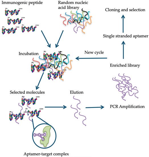
[83]. Briefly, in the case of aptamer designed for gluten detection, specifically the 33-mer immunogenic epitope, a library consisting of 1014-1015 single-stranded DNA oligonucleotides with a portion of the random nucleotide sequence is synthesized by a combinatorial chemical synthesis technique, and incubated with the target [84]. Unconjugated or low-affinity binding molecules are removed, and captured nucleic acid molecules are eluted and amplified by PCR. As a result, double-stranded PCR products are produced, which are later converted to single-stranded aptamers. The whole process is repeated several times until a group of high-affinity binding aptamers is obtained [85] (Figure 1). Aptamers are small molecules typically < 100-mers that fold into three-dimensional structures with their self-annealing properties. Target identification is due to their structure and not by their sequence (see Figure 1). Aptamer-target complexes present dissociation constants (Kd) within the low picomolar (1 × 10⁻¹² M) to nanomolar (1 × 10⁻⁹ M) range, which reflects toward their high binding affinity. Furthermore, target recognition is highly specific because aptamers can clearly distinguish between closely related protein targets [86].
Figure 1. Schematic representation of the different steps in the systematic evolution of ligands by exponential enrichment (SELEX) procedure, modified from Banerjee [87] and Stoltenburg et al. [83][88].
Table 2. Comparison of aptamers and antibodies based on properties.

Properties

Aptamers

Antibodies

Reference

Affinity

Very high target affinity, dissociation constants from micro to picomolar range.

Lower target affinity except for some monoclonal antibodies.

[89]

Immunogenic effect

Independent of immunogenic effect, due to their in vitro production.

Immune response can fail when the target molecule, has a structure similar to an endogenous protein.

[90]

Specificity

High binding specificity, e.g., the Anti-theophyllin aptamer displayed 10,000-fold discrimination against caffeine (Theophyllin differs from caffeine by a single methyl group).

Depends on target type.

[91]

Production

In vitro.

In vivo. Use of animals or cell lines.

[92]

Consistency

Chemical synthesis, extreme accuracy, and reproducibility. Little or no batch-to-batch variation.

May have in vivo variations. Restricted to environmental conditions.

[90]

Properties

Can be optimized on demand for increasing binding affinity and specificity.

Properties cannot be changed on demand.

[84][88]

Stability

Undergo denaturation, but reversible within minutes.

Irreversible denaturation. Stable under physiological conditions

[90]

Range of targets

Combinatorial library can be produced against any type of target, even toxic targets.

Restricted to molecules that produce immunogenic effect.

[93]

Shelf-life

Stable to long-term storage at ambient temperature.

Limited shelf-life.

[92]

Functionalization

Labeling does not affect affinity.

Attachment of molecules can cause loss in affinity.

[84][88][94]

References

  1. Domenico, L.; Donald, K. One and Two Dimensional (two pH) Polyacrilamide Gel Electrophoresis in a Single Gel: Separation of Wheat Proteins. Cereal Chem. 1985, 62, 314–319.
  2. Kasarda, D.D. Can an increase in celiac disease be attributed to an increase in the gluten content of wheat as a consequence of wheat breeding? J. Agric. Food Chem. 2013, 61, 1155–1159.
  3. Arranz-Otaegui, A.; Carretero, L.G.; Ramsey, M.N.; Fuller, D.Q.; Richter, T. Archaeobotanical evidence reveals the origins of bread 14,400 years ago in northeastern Jordan. Proc. Natl. Acad. Sci. USA 2018, 115, 7925–7930.
  4. Elli, L.; Branchi, F.; Tomba, C.; Villalta, D.; Norsa, L.; Ferretti, F.; Roncoroni, L.; Bardella, M.T. Diagnosis of gluten related disorders: Celiac disease, wheat allergy and non-celiac gluten sensitivity. World J. Gastroenterol. WJG 2015, 21, 7110.
  5. Kilian, B.; Martin, W.; Salamini, F. Genetic diversity, evolution and domestication of wheat and barley in the Fertile Crescent. In Evolution in Action; Springer: Berlin, Germany, 2010; pp. 137–166.
  6. Turner, G.D.; Dunne, M.R.; Ryan, A.W. Celiac Disease: Background and Historical Context. In Celiac Disease; Humana Press: New York, NY, USA, 2015; pp. 3–14.
  7. Simpson, R. Gluten-Free Rome: Celiac disease in the bioarchaeological record. COMPASS 2017, 1, 13–24.
  8. Van den Broeck, H.C.; de Jong, H.C.; Salentijn, E.M.; Dekking, L.; Bosch, D.; Hamer, R.J.; Gilissen, L.J.; van der Meer, I.M.; Smulders, M.J. Presence of celiac disease epitopes in modern and old hexaploid wheat varieties: Wheat breeding may have contributed to increased prevalence of celiac disease. TAG. Theor. Appl. Genet. 2010, 121, 1527–1539.
  9. Sapone, A.; Lammers, K.M.; Casolaro, V.; Cammarota, M.; Giuliano, M.T.; De Rosa, M.; Stefanile, R.; Mazzarella, G.; Tolone, C.; Russo, M.I. Divergence of gut permeability and mucosal immune gene expression in two gluten-associated conditions: Celiac disease and gluten sensitivity. BMC Med. 2011, 9, 23.
  10. Freeman, H.J. The Neolithic revolution and subsequent emergence of the celiac affection. Int. J. Celiac. Dis. 2013, 1, 19–22.
  11. Sapone, A.; Bai, J.C.; Ciacci, C.; Dolinsek, J.; Green, P.H.; Hadjivassiliou, M.; Kaukinen, K.; Rostami, K.; Sanders, D.S.; Schumann, M. Spectrum of gluten-related disorders: Consensus on new nomenclature and classification. BMC Med. 2012, 10, 13.
  12. Wen, S.; Wen, N.; Pang, J.; Langen, G.; Brew-Appiah, R.A.; Mejias, J.H.; Osorio, C.; Yang, M.; Gemini, R.; Moehs, C.P.; et al. Structural genes of wheat and barley 5-methylcytosine DNA glycosylases and their potential applications for human health. Proc. Natl. Acad. Sci. USA 2012, 109, 20543–20548.
  13. Shewry, P.R.; Sayanova, O.; Tatham, A.S.; Tamas, L.; Turner, M.; Richard, G.; Hickman, D.; Fido, R.; Halford, N.G.; Greenfield, J.; et al. Structure, Assembly and Targeting of Wheat Storage Proteins. J. Plant Physiol. 1995, 145, 620–625.
  14. Osorio, C.; Wen, N.; Gemini, R.; Zemetra, R.; von Wettstein, D.; Rustgi, S. Targeted modification of wheat grain protein to reduce the content of celiac causing epitopes. Funct. Integr. Genom. 2012, 12, 417–438.
  15. Lebwohl, B.; Ludvigsson, J.F.; Green, P.H. Celiac disease and non-celiac gluten sensitivity. BMJ 2015, 351, h4347.
  16. Biesiekierski, J.R. What is gluten? J. Gastroenterol. Hepatol. 2017, 32, 78–81.
  17. Shan, L.; Molberg, O.; Parrot, I.; Hausch, F.; Filiz, F.; Gray, G.M.; Sollid, L.M.; Khosla, C. Structural basis for gluten intolerance in celiac sprue. Science 2002, 297, 2275–2279.
  18. Shewry, P.R.; Tatham, A.S. Disulphide bonds in wheat gluten proteins. J. Cereal Sci. 1997, 25, 207–227.
  19. Sollid, L.M. Coeliac disease: Dissecting a complex inflammatory disorder. Nat. Rev. Immunol. 2002, 2, 647–655.
  20. Shan, L.; Qiao, S.W.; Arentz-Hansen, H.; Molberg, O.; Gray, G.M.; Sollid, L.M.; Khosla, C. Identification and analysis of multivalent proteolytically resistant peptides from gluten: Implications for Celiac Sprue. J. Proteome Res. 2005, 4, 1732–1741.
  21. Theethira, T.G.; Dennis, M.; Leffler, D.A. Nutritional consequences of celiac disease and the gluten-free diet. Expert Rev. Gastroenterol. Hepatol. 2014, 8, 123–129.
  22. Green, P.H.; Lebwohl, B.; Greywoode, R. Celiac disease. J. Allergy Clin. Immunol. 2015, 135, 1099–1106.
  23. Leonard, M.M.; Sapone, A.; Catassi, C.; Fasano, A. Celiac disease and nonceliac gluten sensitivity: A review. JAMA 2017, 318, 647–656.
  24. Guandalini, S.; Discepolo, V. Celiac disease. In Textbook of Pediatric Gastroenterology, Hepatology and Nutrition; Springer: Cham, Switzerland, 2016; pp. 453–469.
  25. Molberg, O.; McAdam, S.N.; Korner, R.; Quarsten, H.; Kristiansen, C.; Madsen, L.; Fugger, L.; Scott, H.; Noren, O.; Roepstorff, P.; et al. Tissue transglutaminase selectively modifies gliadin peptides that are recognized by gut-derived T cells in celiac disease. Nat. Med. 1998, 4, 713–717.
  26. Dieterich, W.; Ehnis, T.; Bauer, M.; Donner, P.; Volta, U.; Riecken, E.O.; Schuppan, D. Identification of tissue transglutaminase as the autoantigen of celiac disease. Nat. Med. 1997, 3, 797–801.
  27. Bethune, M.T.; Khosla, C. Parallels between pathogens and gluten peptides in celiac sprue. PLoS Pathog. 2008, 4, e34.
  28. Siegel, M.; Strnad, P.; Watts, R.E.; Choi, K.; Jabri, B.; Adler, G.; Ornary, B.; Khosla, C. Extracellular transglutaminase 2 is catalytically inactive, but is transiently activated upon tissue injury in the small intestine. Gastroenterology 2008, 134, A151.
  29. Sollid, L.M.; Jabri, B. Celiac disease and transglutaminase 2: A model for posttranslational modification of antigens and HLA association in the pathogenesis of autoimmune disorders. Curr. Opin. Immunol. 2011, 23, 732–738.
  30. Sollid, L.M.; Jabri, B. Triggers and drivers of autoimmunity: Lessons from coeliac disease. Nat. Rev. Immunol. 2013, 13, 294.
  31. Fasano, A.; Sapone, A.; Zevallos, V.; Schuppan, D. Nonceliac gluten sensitivity. Gastroenterology 2015, 148, 1195–1204.
  32. Tatham, A.S.; Shewry, P.R. Allergens to wheat and related cereals. Clin. Exp. Allergy 2008, 38, 1712–1726.
  33. Czaja-Bulsa, G. Non coeliac gluten sensitivity–A new disease with gluten intolerance. Clin. Nutr. 2015, 34, 189–194.
  34. Bascunan, K.A.; Vespa, M.C.; Araya, M. Celiac disease: Understanding the gluten-free diet. Eur. J. Nutr. 2017, 56, 449–459.
  35. Shepherd, S.; Gibson, P. Nutritional inadequacies of the gluten-free diet in both recently-diagnosed and long-term patients with coeliac disease. J. Hum. Nutr. Diet. 2013, 26, 349–358.
  36. Løvik, A.; Skodje, G.; Bratlie, J.; Brottveit, M.; Lundin, K. Diet adherence and gluten exposure in coeliac disease and self-reported non-coeliac gluten sensitivity. Clin. Nutr. 2017, 36, 275–280.
  37. Verma, A.; Gatti, S.; Galeazzi, T.; Monachesi, C.; Padella, L.; Del, B.G.; Annibali, R.; Lionetti, E.; Catassi, C. Detection of gluten content in the naturally gluten free and gluten free labelled commercially available food products in Italy. Dig. Liver Dis. 2016, 48, e279.
  38. Verma, A.; Gatti, S.; Galeazzi, T.; Monachesi, C.; Padella, L.; Baldo, G.; Annibali, R.; Lionetti, E.; Catassi, C. Gluten contamination in naturally or labeled gluten-free products marketed in Italy. Nutrients 2017, 9, 115.
  39. Catassi, C.; Bai, J.; Bonaz, B.; Bouma, G.; Calabrò, A.; Carroccio, A.; Castillejo, G.; Ciacci, C.; Cristofori, F.; Dolinsek, J. Non-celiac gluten sensitivity: The new frontier of gluten related disorders. Nutrients 2013, 5, 3839–3853.
  40. Allen, P.J. Primary Care Approaches. Gluten-Related Disorders: Celiac Disease, Gluten Allergy, Non-Celiac Gluten Sensitivity. Pediatric Nurs. 2015, 41, 3.
  41. Dominguez-Ortega, G.; Borrelli, O.; Meyer, R.; Dziubak, R.; De Koker, C.; Godwin, H.; Fleming, C.; Thapar, N.; Elawad, M.; Kiparissi, F. Extraintestinal manifestations in children with gastrointestinal food allergy. J. Pediatr. Gastroenterol. Nutr. 2014, 59, 210–214.
  42. Volta, U.; Caio, G.; Tovoli, F.; De Giorgio, R. Non-celiac gluten sensitivity: Questions still to be answered despite increasing awareness. Cell. Mol. Immunol. 2013, 10, 383.
  43. Codex Alimentarius Commission. Codex Standard 118-1979 (rev. 2008), Foods for special dietary use for persons intolerant to gluten. In Codex Alimentarium; FAO: Rome, Italy; WHO: Geneva, Switzerland, 2008; pp. 1–3.
  44. Thompson, T.; Lee, A.R.; Grace, T. Gluten contamination of grains, seeds, and flours in the United States: A pilot study. J. Am. Diet. Assoc. 2010, 110, 937–940.
  45. Thompson, T.; Dennis, M.; Emerson, L. Gluten-Free Labeling: Are Growth Media Containing Wheat, Barley, and Rye Falling through the Cracks? J. Acad. Nutr. Diet. 2018, 118, 2025.
  46. Falcomer, A.L.; Araújo, L.S.; Farage, P.; Monteiro, J.S.; Nakano, E.Y.; Zandonadi, R.P. Gluten contamination in food services and industry: A systematic review. Crit. Rev. Food Sci. Nutr. 2018, 1–15.
  47. Farage, P.; de Medeiros Nóbrega, Y.K.; Pratesi, R.; Gandolfi, L.; Assunção, P.; Zandonadi, R.P. Gluten contamination in gluten-free bakery products: A risk for coeliac disease patients. Public Health Nutr. 2017, 20, 413–416.
  48. Don, C.; Halbmayr-Jech, E.; Rogers, A.; Koehler, P. AACCI Approved Methods Technical Committee report: Collaborative study on the immunochemical quantitation of intact gluten in rice flour and rice-based products using G12 sandwich ELISA. Cereal Foods World 2014, 59, 187–193.
  49. Allred, L.K.; Kupper, C.; Iverson, G.; Perry, T.B.; Smith, S.; Stephen, R. Definition of the “Purity Protocol” for Producing Gluten-Free Oats. Cereal Chem. 2017, 94, 377–379.
  50. Van Eckert, R.; Pfannhauser, W.; Riedl, O. Beitrag zur Qualitätssicherung bei der Herstellung von glutenfreien Lebensmitteln. Ernährung. Nutrition 1992, 16, 511–512.
  51. Fritschy, F.; Windemann, H.; Baumgartner, E. Determination of wheat gliadins in food with ELISA. Zeitschrift fur Lebensmittel-Untersuchung und-Forschung 1985, 181, 379–385.
  52. Janssen, F.; Hägele, G.; de Baaij, J. Gluten-free products, the Dutch experience. In Coeliac Disease; Springer: Dordrecht, The Netherlands, 1991; pp. 95–100.
  53. Olexova, L.; Dovičovičová, L.; Švec, M.; Siekel, P.; Kuchta, T. Detection of gluten-containing cereals in flours and “gluten-free” bakery products by polymerase chain reaction. Food Control 2006, 17, 234–237.
  54. Hernando, A.; Mujico, J.R.; Mena, M.C.; Lombardía, M.; Mendez, E. Measurement of wheat gluten and barley hordeins in contaminated oats from Europe, the United States and Canada by Sandwich R5 ELISA. Eur. J. Gastroenterol. Hepatol. 2008, 20, 545–554.
  55. Geng, T.; Westphal, C.; Yeung, J. Detection of Gluten by Commercial Test Kits: Effects of Food Matrices and Extraction Procedures; American Chemical Society Symposium Series: Washington, DC, USA, 2008; Volume 1001, pp. 462–475.
  56. Kelly, C.P.; Bai, J.C.; Liu, E.; Leffler, D.A. Advances in diagnosis and management of celiac disease. Gastroenterology 2015, 148, 1175–1186.
  57. Rostami, K.; Kerckhaert, J.; Tiemessen, R.; Von Blomberg, B.M.E.; Meijer, J.W.; Mulder, C.J. Sensitivity of antiendomysium and antigliadin antibodies in untreated celiac disease: Disappointing in clinical practice. Am. J. Gastroenterol. 1999, 94, 888.
  58. Catassi, C.; Fasano, A. Celiac disease. In Gluten-Free Cereal Products and Beverages; Academic Press: Cambridge, MA, USA, 2008; p. 1-I.
  59. Singh, P.; Arora, A.; Strand, T.A.; Leffler, D.A.; Catassi, C.; Green, P.H.; Kelly, C.P.; Ahuja, V.; Makharia, G.K. Global prevalence of celiac disease: Systematic review and meta-analysis. Clin. Gastroenterol. Hepatol. 2018, 16, 823–836.e2.
  60. Tanveer, M.; Ahmed, A. Non-Celiac Gluten Sensitivity: A Systematic Review. J. Coll. Physicians Surg. Pak. 2019, 29, 51–57.
  61. Rubio-Tapia, A.; Ludvigsson, J.F.; Brantner, T.L.; Murray, J.A.; Everhart, J.E. The prevalence of celiac disease in the United States. Am. J. Gastroenterol. 2012, 107, 1538.
  62. Barada, K.; Bitar, A.; Mokadem, M.A.-R.; Hashash, J.G.; Green, P. Celiac disease in Middle Eastern and North African countries: A new burden? World J. Gastroenterol. WJG 2010, 16, 1449.
  63. Peña, A.S.; Rodrigo, L. Epidemiology of celiac disease and non-celiac gluten-related disorders. In Advances in the Understanding of Gluten Related Pathology and the Evolution of Gluten-Free Foods; OmniaScience: Barcelona, Spain, 2015; pp. 27–73.
  64. Malekzadeh, R.; Sachdev, A.; Ali, A.F. Coeliac disease in developing countries: Middle East, India and North Africa. Best Pract. Res. Clin. Gastroenterol. 2005, 19, 351–358.
  65. Ramakrishna, B.; Makharia, G.K.; Chetri, K.; Dutta, S.; Mathur, P.; Ahuja, V.; Amarchand, R.; Balamurugan, R.; Chowdhury, S.D.; Daniel, D. Prevalence of adult celiac disease in India: Regional variations and associations. Am. J. Gastroenterol. 2016, 111, 115.
  66. Makharia, G.K.; Verma, A.K.; Amarchand, R.; Bhatnagar, S.; Das, P.; Goswami, A.; Bhatia, V.; Ahuja, V.; Datta Gupta, S.; Anand, K. Prevalence of celiac disease in the northern part of India: A community based study. J. Gastroenterol. Hepatol. 2011, 26, 894–900.
  67. Rajpoot, P.; Makharia, G. Problems and challenges to adaptation of gluten free diet by Indian patients with celiac disease. Nutrients 2013, 5, 4869–4879.
  68. Wu, J.; Xia, B.; von Blomberg, B.; Zhao, C.; Yang, X.; Crusius, J.; Peña, A. Coeliac disease: Emerging in China? Gut 2010, 59, 418–419.
  69. Yuan, J.; Zhou, C.; Gao, J.; Li, J.; Yu, F.; Lu, J.; Li, X.; Wang, X.; Tong, P.; Wu, Z. Prevalence of celiac disease autoimmunity among adolescents and young adults in China. Clin. Gastroenterol. Hepatol. 2017, 15, 1572–1579.e1.
  70. Reese, I.; Schäfer, C.; Kleine-Tebbe, J.; Ahrens, B.; Bachmann, O.; Ballmer-Weber, B.; Beyer, K.; Bischoff, S.C.; Blümchen, K.; Dölle, S.; et al. Non-celiac gluten/wheat sensitivity (NCGS)—A currently undefined disorder without validated diagnostic criteria and of unknown prevalence. Allergo J. Int. 2018, 27, 147–151.
  71. Allred, L.K.; Kupper, C.; Quinn, C. The Use of Visual Examination for Determining the Presence of Gluten-Containing Grains in Gluten Free Oats and Other Grains, Seeds, Beans, Pulses, and Legumes. J. AOAC Int. 2018, 101, 36–44.
  72. Bustamante, M.; Fernández-Gil, M.; Churruca, I.; Miranda, J.; Lasa, A.; Navarro, V.; Simón, E. Evolution of gluten content in cereal-based gluten-free products: An overview from 1998 to 2016. Nutrients 2017, 9, 21.
  73. Sharma, G.M.; Pereira, M.; Williams, K.M. Gluten detection in foods available in the United States–A market survey. Food Chem. 2015, 169, 120–126.
  74. Miranda, J.; Simón, E. Gluten Content Change Over the Two Last Decades. In Nutritional and Analytical Approaches of Gluten-Free Diet in Celiac Disease; Springer: Cham, Switzerland, 2017; pp. 47–57.
  75. Moreno, M.D.; Rodríguez-Herrera, A.; Sousa, C.; Comino, I. Biomarkers to monitor gluten-free diet compliance in celiac patients. Nutrients 2017, 9, 46.
  76. Scherf, K.A.; Poms, R.E. Recent developments in analytical methods for tracing gluten. J. Cereal Sci. 2016, 67, 112–122.
  77. Codex Alimentarius Commission. Codex Standard 234-1999 (amended 2011), Recommended methods of analysis and sampling. Gluten-free foods: Enzyme-linked immunoassay R5 Mendez (ELISA) method. In Codex Alimentarious; FAO: Rome, Italy; WHO: Geneva, Switzerland, 2019; pp. 1–80.
  78. Ellis, H.; Doyle, A.; Wieser, H.; Sturgess, R.; Day, P.; Ciclitira, P. Measurement of gluten using a monoclonal antibody to a sequenced peptide of α-gliadin from the coeliac-activating domain I. J. Biochem. Biophys. Methods 1994, 28, 77–82.
  79. Rumbo, M.; Chirdo, F.G.; Fossati, C.A.; Añón, M.C. Analysis of the effects of heat treatment on gliadin immunochemical quantification using a panel of anti-prolamin antibodies. J. Agric. Food Chem. 2001, 49, 5719–5726.
  80. Doña, V.; Fossati, C.; Chirdo, F. Interference of denaturing and reducing agents on the antigen/antibody interaction. Impact on the performance of quantitative immunoassays in gliadin analysis. Eur. Food Res. Technol. 2008, 226, 591–602.
  81. Song, K.-M.; Lee, S.; Ban, C. Aptamers and their biological applications. Sensors 2012, 12, 612–631.
  82. Berezovski, M.V.; Musheev, M.U.; Drabovich, A.P.; Jitkova, J.V.; Krylov, S.N. Non-SELEX: Selection of aptamers without intermediate amplification of candidate oligonucleotides. Nat. Protoc. 2006, 1, 1359.
  83. Stoltenburg, R.; Reinemann, C.; Strehlitz, B. SELEX—a (r) evolutionary method to generate high-affinity nucleic acid ligands. Biomol. Eng. 2007, 24, 381–403.
  84. Zhou, J.; Battig, M.R.; Wang, Y. Aptamer-based molecular recognition for biosensor development. Anal. Bioanal. Chem. 2010, 398, 2471–2480.
  85. Van Dorst, B.; Mehta, J.; Bekaert, K.; Rouah-Martin, E.; De Coen, W.; Dubruel, P.; Blust, R.; Robbens, J. Recent advances in recognition elements of food and environmental biosensors: A review. Biosens. Bioelectron. 2010, 26, 1178–1194.
  86. Nimjee, S.M.; Rusconi, C.P.; Sullenger, B.A. Aptamers: An emerging class of therapeutics. Annu. Rev. Med. 2005, 56, 555–583.
  87. Banerjee, J.; Nilsen-Hamilton, M. Aptamers: Multifunctional molecules for biomedical research. J. Mol. Med. 2013, 91, 1333–1342.
  88. Strehlitz, B.; Nikolaus, N.; Stoltenburg, R. Protein detection with aptamer biosensors. Sensors 2008, 8, 4296–4307.
  89. Robertson, D.L.; Joyce, G.F. Selection in vitro of an RNA enzyme that specifically cleaves single-stranded DNA. Nature 1990, 344, 467.
  90. Tombelli, S.; Mascini, M. Aptamers as molecular tools for bioanalytical methods. Curr. Opin. Mol. Ther. 2009, 11, 179–188.
  91. Jenison, R.D.; Gill, S.C.; Pardi, A.; Polisky, B. High-resolution molecular discrimination by RNA. Science 1994, 263, 1425–1429.
  92. Ellington, A.D.; Szostak, J.W. In vitro selection of RNA molecules that bind specific ligands. Nature 1990, 346, 818.
  93. Jayasena, S.D. Aptamers: An emerging class of molecules that rival antibodies in diagnostics. Clin. Chem. 1999, 45, 1628–1650.
  94. Wang, J.; Meng, W.; Zheng, X.; Liu, S.; Li, G. Combination of aptamer with gold nanoparticles for electrochemical signal amplification: Application to sensitive detection of platelet-derived growth factor. Biosens. Bioelectron. 2009, 24, 1598–1602.
More
Information
Contributor MDPI registered users' name will be linked to their SciProfiles pages. To register with us, please refer to https://encyclopedia.pub/register :
View Times: 585
Revisions: 2 times (View History)
Update Date: 13 Jul 2021
1000/1000