Submitted Successfully!
To reward your contribution, here is a gift for you: A free trial for our video production service.
Thank you for your contribution! You can also upload a video entry or images related to this topic.
Version Summary Created by Modification Content Size Created at Operation
1 + 4359 word(s) 4359 2021-09-23 05:01:12 |
2 format correct -13 word(s) 4346 2021-09-24 05:57:57 |

Video Upload Options

Do you have a full video?

Confirm

Are you sure to Delete?
Cite
If you have any further questions, please contact Encyclopedia Editorial Office.
Rachakonda, G.; Odiase, P.; Villalta, F. Microbiome Populations in the Nose. Encyclopedia. Available online: https://encyclopedia.pub/entry/14502 (accessed on 20 April 2024).
Rachakonda G, Odiase P, Villalta F. Microbiome Populations in the Nose. Encyclopedia. Available at: https://encyclopedia.pub/entry/14502. Accessed April 20, 2024.
Rachakonda, Girish, Peace Odiase, Fernando Villalta. "Microbiome Populations in the Nose" Encyclopedia, https://encyclopedia.pub/entry/14502 (accessed April 20, 2024).
Rachakonda, G., Odiase, P., & Villalta, F. (2021, September 24). Microbiome Populations in the Nose. In Encyclopedia. https://encyclopedia.pub/entry/14502
Rachakonda, Girish, et al. "Microbiome Populations in the Nose." Encyclopedia. Web. 24 September, 2021.
Microbiome Populations in the Nose
Edit

Acute and chronic upper respiratory illnesses such as asthma, and allergic rhinitis (AR) have been linked to the presence of microorganisms in the nose. Microorganisms can exist in symbiotic or commensal relationships with the human body. However, in certain cases, opportunistic pathogens can take over, leading to altered states (dysbiosis) and causing disease. Thus, the microflora present in a host can be useful to reflect health status. The human body contains 10 trillion to 100 trillion microorganisms. Of these populations, certain pathogens have been identified to promote or undermine wellbeing. Therefore, knowledge of the microbiome is potentially helpful as a diagnostic tool for many diseases. Variations have been recognized in the types of microbes that inhabit various populations based on geography, diet, and lifestyle choices and various microbiota have been shown to modulate immune responses in allergic disease. Interestingly, the diseases affected by these changes are prevalent in certain racial or ethnic populations. These prevalent microbiome variations in these groups suggest that the presence of these microorganisms may be significantly associated with health disparities. 
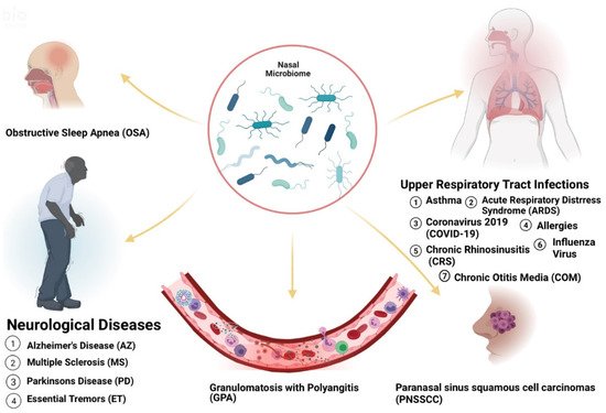
nasal microbiome allergic rhinitis microbial dysbiosis asthma immune response health disparities Moraxella Staphyloccus aureus neurological disease race or ethnicity

1. Introduction

Complex relationships exist between health and biology, genetics and individual behavior. Host lifestyle preferences and hygiene, as well as access to health information and health service, socioeconomic status, environment, discrimination, racism, literacy levels and legislative policies, can impact the health status of an individual or a population group. Disparate geographical surroundings, including physical, chemical and psychosocial factors, impact health choices leading to changes in health outcomes by influencing the severity of disease across racial groups. These aforementioned factors, including the host’s immune system, have also been implicated in observed human microbiome shifts [1]. Health disparities are differences in the health status of groups of people based on factors such as race, ethnicity, age, sex, socio-economic status, geographic location, mental health, disability, citizenship status or other characteristic linked historically to exclusion or discrimination [2]. There are large racial and ethnic differences in health in the United States, and certain health conditions or diseases tend to be prevalent within specific population groups [3]. Despite this, not much is known about agents that spark microbiome alterations across many disease states and not a lot of research has been undertaken to fully understand how racial and ethnic health disparities are linked to variations in the microbiome. This is important as racial and ethnic health differences permeate every social economic stratum. Diversity in diet, and other habits that shape health which are impacted by cultural, ancestral and social frameworks may determine microbiome differences and cause health disparities. Previous research studies have categorized environmental factors that influence health disparities and even certain microbiome variations in particular ethnic groups. For example, single gene mutations in sickle cell disease (SCD) are most common in Blacks and African Americans (AAs) owing to ancestral linkage to genetic adaptation to environmental factors such as malaria-causing anopheles’ mosquitos in Africa [4]. Similarly, SCD is also observed among Indian and Middle Eastern Arab populations owing to the endemicity of malaria [5][6]. SCD patients, when compared with carriers of the sickle cell trait, showed differences in microbiota composition in the top 15 genera that accounted for 84% of the taxonomic abundance across all samples. Pseudobutyrivibrio (p = 0.05), Faecalibacterium (p = 0.11), Subdoligranulum (p = 0.16) (all phylum Firmicutes), Prevotella 9 (p = 0.07), and Alistipes (p = 0.04) (both phylum Bacteroidetes) were lower in abundance, while Escherichia-Shigella (p = 0.11) (phylum Proteobacteria) was higher in abundance in SCD [7]. Vaginal characterization amongst women of different ethnic backgrounds revealed variations in vaginal pH and the microbiota in European women are more likely than AA women to be Lactobacillus-dominated, which appeared to maintain vaginal health [8]. These highlighted studies reveal interesting features of the microbiome in various ethnic populations and show how understanding social and environmental factors can influence our understanding of disease. Very little is known about the impact of nasal microbiota on immune responses in the host. Yet, variations in nasal microbiota are observed among ethnic groups with associated disparities in the prevalence of diseases. For example, rheumatoid arthritis, an autoimmune disorder, has been associated with changes in the oral and gut microbiomes which influence the loss of tolerance against self-antigens and impact the inflammatory events that aid the damage of joints. Interestingly, rheumatoid arthritis occurs in varying levels among various ethnic groups in the United States. Significant differences of mean disease activity level (p < 0.001) were observed across racial and ethnic groups and these differences persisted (p < 0.046) even though improvements in disease activity were observed in all groups over a 5-year period. Remission rates also remained significantly different across racial/ethnic groups across all models ranging from 22.7 (95% Confidence Interval (CI) 19.5–25.8) in AAs to 27.4 (95% CI 24.9–29.8) in CAs [9]. Since the microbiome has immunomodulatory functions, an imbalance or dysbiosis in microbial community structure could be the driving force behind diseases [10]. Thus, we aim to analyze the information from the current/existing literature on nasal biome variations and investigate its role in health disparities by considering its impacts on the physiological and biological processes. Furthermore, we aim to evaluate the social and physical environmental factors that influence the genesis of such microbiome variations, especially within ethnic groups. Revealing the influence that the microbiome has on the existence of health disparities could shed light on the increased prevalence of certain diseases in some populations and improve our comprehension of why certain ethnic groups have greater disease risk and fatality compared to others.

2. Microbiome Populations in the Nose and Impacts of Dysbiosis in Disease States

The human body hosts a variety of microorganisms that reside in helpful, harmful, or otherwise commensal relationships within the body. The human microbiome refers specifically to the complex community of over 100 trillion microorganisms, living in human microhabitats [11]. Due to microbial niche specificity, the composition of microbes within a microenvironment differs based on the location on the human body, such as the gastrointestinal tract, skin and airways. Analysis of bacterial populations in the anterior nares from metagenomic samples revealed the presence of four dominant bacterial communities: Moraxella, Staphylococcus, Propionibacterium and Corynebacterium [12]. In Finland, five nasal microbiota profiles were observed in infants namely Moraxella, Streptococcus, Dolosigranulum, Staphylococcus and Corynebacteriaceae [13]. Other bacteria detected in the sinuses included Firmicutes, Proteobacteria and Actinobacteria phyla. On the species level, Staphylococcus epidermidis, Propionibacterium acnes and Staphylococcus aureus were the most prevalent. Corynebacterium tuberculostearicum was the most dominant of the Corynebacterium phyla. Opportunistic pathogens including Streptococcus pneumoniae, Haemophilus influenzae, Moraxella catarrhalis, Stenotrophomonas maltophilia, Enterobacter species were also found in healthy individuals [12]. Many studies report that the nasal microbiome of healthy humans is primarily composed of the phyla Actinobacteria, Bacteroidetes, Firmicutes and Proteobacteria with representatives of genera Bifidobacterium, Corynebacterium, Staphylococcus, Streptococcus, Dolosigranulum and Moraxella predominating. Significant alterations in the microbial community structure have been linked to the development and progression of disease [14] (Figure 1).
Figure 1. Several diseases are linked to dysbiosis in nasal microbiota.
Nasal bacterial communities also show spatial variation, dependent on epithelium type. In adenoid and tonsillar tissue, Haemophilus influenzae primarily diffusely infiltrated the tissue, Streptococcus and Bacteroides were chiefly found in fissures, and Fusobacteria, Pseudomonas and Burkholderia were exclusively located within adherent bacterial layers and infiltrates [15]. In terms of the pathology of infection, microbial dominance plays a chief role (Table 1).
Table 1. Disparities in the abundance of microbiota are linked to disease and may vary across ethnic groups.
Disease Greater Abundance Linked to Disease Affected Group Known Risk Factors
Chronic otitis media with effusion (COME) Corynebacterium, Streptococcus, Moraxella [16] Caucasian Americans (CAs) [17] allergy or atopy, upper respiratory tract infection, snoring, acute otitis media, passive smoke exposure and low social status [17][18]
Pediatric Asthma Corynebacterium [19] Puerto-Ricans (PRs), African Americans (AAs) [20] parental asthma, prenatal environmental tobacco smoke, having cats and prematurity [21][22]
Chronic Rhinosinusitis (CRS) Corynebacterium, Burkholderia [23][24] Latino Americans (LAs) [24] Asthma, genetics, GERD, rheumatoid arthritis, migraine, cigarette smoking [25]
COVID-19 Salmonella, Scardovia, Serratia and Pectobacteriaceae [26][27] AAs and Asian Americans (AS) [28] Hypertension, coronary artery disease, history of stroke, diabetes, obesity, severe obesity, chronic kidney disease, asthma, and chronic obstructive pulmonary disease [29]
Granulomatosis with Polyangiitis (GPA) Staphylococcus aureus [30] Not Distinguishable [31] Animal (horses) exposure, history of bronchiectasis, autoimmune disease, chronic renal impairment, Pulmonary fibrosis [32][33]
Acute Otitis Media (AOM) Haemophilus, Moraxella, and Neisseria [16] CAs [34] Out-of-home daycare, multiple children living in the home, and mother’s multiparity [34]
Influenza B virus (IBV) Streptococcus species and Prevotella salivae [35]   Diabetes, chronic respiratory disease [36]
Influenza A virus (IAV) Staphylococcus aureus, Staphylococcu pneumoniae, Klebsiella and Aerococcus [37]  
Acute Respiratory Distress Syndrome (ARDS)   CAs [38] abuse of alcohol and tobacco, malnutrition and obesity [39]
Obstructive Sleep Apnea (OSA) Streptococcus, Prevotella and Veillonella [40]   Obesity, rhinitis, adenoid hypertrophy [41]
Parkinson’s Disease (PD) Actinobacter [42][43]   OSA, Head injury, family history of trauma and depression, family history of PD [41][44]
Alzheimer’s Disease (AZ)     OSA, diet, the immune system, mitochondrial function, metal exposure, and infection [41][45]
Essential Tremors (ET)   AAs, LAs [46] Exposure to neurotoxic compounds such as β-carboline alkaloids and ethanol. Exposure to pesticide and lead. Tobacco exposure [47][48]
Atopic Dermatitis (AD) AAs [49] Viral and bacterial skin infections, neuropsychiatric diseases, family history, smoking, allergy, maternal asthma, and dogs [50]
Psoriasis CAs, Eastern African [51][52] Stress, diabetes mellitus, obesity, smoking, air pollution arthritis, inflammatory bowel disease, alcohol, drugs, cardiovascular disease, infection, sun exposure, hypertension [53][54]
Interestingly, unique microbiota signatures exist within ethnic groups in the United States. Nasal microbiota has not yet been successfully characterized within ethnic groups in the United States, however, Mason et al., undertook a study using a machine learning classifier to distinguish between ethnicity of individuals based on their salivary samples. The classifier identified African Americans with a 100% sensitivity and 74% specificity and Caucasians with a 50% sensitivity and 91% specificity signifying that our microbiome may be genetically influenced and can act as an ethnic fingerprint [55]. Although nasal and oral microbiota have obvious discrepancies due to distinct environments for bacterial adhesion, survival, and growth [56], the differences observed are broadly mirrored disparities in each taxon’s signal abundance at each site. Since both the oropharynx and nostril collect drainage from shared sources (nasopharynx, sinuses, and nasal cavity), there is a major overlap in the taxa seen [57]. Mason et al. discovered that ethnicity was a crucial determinant of oral microbial colonization and that there exists a significant association between ethnicity and the composition of the oral microbiome among AAs, CAs, Chinese (CH) and LAs living within the United States. The abundances of four species level operational taxonomic units (OTUs) and several genus level-OTUs were significantly different between the ethnicities (* p < 0.05, ** p < 0.01, *** p < 0.001, **** p < 0.0001). Importantly, AAs demonstrated significantly lower bacterial diversity (*** p < 0.001, ANOVA) and equitability (*** p < 0.001, ANOVA) compared to the other ethnicities [55]. This indicates that AAs have fewer subgingival species and that a few of these species are numerically dominant members of the community when compared to the other ethnicities. Decreased diversity is considered an indicator of an unhealthy microbiome and has been linked to different chronic conditions such as decreased gut microbiota diversity in obesity and type 2 diabetes [58]. Thus, AAs may be more susceptible to respiratory illnesses in the United States.

2.1. Asthma

In children diagnosed with asthma, at least 95% hosted pathogenic genera Moraxella, Staphylococcus, Streptococcus, and Haemophilus in the nose. Different asthma phenotypes were significantly associated with variations in phyla (Proteobacteria, Actinobacteria and Bacteroidetes) abundances, community composition, structure, and co-occurrence of bacterial genera (Moraxella, Corynebacterium, Dolosigranulum and Prevotella) [59]. A predominant Corynebacterium population led to better asthma control compared to those with Moraxella. As exacerbation rates worsened, the microbiome population switched from Corynebacterium to a higher relative abundance of Moraxella [19]. In older (7.5 years old) children, the development of asthma was associated with an increase in H.influenzae colonization at 13 months of age. This association was not observed with H.influenzae colonization at 2 months of age [60]. The dominance of Moraxella during ages 2 to 13 months was also associated with a higher risk of developing childhood asthma [61]. In the United States, childhood asthma, affects one group of people differently than another group. Although childhood asthma is prevalent across all ethnic groups, the disease burden is disproportionately shared by Puerto Ricans (PRs) (19.2%) or African Americans (12.7%) compared to Caucasian Americans (CAs) (8%) or Mexican Americans (MAs) (6.4%) [20]. Ethnic disparities in asthma mortality rate are even greater with asthma mortality rates in children and adults in AAs being nearly eightfold and threefold higher, respectively, than in CAs [62]. The saliva of AA children with or without asthma was analyzed to compare bacterial diversity within the same demographic due to difficulty in obtaining nasal samples. Saliva samples from asthma cases were more enriched in pathogenic species compared to healthy controls. Differences between cases and controls were revealed in terms of diversity, as well as in relative abundance for Streptococcus genus (13.0% in cases vs. 18.3% in controls, p = 0.003) and Veillonella genus (11.1% in cases vs. 8.0% in controls, p = 0.002) [63]. Interestingly, no correlation with Moraxella, Neisseria or Haemophilus genera was found. The phylum in which these genera, Proteobacteria, were, however, included, and showed a trend to be more abundant in cases (27.7%) than in controls (25.4%) [63]. Risk factors include allergy or atopy, upper respiratory tract infection, snoring, acute otitis media, passive smoke exposure and low social status [21][22].

2.2. Acute Respiratory Illnesses

Influenza and other respiratory viral infections are the most common type of acute respiratory infection with great effects on host immune mechanisms. Acute respiratory disease (ARI) was shown to develop more frequently in children with early Moraxella-dominant profiles and less in Corynebacteriaceae-dominant profiles [13]. Streptococcus species and Prevotella salivae were additionally associated with the greater susceptibility to influenza B viral (IBV) infection and decreased susceptibility to influenza A viral (IAV) infection [35] ARIs such as Influenza induce antiviral immune responses that are associated with changes in microbial composition and function. These changes in the immune response also predispose patients to secondary bacterial infections, which are typically clinically more severe [64]. After adjusting for sex, age, race, disease severity, type of hospital and median household income for patient ZIP code, AAs had a greater odds ratio of in-hospital death for sepsis-related respiratory failure when compared with CAs (odds ratio [OR], 1.13; 95% CI, 1.11–1.14; p < 0.001), and LAs also had a greater odds ratio of in-hospital death when compared with CAs (OR, 1.17; 95% CI, 1.15–1.19; p < 0.001), and so did Asian and Pacific Islanders (OR, 1.15; 95% CI, 1.12–1.18; p < 0.001) and Native Americans (OR, 1.08; 95% CI, 1.00–1.15; p < 0.001) when compared with CAs (OR, 1.0) [22]. These variations may be related to complex interactions between an altered microbiome, virus-induced changes in immune response and growth of pathogenic bacteria as microbial diversity decreases [65]. Diabetes and Chronic respiratory disease also influence the occurrence of Influenza [37] Differences in Acute Respiratory Distress Syndrome (ARDS) incidence and associated mortality were also observed among different racial/ethnic affiliations. Among 96,350 patients studied, discrepancies were found among AAs and CAs for ARDS incidence (0.70% vs. 0.93%) and between LAs and CAs for ARDS-associated mortality (0.27% vs. 0.17%). There appeared to be a protective effect of AA race/ethnicity for ARDS incidence (OR, 0.73; 95% CI, 0.58–0.91) [38]. Risk factors for ARDS include abuse of alcohol and tobacco, malnutrition, and obesity [39].
Coronavirus disease 2019 (COVID-19), an infectious disease caused by binding of the viral spike (S) protein of the coronavirus to ACE2 receptors highly expressed in nasal goblet and ciliated cells, also correlated with microbial disparities in patients. In a study, Deinococcus thermus showed exclusive presentation only in controls when compared to COVID-19 patients admitted to intensive care unit (ICU), paucisymptomatic or affected by other coronaviruses. Candidatus saccharibacteria (formerly known as TM7) was strongly increased in negative controls and COVID-19 paucisymptomatic patients as compared to COVID-19 ICU patients. Bifidobacterium and Clostridium were completely depleted only in ICU COVID-19 patients and, Salmonella, Scardovia, Serratia and Pectobacteriaceae were observed only in ICU COVID-19 patients [26]. Likewise, there was a differential increase in species in patients with COVID-19 infection. Peptoniphilus lacrimalis, Campylobacter hominis, Prevotella 9 copri and Anaerococcus were more abundant in those with high viral load and COVID-19 infection whereas Staphylococcus haemolyticus, Prevotella disiens and Corynebacterium variants were more abundant in those with low viral load and without COVID-19 infection [27]. Ethnic disparities were also recorded in the United States. Using a large database containing 18,728,893 patients from 50 studies, AAs and AS ethnicities had a higher risk of COVID-19 infection compared to CAs individuals (pooled adjusted relative risk (RR) for AAs: 2.02, 95% CI 1.67–2.44; pooled adjusted RR for AS: 1.50, 95% CI 1.24–1.83) and sensitivity analyses examining peer-reviewed studies only (pooled adjusted RR for AA: 1.85, 95%CI: 1.46–2.35; pooled adjusted RR for AS: 1.51, 95% CI 1.22–1.88 [28]. Risk factors include hypertension, coronary artery disease, history of stroke, diabetes, obesity, severe obesity, chronic kidney disease, asthma and chronic obstructive pulmonary disease [29].

2.3. Rhinitis and Chronic Rhinosinusitis

Early childhood studies reveal that a decrease in bacterial diversity in infants over the period of 18 months, was associated with the development of rhinitis. Furthermore, symptoms associated with rhinitis were observed in infants with a depleted abundance of Corynebacterium [66]. Chronic rhinosinusitis (CRS) patients, however, showed an increased abundance of Corynebacterium [23]. Postoperatively patients with CRS who had better outcomes presented with greater bacterial diversity with higher relative abundances of Actinobacteria at the time of surgery [67]. LAs with CRS have greater disease severity and morbidity compared with CAs. Analysis of the nasal microbiota of CRS patients revealed that the nares of Latino patients were significantly less diverse compared to CAs (adjusted p-value = 0.03) and had a significantly higher relative abundance of Burkholderia genus compared to CAs (adjusted p-value = 0.04) [24]. Risk factors associated with CRS include asthma, gastroesophageal reflux disease (GERD), genetic polymorphisms, rheumatoid arthritis, migraine and cigarette smoking [25].

2.4. Otitis Media

Otitis media (OM) is a common pediatric diagnosis. Several risk factors have been associated with OM including lack of breast-feeding practices, daycare, mother multiparity and ethnic affiliation [34]. The association between OM diagnosis and race/ethnicity in 11,349 non-low-birthweight infants was measured. After adjustment for relevant confounders, AA (OR 0.74; 95% CI 0.61–0.89) and AS infants (OR 0.77; 95% CI 0.57–1.0) were less likely to be diagnosed with OM than CA infants [34].
Children with chronic otitis media with effusion (COME) had profiles that were Corynebacterium-dominated, Streptococcus-dominated or Moraxella-dominated when compared with healthy candidates. Haemophilus influenzae, Streptococcus pneumoniae, Moraxella catarrhalis and to a lesser extent Staphylococcus aureus were identified as principal composites involved in otitis. In contrast, higher abundances of Haemophilus, Moraxella and Neisseria in the nose have been found to be associated with acute OM (AOM) with lower abundances of Staphylococcus, Corynebacterium, Propionibacterium, Streptococcus (usually not S. pneumoniae) and Lactococcus [16]. There are several risk factors recorded for chronic OM (COM) such as the presence of allergy or atopy (OR, 1.36; 95% CI, 1.13–1.64; p = 0.001), an upper respiratory tract infection (OR, 6.59; 95% CI, 3.13–13.89; p < 0.00001), Snoring (OR, 1.96; 95% CI, 1.78–2.16; p < 0.00001), patient history of AOM (OR, 11.13; 95% CI, 1.06–116.44; p = 0.04), passive smoke exposure (OR, 1.39; 95% CI, 1.02–1.89 p = 0.04) and low social status (OR, 3.82; 95% CI, 1.11–13.15; p = 0.03) [18]. Chronic suppurative otitis media have also been shown to appear peculiar in West Africa [17].

2.5. Granulomatosis with Polyangiitis

Similarly, higher abundance of S.aureus was found in patients with active granulomatosis with polyangiitis (GPA) compared to healthy controls who had a higher abundance of S.epidermidis and Propionibacterium acnes (p = 0.04) [30]. Patients with active GPA (66.7%) had more S.aureus colonization compared with inactive GPA (34.1%) and nasal microbiota composition differed substantially between GPA patients and healthy controls (p = 0.039). Interestingly, the incidence was not significantly different in the AA/Minority Ethnic population from that in the CA population in the United Kingdom (adjusted odds ratio = 0.78, 95% CI: 0.53, 1.13, p = 0.13) [31]. Risk factors strongly associated with GPA included exposure to animals, especially horses (OR 3.08, 95% CI 1.34–7.08), a history of bronchiectasis up to 5 years before GPA diagnosis (OR 5.1; p < 0.0001), pulmonary fibrosis in the previous 3 years (OR 5.7; p = 0.01) and a previous diagnosis of an autoimmune disease or chronic renal impairment [32][33].

2.6. Atopic Dermatitis and Psoriasis

A Moraxella-dominant profile was also associated with increased severity in pediatric atopic dermatitis (AD). Correlations between nasal microbiota and skin microbiota were also found although the nose and skin harbor distinct microbial communities (n = 48 paired samples; p < 0.001) [49]. Persistent S. aureus colonization was also recorded in adult patients with AD with the same protein A gene type expressed both nasally and on the skin [49]. AD is a risk factor for colonization of nasal mucous membranes and the skin by methicillin-resistant S. aureus [68]. Higher overall rates of AD were found in Africa and Oceania compared to India and Northern and Eastern Europe. In the United States, AD prevalence was higher in AA (19.3%) compared with CA (16.1%) children. The immune phenotype of all ethnic groups was characterized by strong TH2 activation, however, important differences in immune polarization exist among the different ethnicities. AS patients with AD had stronger TH17/TH22 activation than AA and CA patients with AD, whereas AA patients had the highest serum IgE levels among all groups, while largely lacking TH1 and TH17 activation [50]. Risk factors associated with AD include viral and bacterial skin infections [69]. Interestingly, AD was also shown to be a risk factor for developing attention-deficit hyperactivity disorder (hazard ratio (HR) = 2.92, 95% CI = 2.48–3.45) or autistic spectrum disorder in children (HR = 8.90, 95% CI = 4.98–15.92) when aged 3 years or older [70].
Compared to healthy skin bacterial communities, psoriatic lesion and non-lesion skin also showed unique microbial signatures, with higher diversity and more heterogeneity compared to healthy skin bacterial communities. S. aureus was relatively more abundant while S. epidermidis and Propionibacterium acnes were diminished. Newborn mice colonized with S. aureus demonstrated strong Th17 polarization probably due to cutaneous inflammation [51]. Psoriasis arises as a result of the interaction between hyperproliferative keratinocytes and infiltrating immune cells. Immune responses on the skin are regulated by commensal microbiota that reside on the skin surface. For instance, S.epidermis colonization shapes the skin’s T cell network, bolstering cutaneous CD8+ T cells to produce interferon gamma, (IFNγ) and interleukin-17A (IL-17) effector functions which protect against pathogenic bacteria, Leishmania and C. albicans and also prevents exposure to S.aureus, hindering inflammatory diseases, which are strongly associated with psoriasis [54]. Corynebacterium accolens, in turn protects against S. epidermidis and C. albicans through the expansion of IL-17-producing dermal γδ T cells thus maintaining cutaneous homeostasis [54]. Disruption of this homeostasis, resulting in S.aureus abundance and S.epidermis attenuation is characteristic of psoriasis [51]. Interestingly, psoriatic patients tend to develop metabolic syndromes such as cardiometabolic diseases, abdominal obesity, diabetes mellitus and dyslipidemia. Researchers have identified common inflammatory processes, including genetic and immune responses that drive the pathology of psoriasis, cardiovascular comorbidities and immunological diseases [71]. Nevertheless, there is strong evidence that rectifying microbial composition can have a therapeutic effect, in the improvement of psoriasis and associated diseases. For example, fecal transplant to a 36-year-old male patient diagnosed with severe psoriasis for 10 years and inflammatory bowel syndrome for 15 years improved the two conditions significantly [72].
Likewise, oral intake of Lactobacillus pentosus GMNL-77 significantly decreased erythematous scaling lesions in imiquimod-treated mice with epidermal hyperplasia and psoriasis-like skin inflammation, with decreased tumor necrosis factor-alpha (TNF-α), interleukin (IL)-6, and IL-23/IL-17A axis-associated cytokine (IL-23, IL-17A/F and IL-22) levels in the skin and reduced IL-17- and IL-22-producing CD4+ T cells in the spleen. Additionally, ethanol extract (SEL001), isolated from Lactobacillus sakei proBio-65, exhibited protective effects on imiquimod-treated psoriasis-like skin inflammation in a mouse model, with decreased gene expression levels of IL-19, IL-17A and IL-23 [71].
Psoriasis was most common in CAs (3.6%), followed by AAs (1.9%), and LAs (1.6%) [51]. Though population-based studies on the prevalence of psoriasis have not been conducted in Africa, clinic-based studies have found differing prevalence rates dependent upon location, with higher prevalence (2.6–3.3%) in eastern African countries (Kenya, Uganda and Tanzania), compared to western African countries (0.05–0.3% in Nigeria, Mali, Angola) [52]. Risk factors may be extrinsic or intrinsic including stress, obesity diabetes mellitus, smoking, air pollution, arthritis, inflammatory bowel disease, alcohol, drugs, cardiovascular disease, infection, sun exposure and hypertension. Depending on genetic disposition, the aforementioned factors may trigger or exacerbate psoriasis [53][54].

2.7. Neurological Illness: Alzheimer’s, Parkinson’s and Multiple Sclerosis

Nasal microbiota dysbiosis has also been implicated in neurological diseases such as Alzheimer’s Disease (AZ), Parkinson’s disease (PD) and Multiple Sclerosis (MS). Some suggested mechanisms include transport of bacteria and their products from the nose to the brain, alteration of systemic or CNS-specific immunity, and reactivation of non-degradable spores from Actinobacteria, escorted by α-synuclein via retrograde axonal transport to hibernate in the associated cerebral nuclei [42][43]. Ethnically, there were differences in the total tremor score (F = 3.68, p = 0.03) found among subjects with Essential Tremors (ET). The CA group had a mean total tremor score that was 6.1 points lower than that of the Hispanic group (p = 0.07) and 7.2 points lower than that of the AA group (p = 0.05) [46]. These phenotypic differences may be reflective of genotypic differences or differences in exposure to environmental factors that influence tremor as well as variability in microbiota composition. Risk Factors for ET include exposure to neurotoxic compounds such as β-carboline alkaloids and ethanol, exposure to pesticide, lead, and tobacco [47][48] Multiple members of the microbiota such as Escherichia, Lactobaccillus, Bifidobacterium, Enterococcus and Truchuris produce neurotransmitters and neuropeptides such as dopamine, acetylcholine, gamma-aminobutyric acid, serotonin (5-hydroxytryptamine) and brain-derived neurotrophic factor [73]. These metabolites are known to be vital for brain development and functioning. A bidirectional relationship between the body microbiota and the brain suggests that microbiota-dependent signals can stimulate the nervous system and there may be a link between neuronal activation and Treg cell differentiation. This may explain the immunoregulatory impacts of the microbiome. In a mouse study, disruption or absence of the microbiota impaired the function of the blood–brain barrier, altered cortical myelination and hippocampal neurogenesis, decreased cognitive function and memory formation, and decreased social and anxiety-like behavior. Microbiota-derived short-chain fatty acids have also been shown to promote the differentiation and function of microglia and macrophages in the brain [74]. They also play a significant role in the appearance of motor deficits mediated by the neuronal protein α-synuclein in a mouse model of Parkinson’s disease [75]. Risk factors for Parkinson’s Disease include head injury, family history of Parkinson’s disease, depression, and trauma [44]. In addition, patients diagnosed with obstructive sleep apnea (OSA) had associated nasal microbiome diversity correlated with the severity of the disease. Streptococcus, Prevotella and Veillonella were more prevalent in severe OSA cases and despite treatment, the composition of the nasal microbiota remained unchanged [40]. OSA has been suggested to accelerate the onset of mild cognitive impairment and AZ and could be an independent risk factor PD. In the early stages of AZ, continuous positive airway pressure (CPAP) treatment may slow down disease progression, thus OSA screening can be a timely intervention in these patients [76]. Risk factors of OSA include obesity, rhinitis, and adenoid hypertrophy [41] while diet, the immune system, mitochondrial function, metal exposure, and infection impacts the risk of AZ occurring [45].

References

  1. Findley, K.; Williams, D.R.; Grice, E.A.; Bonham, V.L. Health Disparities and the Microbiome. Trends Microbiol. 2016, 24, 847–850.
  2. Braveman, P. What are health disparities and health equity? We need to be clear. Public Health Rep. 2014, 129 (Suppl. 2), 5–8.
  3. Williams, D.R.; Mohammed, S.A.; Leavell, J.; Collins, C. Race, socioeconomic status, and health: Complexities, ongoing challenges, and research opportunities. Ann. N. Y. Acad. Sci. 2010, 1186, 69–101.
  4. Solovieff, N.; Hartley, S.W.; Baldwin, C.T.; Klings, E.S.; Gladwin, M.T.; Taylor, J.G.; Kato, G.J.; Farrer, L.A.; Steinberg, M.H.; Sebastiani, P. Ancestry of African Americans with sickle cell disease. Blood Cells Mol. Dis. 2011, 47, 41–45.
  5. El-Hazmi, M.A.; Al-Hazmi, A.M.; Warsy, A.S. Sickle cell disease in Middle East Arab countries. Indian J. Med. Res.. 2011, 134, 597–610.
  6. Purohit, P.; Dehury, S.; Patel, S.; Patel, D.K. Prevalence of deletional alpha thalassemia and sickle gene in a tribal dominated malaria endemic area of eastern India. ISRN Hematol. 2014, 2014, 745245.
  7. Lim, S.H.; Morris, A.; Li, K.; Fitch, A.C.; Fast, L.; Goldberg, L.; Quesenberry, M.; Sprinz, P.; Methé, B. Intestinal microbiome analysis revealed dysbiosis in sickle cell disease. Am. J. Hematol. 2018, 93, E91–E93.
  8. Fettweis, J.M.; Brooks, J.P.; Serrano, M.G.; Sheth, N.U.; Girerd, P.H.; Edwards, D.J.; Strauss, J.F., III; Jefferson, K.K.; Buck, G.A. Differences in vaginal microbiome in African American women versus women of European ancestry. Microbiology 2014, 160, 2272–2282.
  9. Greenberg, J.D.; Spruill, T.M.; Shan, Y.; Reed, G.; Kremer, J.M.; Potter, J.; Yazici, Y.; Ogedegbe, G.; Harrold, L.R. Racial and ethnic disparities in disease activity in patients with rheumatoid arthritis. Am. J. Med. 2013, 126, 1089–1098.
  10. Espina, M.d.T.; Gabarrini, G.; Harmsen, H.J.M.; Westra, J.; jan van Winkelhoff, A.; van Dijl, J. Talk to your gut: The oral-gut microbiome axis and its immunomodulatory role in the etiology of rheumatoid arthritis. FEMS Microbiol. Rev. 2019, 43, 1–18.
  11. Gotschlich, E.C.; Colbert, R.A.; Gill, T. Methods in microbiome research: Past, present, and future. Best Pract. Res. Clin. Rheumatol. 2019, 33, 101498.
  12. Zhou, Y.; Mihindukulasuriya, K.A.; Gao, H.; La Rosa, P.S.; Wylie, K.M.; Martin, J.C.; Kota, K.; Shannon, W.D.; Mitreva, M.; Sodergren, E.; et al. Exploration of bacterial community classes in major human habitats. Genome Biol. 2014, 15, R66.
  13. Toivonen, L.; Hasegawa, K.; Waris, M.; Ajami, N.J.; Petrosino, J.F.; Camargo, C.A., Jr.; Peltola, V. Early nasal microbiota and acute respiratory infections during the first years of life. Thorax 2019, 74, 592–599.
  14. Lee, J.T.; Frank, D.N.; Ramakrishnan, V. Microbiome of the paranasal sinuses: Update and literature review. Am. J. Rhinol. Allergy 2016, 30, 3–16.
  15. Swidsinski, A.; Göktas, O.; Bessler, C.; Loening-Baucke, V.; Hale, L.P.; Andree, H.; Weizenegger, M.; Hölzl, M.; Scherer, H.; Lochs, H. Spatial organisation of microbiota in quiescent adenoiditis and tonsillitis. J. Clin. Pathol. 2007, 60, 253–260.
  16. Walker, R.E.; Walker, C.G.; Camargo, C.A.; Bartley, J.; Flint, D.; Thompson, J.M.D.; Mitchell, E.A. Nasal microbial composition and chronic otitis media with effusion: A case-control study. PLoS ONE. 2019, 14, e0212473.
  17. Lasisi, A.O.; Olaniyan, F.A.; Muibi, S.A.; Azeez, I.A.; Abdulwasiu, K.G.; Lasisi, T.J.; Imam, Z.O.; Yekinni, T.O.; Olayemi, O. Clinical and demographic risk factors associated with chronic suppurative otitis media. Int. J. Pediatr. Otorhinolaryngol. 2007, 71, 1549–1554.
  18. Zhang, Y.; Xu, M.; Zhang, J.; Zeng, L.; Wang, Y.; Zheng, Q.Y. Risk factors for chronic and recurrent otitis media-a meta-analysis. PLoS ONE 2014, 9, e86397.
  19. Zhou, Y.; Jackson, D.; Bacharier, L.B.; Mauger, D.; Boushey, H.; Castro, M.; Durack, J.; Huang, Y.; Lemanske, R.F.; Storch, G.A.; et al. The upper-airway microbiota and loss of asthma control among asthmatic children. Nat. Commun. 2019, 10, 5714.
  20. Forno, E.; Celedón, J.C. Health disparities in asthma. Am. J. Respir. Crit Care Med. 2012, 15, 1033–1035.
  21. Castro-Rodriguez, J.A.; Forno, E.; Rodriguez-Martinez, C.E.; Celedón, J.C. Risk and Protective Factors for Childhood Asthma: What Is the Evidence? J. Allergy Clin. Immunol. Pract. 2016, 4, 1111–1122.
  22. Aligne, C.A.; Auinger, P.; Byrd, R.S.; Weitzman, M. Risk factors for pediatric asthma. Contributions of poverty, race, and urban residence. Am. J. Respir. Crit. Care Med. 2000, 162, 873–877.
  23. Wagner Mackenzie, B.; Waite, D.W.; Hoggard, M.; Taylor, M.W.; Biswas, K.; Douglas, R.G. Moving beyond descriptions of diversity: Clinical and research implications of bacterial imbalance in chronic rhinosinusitis. Rhinology 2017, 55, 291–297.
  24. Soto, P.F.; Padhye, L.; Moore, D.; Codispoti, C.; Tobin, M.; Batra, P.; Mahdavinia, M. Latino Ethnicity Is Associated with Variations in the Nasal Microbiome in Patients With CRS. J. Allergy Clin. Immunol. 2020, 145, AB64.
  25. Min, J.Y.; Tan, B.K. Risk factors for chronic rhinosinusitis. Curr. Opin. Allergy Clin. Immunol. 2015, 15, 1–13.
  26. Rueca, M.; Fontana, A.; Bartolini, B.; Piselli, P.; Mazzarelli, A.; Copetti, M.; Binda, E.; Perri, F.; Gruber, C.E.M.; Nicastri, E.; et al. Investigation of Nasal/Oropharyngeal Microbial Community of COVID-19 Patients by 16S rDNA Sequencing. Int. J. Environ. Res. Public Health 2021, 18, 2174.
  27. Rosas-Salazar, C.; Kimura, K.S.; Shilts, M.H.; Strickland, B.A.; Freeman, M.H.; Wessinger, B.C.; Gupta, V.; Brown, H.M.; Rajagopala, S.V.; Turner, J.H.; et al. SARS-CoV-2 infection and viral load are associated with the upper respiratory tract microbiome. J. Allergy Clin. Immunol. 2021, 147, 1226–1233.
  28. Ko, J.Y.; Danielson, M.L.; Town, M.; Derado, G.; Greenlund, K.J.; Kirley, P.D.; Alden, N.B.; Yousey-Hindes, K.; Anderson, E.J.; Ryan, P.A.; et al. COVID-NET Surveillance Team. Risk Factors for Coronavirus Disease 2019 (COVID-19)-Associated Hospitalization: COVID-19-Associated Hospitalization Surveillance Network and Behavioral Risk Factor Surveillance System. Clin Infect Dis. 2021, 72, e695–e703.
  29. Sze, S.; Pan, D.; Nevill, C.; Gray, L.; Martin, C.; Nazareth, J.; Minhas, J.; Divall, P.; Khunti, K.; Abrams, K.; et al. Ethnicity and clinical outcomes in COVID-19: A systematic review and meta-analysis. EclinicalMedicine 2021, 100630.
  30. Rhee, R.L.; Sreih, A.G.; Najem, C.E.; Grayson, P.C.; Zhao, C.; Bittinger, K.; Collman, R.G.; Merkel, P.A. Characterisation of the nasal microbiota in granulomatosis with polyangiitis. Ann. Rheum Dis. 2018, 77, 1448–1453.
  31. Fiona, A.; Pearce, M.J.; Lanyon, P.C.; Watts, R.A.; Hubbard, R.B. The incidence, prevalence and mortality of granulomatosis with polyangiitis in the UK Clinical Practice Research Datalink. Rheumatology 2017, 56, 589–596.
  32. Lindberg, H.; Colliander, C.; Nise, L.; Dahlqvist, J.; Knight, A. Are Farming and Animal Exposure Risk Factors for the Development of Granulomatosis With Polyangiitis? Environmental Risk Factors Revisited: A Case-Control Study. J. Rheumatol. 2021, 48, 894–897.
  33. Onuora, S. New insights into risk factors for GPA. Nat. Rev. Rheumatol. 2018, 14, 248.
  34. Vernacchio, L.; Lesko, S.M.; Vezina, R.M.; Corwin, M.J.; Hunt, C.E.; Hoffman, H.J.; Mitchell, A.A. Racial/ethnic disparities in the diagnosis of otitis media in infancy. Int. J. Pediatr. Otorhinolaryngol. 2004, 68, 795–804.
  35. Tsang, T.K.; Lee, K.H.; Foxman, B.; Balmaseda, A.; Gresh, L.; Sanchez, N.; Ojeda, S.; Lopez, R.; Yang, Y.; Kuan, G.; et al. Association Between the Respiratory Microbiome and Susceptibility to Influenza Virus Infection. Clin. Infect. Dis. 2020, 71, 1195–1203.
  36. Ezzine, H.; Cherkaoui, I.; Rguig, A.; Oumzil, H.; Mrabet, M.; Bimouhen, A.; Falaki, F.E.; Regragui, Z.; Tarhda, Z.; Youbi, M.; et al. Influenza epidemiology and risk factors for severe acute respiratory infection in Morocco during the 2016/2017 and 2017/2018 seasons. Pan. Afr. Med. J. 2020, 36, 159.
  37. Planet, P.J.; Parker, D.; Cohen, T.S.; Smith, H.; Leon, J.D.; Ryan, C.; Hammer, T.J.; Fierer, N.; Chen, E.I.; Prince, A.S. Lambda Interferon Restructures the Nasal Microbiome and Increases Susceptibility to Staphylococcus aureus Superinfection. mBio 2016, 7, e01939-15.
  38. Ryb, G.E.; Cooper, C. Race/ethnicity and acute respiratory distress syndrome: A National Trauma Data Bank study. J. Natl. Med. Assoc. 2010, 102, 865–869.
  39. Odeyemi, Y.; Moraes, A.G.D.; Gajic, O. What factors predispose patients to acute respiratory distress syndrome? Evid.-Based Pract. Crit. Care 2020, 103–108.e1.
  40. Wu, B.G.; Sulaiman, I.; Wang, J.; Shen, N.; Clemente, J.C.; Li, Y.; Laumbach, R.J.; Lu, S.E.; Udasin, I.; Le-Hoang, O.; et al. Severe Obstructive Sleep Apnea Is Associated with Alterations in the Nasal Microbiome and an Increase in Inflammation. Am. J. Respir. Crit. Care Med. 2019, 199, 99–109.
  41. Gulotta, G.; Iannella, G.; Vicini, C.; Polimeni, A.; Greco, A.; de Vincentiis, M.; Visconti, I.C.; Meccariello, G.; Cammaroto, G.; De Vito, A.; et al. Risk Factors for Obstructive Sleep Apnea Syndrome in Children: State of the Art. Int. J. Environ. Res. Public Health 2019, 16, 3235.
  42. Bell, J.S.; Spencer, J.I.; Yates, R.L.; Yee, S.A.; Jacobs, B.M.; DeLuca, G.C. Invited Review: From nose to gut—The role of the microbiome in neurological disease. Neuropathol. Appl. Neurobiol. 2019, 45, 195–215.
  43. Berstad, K.; Berstad, J.E.R. Parkinson’s disease; the hibernating spore hypothesis. Med Hypotheses. 2017, 104, 48–53.
  44. Taylor, C.A.; Saint-Hilaire, M.H.; Cupples, L.A.; Thomas, C.A.; Burchard, A.E.; Feldman, R.G.; Myers, R.H. Environmental, medical, and family history risk factors for Parkinson’s disease: A New England-based case control study. Am. J. Med. Genet. 1999, 88, 742–749.
  45. Armstrong, R. Risk factors for Alzheimer’s disease. Folia Neuropathol. 2019, 57, 87–105.
  46. Louis, E.D.; Barnes, L.F.; Ford, B.; Pullman, S.L.; Yu, Q. Ethnic differences in essential tremor. Arch. Neurol. 2000, 57, 723–727.
  47. Ong, Y.L.; Deng, X.; Tan, E.K. Etiologic links between environmental and lifestyle factors and Essential tremor. Ann. Clin. Transl. Neurol. 2019, 6, 979–989.
  48. Jiménez-Jiménez, F.J.; de Toledo-Heras, M.; Alonso-Navarro, H.; Ayuso-Peralta, L.; Arévalo-Serrano, J.; Ballesteros-Barranco, A.; Puertas, I.; Jabbour-Wadih, T.; Barcenilla, B. Environmental risk factors for essential tremor. Eur. Neurol. 2007, 58, 106–113.
  49. Totté, J.E.E.; Pardo, L.M.; Fieten, K.B.; Vos, M.C.; van den Broek, T.J.; Schuren, F.H.J.; Pasmans, S.G. Nasal and skin microbiomes are associated with disease severity in paediatric atopic dermatitis. Br. J. Dermatol. 2019, 181, 796–804.
  50. Wollina, U. Microbiome in atopic dermatitis. Clin. Cosmet Investig. Dermatol. 2017, 10, 51–56.
  51. Chang, H.W.; Yan, D.; Singh, R.; Liu, J.; Lu, X.; Ucmak, D.; Lee, K.; Afifi, L.; Fadrosh, D.; Leech, J.; et al. Alteration of the cutaneous microbiome in psoriasis and potential role in Th17 polarization. Microbiome 2018, 6, 154.
  52. Alexis, A.F.; Blackcloud, P. Psoriasis in skin of color: Epidemiology, genetics, clinical presentation, and treatment nuances. J. Clin. Aesthet Dermatol. 2014, 7, 16–24.
  53. Kamiya, K.; Kishimoto, M.; Sugai, J.; Komine, M.; Ohtsuki, M. Risk Factors for the Development of Psoriasis. Int. J. Mol. Sci. 2019, 20, 4347.
  54. Chen, L.; Li, J.; Zhu, W.; Kuang, Y.; Liu, T.; Zhang, W.; Chen, X.; Peng, C. Skin and Gut Microbiome in Psoriasis: Gaining Insight Into the Pathophysiology of It and Finding Novel Therapeutic Strategies. Front. Microbiol. 2020, 11, 589726.
  55. Mason, M.R.; Nagaraja, H.N.; Camerlengo, T.; Joshi, V.; Kumar, P.S. Deep sequencing identifies ethnicity-specific bacterial signatures in the oral microbiome. PLoS ONE. 2013, 8, e77287.
  56. Fan, C.; Guo, L.; Gu, H.; Huo, Y.; Lin, H. Alterations in Oral-Nasal-Pharyngeal Microbiota and Salivary Proteins in Mouth-Breathing Children. Front. Microbiol. 2020, 11, 575550.
  57. Lemon, K.P.; Klepac-Ceraj, V.; Schiffer, H.K.; Brodie, E.L.; Lynch, S.V.; Kolter, R. Comparative analyses of the bacterial microbiota of the human nostril and oropharynx. mBio 2010, 1, e00129-10.
  58. Deng, F.; Li, Y.; Zhao, J. The gut microbiome of healthy long-living people. Aging 2019, 11, 289–290.
  59. Pérez-Losada, M.; Authelet, K.J.; Hoptay, C.E.; Kwak, C.; Crandall, K.A.; Freishtat, R.J. Pediatric asthma comprises different phenotypic clusters with unique nasal microbiotas. Microbiome 2018, 6, 179.
  60. Teräsjärvi, J.T.; Toivonen, L.; Vuononvirta, J.; Mertsola, J.; Peltola, V.; He, Q. Polymorphism, Nasopharyngeal Bacterial Colonization, and the Development of Childhood Asthma: A Prospective Birth-Cohort Study in Finnish Children. Genes 2020, 11, 768.
  61. Toivonen, L.; Karppinen, S.; Schuez-Havupalo, L.; Waris, M.; He, Q.; Hoffman, K.L.; Petrosino, J.F.; Dumas, O.; Camargo, C.A.; Hasegawa, K.; et al. Longitudinal Changes in Early Nasal Microbiota and the Risk of Childhood Asthma. Pediatrics 2020, 146.
  62. Leong, A.B.; Ramsey, C.D.; Celedón, J.C. The challenge of asthma in minority populations. Clin. Rev. Allergy Immunol. 2012, 43, 156–183.
  63. Espuela-Ortiz, A.; Lorenzo-Diaz, F.; Baez-Ortega, A.; Eng, C.; Hernandez-Pacheco, N.; Oh, S.S.; Lenoir, M.; Burchard, E.G.; Flores, C.; Pino-Yanes, M. Bacterial salivary microbiome associates with asthma among african american children and young adults. Pediatr. Pulmonol. 2019, 54, 1948–1956.
  64. Bime, C.; Poongkunran, C.; Borgstrom, M.; Natt, B.; Desai, H.; Parthasarathy, S.; Garcia, J.G. Racial Differences in Mortality from Severe Acute Respiratory Failure in the United States, 2008–2012. Ann. Am. Thorac. Soc. 2016, 13, 2184–2189.
  65. Hanada, S.; Pirzadeh, M.; Carver, K.Y.; Deng, J.C. Respiratory Viral Infection-Induced Microbiome Alterations and Secondary Bacterial Pneumonia. Front. Immunol. 2018, 9, 2640.
  66. Ta, L.D.H.; Yap, G.C.; Tay, C.J.X.; Lim, A.S.M.; Huang, C.H.; Chu, C.W.; De Sessions, P.F.; Shek, L.P.; Goh, A.; Van Bever, H.P.; et al. Establishment of the nasal microbiota in the first 18 months of life: Correlation with early-onset rhinitis and wheezing. J. Allergy Clin. Immunol. 2018, 142, 86–95.
  67. Ramakrishnan, V.R.; Hauser, L.J.; Feazel, L.M.; Ir, D.; Robertson, C.E.; Frank, D.N. Sinus microbiota varies among chronic rhinosinusitis phenotypes and predicts surgical outcome. J. Allergy Clin. Immunol. 2015, 136, 334–342.
  68. van Mierlo, M.M.F.; Pasmans, S.G.M.A.; Totté, J.E.E.; de Wit, J.; Herpers, B.L.; Vos, M.C.; Klaassen, C.H.W.; Pardo, L.M. Temporal Variation in Staphylococcus aureus Protein A Genotypes from Nose and Skin in Atopic Dermatitis Patients. Dermatology 2021, 237, 506–512.
  69. Chiesa Fuxench, Z.C. Atopic Dermatitis: Disease Background and Risk Factors. Adv. Exp. Med. Biol. 2017, 1027, 11–19.
  70. Brunner, P.M.; Guttman-Yassky, E. Racial differences in atopic dermatitis. Ann. Allergy Asthma Immunol. 2019, 122, 449–455.
  71. Wang, W.M.; Jin, H.Z. Skin Microbiome: An Actor in the Pathogenesis of Psoriasis. Chin. Med. J. 2018, 131, 95–98.
  72. Krahel, J.A.; Baran, A.; Kamiński, T.W.; Flisiak, I. Proprotein Convertase Subtilisin/Kexin Type 9, Angiopoietin-Like Protein 8, Sortilin, and Cholesteryl Ester Transfer Protein-Friends of Foes for Psoriatic Patients at the Risk of Developing Cardiometabolic Syndrome? Int. J. Mol. Sci. 2020, 21, 3682.
  73. Yin, G.; Li, J.F.; Sun, Y.F.; Ding, X.; Zeng, J.Q.; Zhang, T.; Peng, L.H.; Yang, Y.S.; Zhao, H. Fecal microbiota transplantation as a novel therapy for severe psoriasis. Zhonghua Nei Ke Za Zhi 2019, 58, 782–785. (In Chinese)
  74. Strandwitz, P. Neurotransmitter modulation by the gut microbiota. Brain Res. 2018, 1693, 128–133.
  75. Erny, D.; Hrabě de Angelis, A.L.; Jaitin, D.; Wieghofer, P.; Staszewski, O.; David, E.; Keren-Shaul, H.; Mahlakoiv, T.; Jakobshagen, K.; Buch, T.; et al. Host microbiota constantly control maturation and function of microglia in the CNS. Nat. Neurosci. 2015, 18, 965–977.
  76. Sampson, T.R.; Debelius, J.W.; Thron, T.; Janssen, S.; Shastri, G.G.; Ilhan, Z.E.; Challis, C.; Schretter, C.E.; Rocha, S.; Gradinaru, V.; et al. Gut Microbiota Regulate Motor Deficits and Neuroinflammation in a Model of Parkinson’s Disease. Cell 2016, 167, 1469–1480.
More
Information
Subjects: Microbiology
Contributors MDPI registered users' name will be linked to their SciProfiles pages. To register with us, please refer to https://encyclopedia.pub/register : , ,
View Times: 375
Revisions: 2 times (View History)
Update Date: 24 Nov 2021
1000/1000