Submitted Successfully!
To reward your contribution, here is a gift for you: A free trial for our video production service.
Thank you for your contribution! You can also upload a video entry or images related to this topic.
Version Summary Created by Modification Content Size Created at Operation
1 + 3048 word(s) 3048 2021-07-09 07:45:33 |
2 format correct Meta information modification 3048 2021-07-27 07:55:50 |

Video Upload Options

Do you have a full video?

Confirm

Are you sure to Delete?
Cite
If you have any further questions, please contact Encyclopedia Editorial Office.
Duszka, K. Molecular Pathways Affected by CR. Encyclopedia. Available online: https://encyclopedia.pub/entry/12449 (accessed on 19 April 2024).
Duszka K. Molecular Pathways Affected by CR. Encyclopedia. Available at: https://encyclopedia.pub/entry/12449. Accessed April 19, 2024.
Duszka, Kalina. "Molecular Pathways Affected by CR" Encyclopedia, https://encyclopedia.pub/entry/12449 (accessed April 19, 2024).
Duszka, K. (2021, July 26). Molecular Pathways Affected by CR. In Encyclopedia. https://encyclopedia.pub/entry/12449
Duszka, Kalina. "Molecular Pathways Affected by CR." Encyclopedia. Web. 26 July, 2021.
Molecular Pathways Affected by CR
Edit

The circadian rhythm plays a chief role in the adaptation of all bodily processes to internal and environmental changes on the daily basis. Next to light/dark phases, feeding patterns constitute the most essential element entraining daily oscillations, and therefore, timely and appropriate restrictive diets have a great capacity to restore the circadian rhythm. One of the restrictive nutritional approaches, caloric restriction (CR) achieves stunning results in extending health span and life span via coordinated changes in multiple biological functions from the molecular, cellular, to the whole–body levels. The main molecular pathways affected by CR include mTOR, insulin signaling, AMPK, and sirtuins. Members of the family of nuclear receptors, the three peroxisome proliferator–activated receptors (PPARs), PPARα, PPARβ/δ, and PPARγ take part in the modulation of these pathways. 

caloric restriction nuclear receptors circadian rhythm metabolism

1. Insulin Signaling in Metabolism and Circadian Rhythm Regulation

Insulin/IGF–1 pathway activity plays a major role in the control of aging [1][2][3][4][5] and in the beneficial effects of CR [6][7]. Insulin is a key regulator of glucose uptake and utilization in insulin–responsive tissues. Following food intake, increased blood glucose levels trigger pancreatic β–cells to secrete insulin. Free circulating insulin activates insulin receptors on the surface of target cells eliciting a signaling cascade initiated by the activation of insulin receptor substrates (IRS 1–4) followed by phosphorylation of phosphoinositide 3–kinase (PI3K), which manages metabolic response including PDK1 and Akt stimulation by phosphorylation. Akt signaling prompts glucose transporter 4 (GLUT4) to translocate to the cell membrane where it initiates cellular glucose uptake. Akt also stimulates the production of glycogen and inhibits gluconeogenesis. Moreover, Akt activates mTOR, which facilitates anabolic processes, while mTORC2 feeds back to regulate Akt [8]. Insulin signaling affects multiple downstream pathways including mitogen–activated protein kinase (MAPK), which controls growth, sterol regulatory element–binding protein 1 (SREBP–1), which stimulates the synthesis of lipid and cholesterol as well as the family of Forkhead (FOXO) transcription factors regulating metabolism and autophagy [9][10]. Inhibition of IGF–1/PI3K/Akt signaling contributes to the anti–cancer and DNA–repair activity of CR [11][12][13].
The direct interconnection between CR and circadian rhythms has been evinced by the fact that Igf–1 expression is regulated by both CR and the circadian clock [14]. Interestingly, Igf–1 expression is rhythmic and shows sexual dimorphism [14]. Furthermore, a genome–wide RNAi screen for genes that regulate cellular clock functions in human cells identified the insulin signaling pathway as the most overrepresented pathway [15]. Accordingly, the impact of CR on plasma IGF–1 and insulin level is compromised in mice deficient for Bmal1 [16]. Further, the postprandial release of insulin resets peripheral clocks by regulating the expression of core circadian genes. Insulin rapidly increases the expression of Per2 in insulin–sensitive tissues like the liver, muscle, or adipose tissue, but not the lung or brain [17]. Insulin secreted upon refeeding–after–fasting stimulates Per2 and reduces Rev–erbα expression in hepatocytes [18]. The capacity of insulin to initiate entrainment of the liver clock was demonstrated by the administration of insulin in cultured rat hepatocytes which acutely induced expression of Per1Per2, and Dec1 [17]. Accordingly, inhibition of pathways downstream of insulin signaling, such as MAPK and PI3K, blocks the induction of the Per1 and Per2 clock genes [17].
Next to insulin, glucose is a circadian clock regulator. Glucose levels control AMPK activity, which phosphorylates and controls the stability of the CRY proteins [19]. In rats, glucose infusion during the light phase strongly induces expression of Per2 in the SCN and reduces Per2 expression in the liver [20]. Accordingly, a reduced level of glucose availability delays the light–induced phase shift [21]. Glucose also reduces the expression of Per1 and Per2 in fibroblasts in vitro [22].
Reciprocally, the circadian clock in the pancreas regulates insulin and glucagon production and secretion and their signaling in SCN via the autonomic nervous system [23][24][25][26]. Glucagon secretion is also regulated by Rev–Erbα [24]. In vitro islet β–cells exhibit robust rhythms of both Bmal1 and Per1 [25][27], while disruption of circadian clock functions in pancreas–specific Bmal1 KO mice leads to glucose intolerance, defective insulin production and secretion [25]. BMAL1 and CLOCK contribute to the regulation of the recovery from the hypoglycemic response to insulin [28]. Mice deficient in BMAL1 and CLOCK exhibit dysregulated glucose homeostasis, impaired glucose tolerance, and reduced insulin sensitivity [23]. On the contrary, KO of the negative arm of the circadian machinery, that is invalidation of CrysPers, or Rev–erbα leads to increased insulin levels [29][30][31]. In the Per2 KO mouse, insulin secretion is more effectively stimulated by glucose and its analogs compared to WT animals. At the same time, the circadian rhythm of hepatic insulin–degrading enzyme (Ide) is disrupted leading to decreased insulin clearance. Consequently, Per2 KO animals suffer from hyperglycemia [30].
Control of glucose homeostasis requires both the central and peripheral clocks, and disruption of synchronization between them affects glucose metabolism negatively [32]. CLOCK drives the transcriptional stimulation of glycogen synthase 2 (Gys–2) and therefore modulates the circadian rhythms of hepatic glycogen synthesis [33]. BMAL1 and CLOCK stimulate gluconeogenesis that consequently is reduced in the KO models of these proteins [28]. Similarly, RORα directly induces phosphoenolpyruvate–carboxykinase (Pepck) expression [34] and thus, RORα deficiency, as well as treatment with RORα antagonists, inhibits PEPCK expression and glucose production [35]. Accordingly, glucose–6–phosphatase (G6Pase) and PEPCK are suppressed in HepG2 cells overexpressing Rev–erbα, encoding the physiological repressor of RORα. Accordingly, silencing Rev–erbα significantly increases the expression of G6Pase and PEPCK [34][36][37]. Alike, during fasting, rhythmically expressed CRY proteins in the liver reduce gluconeogenesis through phosphorylation of cAMP response element–binding protein (CREB) [38], and phosphoenolpyruvate carboxykinase 1 (PCK1) by direct interaction of CRY with the Pck1 promoter [39]. Furthermore, during feeding and acute fasting, PER2 dampers gluconeogenesis and enhances glycogen storage by decreasing the activity of glycogen phosphorylase (GP) [40].

2. mTOR Signaling and the Circadian Rhythm

The mTOR pathway is a key effector pathway of CR and it is known for monitoring the availability of nutrients and regulating longevity. TOR is one of the Ser/Thr protein kinases from the family of phosphatidylinositol 3 (PI3) kinase–related kinases [41][42] and it functions as a key component of two complexes, mTORC1 and mTORC2 [41]. mTORC1 is sensitive to cellular energy levels, nutrient status, mitogenic signals, and oxygen levels and it is inhibited by rapamycin. mTORC1 signaling leads to the regulation of mRNA translation and autophagy. mTORC2 is not rapamycin sensitive and operates as a regulator of the cellular actin cytoskeleton [43][44]. The mTOR pathway integrates intracellular and extracellular physiological stimuli. In this pathway, the protein complex of tuberous sclerosis proteins 1 and 2 (TSC1 and TSC2) mediates upstream signals from growth factors, such as insulin and IGF–1 to mTORC1. Activation of mTOR mediates the phosphorylation of several executor proteins involved in mRNA translation and ribosome biogenesis, such as ribosomal S6 kinase 1 (S6K1) and eukaryotic initiation factor 4E–binding protein (4E–BP) [45][46][47]. mTORC1 downstream signaling controls autophagy and metabolism, including the glycolytic turnover and anabolic processes associated with the fed state including lipogenesis [48][49][50][51], cholesterol synthesis via activation of SREBP–1/2 [48][52][53], and protein synthesis [54][55].
mTOR activity in vivo is induced by the abundance of nutrients and gradually decreases during fasting. However, food–independent rhythmicity in activity and expression of the mTORC1 complex members has been observed in the SCN and liver, cardiac and skeletal muscles, adipocytes, and retinal photoreceptors but not in the intestine or lung [56][57][58][59][60][61][62][63][64]. In the mouse brain, mTORC1 activities exhibit daily alterations in the arcuate nucleus, hippocampus, and the frontal cortex [60][65][66], all regions that manage circadian rhythms, feeding, learning, memory, and emotions. The oscillations of mTOR activity are delimited by internal and external regulators [67]. In the brain of Drosophila, TOR rhythms are found particularly in the ventral lateral neurons [68][69]. Neuronal TORC1 and AKT signaling have been shown to drive behavior [68]. Also, in the SCN, mTORC1 signaling is activated by light and controls behavior in a circadian manner [56][57][70]. Brief light exposure of mice during the night, but not during the day, triggers instant phosphorylation of the mTOR translation effectors S6K1, S6 ribosomal protein (S6), and translational repressor 4E–BP1 [56]. A KO of 4E–BP1 in mice leads to a higher amplitude of molecular rhythms in the SCN, increased capacity for re–entrainment to a shifted light/dark cycle, and higher resistance to the disruption of rhythm by constant light [70]. In vivo, infusion of the inhibitor of mTOR1 rapamycin leads to an attenuation of the phase–delaying effect of early–night light. Equally, disruption of mTOR during the late–night augments the phase–advancing effect of light. At both the early– and late–night time points, abrogation of mTOR signaling leads to a significant attenuation of light–induced PER protein expression [71]. Conversely, constitutive activation of mTOR in Tsc2–deficient fibroblasts alters the dynamics of clock gene rhythmicity and raises levels of principal clock proteins, including CRY1, BMAL1, and CLOCK [72]. Heterozygous mTor KO mice present a lengthened circadian period of locomotor activity rhythms both in constant darkness and constant light [72]. mTOR inhibition lengthens the period and dampens the amplitude of circadian clock proteins, whereas mTOR activation shortens the period and augments amplitude in hepatocytes, adipocytes, and human U2OS cells [72][73]. In Drosophila, TOR modulates the circadian period in opposite direction compared to mice. Overexpressing S6K in the ventral lateral neurons, the central pacemaker cells, extends the circadian period [68]. Consistently, KO of Tor in Per expressing cells shortens the period of locomotor rhythms in Drosophila [69].
Importantly, the intracellular concentration of Mg2+ ions act as a cell–autonomous timekeeping component that controls key clock properties from unicellular algae to human. Furthermore, Mg2+ ions oscillations regulate circadian control of translation by mTOR [73].
In the liver, fasting/feeding cycles regulate the energy sensor AMPK and mTORC1 [74][59][60] independently of CRY1 and CRY2 but partly relying on BMAL1 [67]. S6K1 rhythmically phosphorylates BMAL1 at an evolutionarily conserved site to activate it for modulation of mRNA translation and protein synthesis. Therefore, protein synthesis rates show circadian oscillations that are dependent on BMAL1 [61], which deficiency increases mTORC1 activity both in vivo and in vitro [60]. Increased mTOR signaling correlates with faster aging while treatment with rapamycin extends the lifespan of prematurely–aging Bmal1 KO mice by 50% [60]. Therefore, increased BMAL1 activity likely contributes to lower mTORC1 activity in CR. In summary, the regulation of mTOR downstream and upstream of the circadian rhythm connects daily oscillations with sensing of the nutrient status (
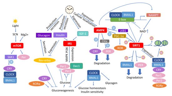
Figure 1).
Figure 1. Circadian rhythm affects the main molecular pathways mediating the outcome of caloric restriction (CR). Various types of interactions connect the CR–associated pathways mTOR, insulin signaling, AMPK, and sirtuins with circadian rhythm. Abbreviations: AMPK—adenosine monophosphate (AMP) activated protein kinase; CKIε—casein kinase ε; CLOCK—circadian locomotor output cycles kaput; Dec1—differentially expressed In chondrocytes 1; IGF–1—insulin–like growth factor 1; IRS—insulin receptor substrates; LKB1—liver kinase B1; mTOR—mammalian target of rapamycin; NAD—nicotinamide adenine dinucleotide; NAMPT—nicotinamide phosphoribosyltransferase; SIRT1—Sirtuin 1; SCN—suprachiasmatic nucleus, S6K1—ribosomal protein S6 kinase 1. The circled Ac indicates acetylation and circled P phosphorylation.

3. AMPK as an Energy Sensor and Executor of Circadian Metabolic Activities

AMPK serves as a key energy sensor of the AMP:ATP and ADP:ATP ratios. AMPK, activated at low levels of ATP, suppresses energy–consuming anabolic pathways and promotes catabolism resulting in the recruitment of stored energy to reestablish the ATP levels [75]. AMPK impacts metabolism via several downstream effectors including CREB–regulated transcriptional coactivator–2 (CRTC2) [76], TBC1D1/AS160 [77][78], peroxisome proliferator–activated receptor γ coactivator 1 α (PGC–1α) [79], and histone deacetylase 5 (HDAC5) [80]. Functionally, AMPK inhibits fatty acid (FA), TG and cholesterol synthesis, while stimulating FA uptake and β–oxidation [81][82][83][84][85]. Furthermore, AMPK reduces protein synthesis by inhibiting mTOR [86]. AMPK also affects the metabolism of glucose by stimulating glycolysis [87] and inhibiting glycogen synthesis [88] and gluconeogenesis [89][90][91]. It also drives nutrient–induced insulin secretion from islet β–cells [92] and glucose uptake by GLUT4 [93]. Besides metabolism, AMPK plays a role in inflammation, cell growth, autophagy, and apoptosis [94].
The circadian clock is paired with cellular metabolic fluctuations via nutrient–sensing pathways. While the expression of six of the transcripts encoding subunits of mammalian AMPK does not vary during the day, that of the AMPKβ2 subunit is eight times higher in the middle of the day than it is at night. AMPKβ2 determines the cellular localization of the heterotrimeric AMPK complex [95]; therefore, the presence of the AMPKα1 subunit in the nucleus increases at the time of day when AMPKβ2 expression peaks [19]. The rhythmic hepatic activity and nuclear localization of AMPK correlate inversely with CRY1 levels, which is likely due to the fact that stimulation of AMPK leads to phosphorylation and degradation of CRY [19]. Further, activation of AMPK by 5–aminoimidazole–4–carboxamide ribonucleoside (AICAR) or metformin in mouse livers causes a phase shift of the clock, and animals in which the AMPK pathway is genetically disrupted show alterations in peripheral clocks [19][96]. Moreover, acute AICAR stimulation alters the expression of clock genes in WT mice but not in mice lacking the AMPKγ3 regulatory subunit [97]. Genetic disruption of either AMPKα1 or AMPKα2 subunit dampens the rhythm of body temperature, the free–running activity, changes the circadian pattern of core clock gene expression in mice in an isoform– and tissue–specific manners [96]. In mouse muscles, AMPK regulates the expression patterns of the circadian genes Cry2Rev–erbα, and Bhlhb2 (basic helix–loop–helix domain containing class B 2) [97]. Moreover, AMPK is capable of phosphorylating casein kinase ε (CKIε) and thereby increases its enzymatic activity, indirectly leading to a destabilization of PER2 [98]. PGC–1α, which co–activates the RORs and consequently stimulates the expression of Bmal1 and Rev–erbα, is phosphorylated by AMPK [79][99][100]. PGC–1α is required for cell–autonomous clock function [100] and PGC–1α KO mice show an abnormal diurnal rhythm of physical activity, body temperature, and metabolic rate, due to disrupted expression of clock genes and genes involved in energy metabolism. Besides the direct impact on ATP levels, AMPK affects the energy status by promoting feeding through signaling in the hypothalamus as well as by adjusting circadian metabolism [101][102]. AMPK may thereby mediate the influence of fasting/feeding cycles on the circadian clock (Figure 1).

4. SIRT Energy Sensors in the Context of CR and Daily Rhythmicity

SIRTs serve as energy sensors by detecting the ratio of reduced to oxidized nicotinamide adenine dinucleotide NAD+:NADH and react as transcriptional effectors mostly through their HDAC activity. SIRTs are class III HDACs that manage processes connected with nucleic acid biology including DNA repair, homologous recombination, and histone deacetylation, as well as transcriptional gene silencing [103][104]. There are seven subtypes of SIRTs (SIRT1–7) in mice and humans which differ in their cellular localization and function. SIRT1–SIRT3, SIRT5, SIRT6, and SIRT7 act as deacetylases, while SIRT4 and SIRT6 have ADP–ribosylation activity. Besides histones, SIRTs modify also several transcriptional regulators including the nuclear factor kappa–light–chain enhancer of activated B cells (NF–κB), p53, FOXO, PGC–1α, as well as enzymes, including acetyl coenzyme A (CoA) synthetase 2 (AceCS2), long–chain acyl–coenzyme A dehydrogenase (LCAD), 3–hydroxy–3–methylglutaryl–CoA synthase 2 (HMGCS2), superoxide dismutase 2 (SOD–2), and structural proteins, such as α–tubulin [105][106][107][108][109]. Therefore, SIRTs impacts multiple processes and pathways including circadian clocks, cell cycle, mitochondrial biogenesis, and energy homeostasis, consequently influencing aging, apoptosis, inflammation, and stress resistance [110][111]. SIRT1 is mostly associated with metabolism. In S. cerevisiae an extra copy of the Sir2 gene, a yeast homolog of mammalian Sirt1, increases lifespan [112][113], whereas the deletion of Sir2 shortens it [112]. In yeast and Drosophila, lack of the Sirt1 homolog offsets CR–triggered life extension [114][115][116]. A yeast analog of Sirt1 takes part in DNA repair and regulates aging–related gene expression [117].
PGC–1α, one of the main metabolic effectors of SIRT1, is activated by SIRT1–mediated deacetylation [118][119]. Activated PGC–1α enhances hepatic gluconeogenesis [118], mitochondrial activity in muscle and BAT leading to increased exercise capacity and thermogenesis; consequently, PGC–1α promotes protection against obesity and metabolic dysfunction [120]. SIRTs interact also with factors involved in response to CR including the FOXO family of transcription factors [121][122][123], which affects gluconeogenesis and glucose release from hepatocytes [124], cell differentiation, metabolism as well as longevity regulation [125][126][127]. Further, AMPK enhances SIRT1 activity by increasing cellular NAD+ levels [128][129] and activation of SIRT1 may cause AMPK phosphorylation via deacetylation–dependent activation of the AMPK–activating kinase liver kinase B1 (LKB1) [130][131].
SIRT1 controls the circadian expression of the core clock genes Bmal1RorγPer2, and Cry1. Also, SIRT1 is recruited by CLOCK:BMAL1 chromatin at circadian promoters [132]. Further, the levels of NAD+, NADP+, NADH, and NADPH affect the binding capacity of CLOCK–BMAL1 heterodimers to E–box elements [133]. Similarly, resveratrol, a polyphenolic SIRT activator, regulates the expression of clock genes Per1Per2, and Bmal1 in Rat–1 fibroblast cells [134]. It also modifies the rhythmic expression of clock genes (ClockBmal1, and Per2) and lipid metabolism–related genes controlled by the clock (PparαSrebp–1cAcc1, and Fas) in HFD–fed mice [135] and reverses the change induced by high–fat feeding in the expression of Rev–Erbα in adipose tissue of rats [136]. SIRT1 also promotes the deacetylation and subsequent degradation of PER2 in a circadian manner [132].
The rhythmic acetylation of BMAL1 and acetyl–histone H3 Lys9/Lys14 at circadian promoters correlates with SIRT1 HDAC activity that is regulated in a circadian manner. Therefore, genetic or pharmacological inhibition of SIRT1 activity causes disturbances in the acetylation of H3 and BMAL1 and the circadian rhythm [137]. Moreover, the circadian transcription factor CLOCK has histone acetyltransferase (HAT) activity, and SIRT1 HDAC activity counteracts the HAT activity of CLOCK [132][137]. Another circadian protein, REV–ERBα regulates pancreatic glucagon secretion via the AMPK/nicotinamide phosphoribosyltransferase (NAMPT)/SIRT1 pathway [24]. Further, the expression of NAMPT, a rate–limiting enzyme involved in NAD+ production through the salvage pathway, is regulated by the BMAL/CLOCK heterodimer. Consequently, NAD+ levels exhibit rhythmic daily oscillations [138][139]. Moreover, by being recruited to the Nampt promoter, SIRT1 contributes to the circadian synthesis of its own coenzyme [139]. Inhibition of NAMPT promotes fluctuations of Per2 by releasing CLOCK:BMAL1 from suppression by SIRT1. In turn, CLOCK binds to the Nampt promoter and stimulates its activity, thereby contributing to a feedback loop comprising NAMPT/NAD+ and SIRT1/CLOCK:BMAL1 [138].
SIRT3 also interacts with the circadian rhythm. It sets the pace in the acetylation and activity of oxidative enzymes and consequently respiration in isolated mitochondria. Bmal1 KO mice have significantly decreased SIRT3 activity, which affects mitochondrial oxidative function, and supplementation with nicotinamide mononucleotide (NMN), a NAD+ precursor, restores SIRT3 function and enhances oxygen consumption in these animals [140]. Importantly, the rhythm of cyclic global protein acetylation dampens with aging in mice [141]. CR regulates SIRT1 activity and therefore it modulates the circadian acetylation of AceCS1, a pathway controlling rhythmic nucleocytoplasmic acetyl–CoA production [142][143]. Consequently, CR rescues the hepatic protein acetylation rhythm over the day/night cycle in mice. Accordingly, the circadian transcriptome of CR–mediated effects on circadian reprogramming and SIRT1–specific transcriptome overlaps [141] indicating a pivotal role of SIRT1 in connecting CR and the circadian rhythm (Figure 1).

References

  1. Bluher, M.; Kahn, B.B.; Kahn, C.R. Extended longevity in mice lacking the insulin receptor in adipose tissue. Science 2003, 299, 572–574.
  2. Tatar, M.; Bartke, A.; Antebi, A. The endocrine regulation of aging by insulin-like signals. Science 2003, 299, 1346–1351.
  3. Kenyon, C.; Chang, J.; Gensch, E.; Rudner, A.; Tabtiang, R. A C. elegans mutant that lives twice as long as wild type. Nature 1993, 366, 461–464.
  4. Taguchi, A.; Wartschow, L.M.; White, M.F. Brain IRS2 signaling coordinates life span and nutrient homeostasis. Science 2007, 317, 369–372.
  5. Selman, C.; Lingard, S.; Choudhury, A.I.; Batterham, R.L.; Claret, M.; Clements, M.; Ramadani, F.; Okkenhaug, K.; Schuster, E.; Blanc, E.; et al. Evidence for lifespan extension and delayed age-related biomarkers in insulin receptor substrate 1 null mice. FASEB J. 2008, 22, 807–818.
  6. Weindruch, R. The retardation of aging by caloric restriction: Studies in rodents and primates. Toxicol. Pathol. 1996, 24, 742–745.
  7. Kim, D.H.; Park, M.H.; Lee, E.K.; Choi, Y.J.; Chung, K.W.; Moon, K.M.; Kim, M.J.; An, H.J.; Park, J.W.; Kim, N.D.; et al. The roles of FoxOs in modulation of aging by calorie restriction. Biogerontology 2015, 16, 1–14.
  8. Oh, W.J.; Jacinto, E. mTOR complex 2 signaling and functions. Cell Cycle 2011, 10, 2305–2316.
  9. Rowland, A.F.; Fazakerley, D.J.; James, D.E. Mapping insulin/GLUT4 circuitry. Traffic 2011, 12, 672–681.
  10. Siddle, K. Signalling by insulin and IGF receptors: Supporting acts and new players. J. Mol. Endocrinol. 2011, 47, R1–R10.
  11. Piscitello, D.; Varshney, D.; Lilla, S.; Vizioli, M.G.; Reid, C.; Gorbunova, V.; Seluanov, A.; Gillespie, D.A.; Adams, P.D. AKT overactivation can suppress DNA repair via p70S6 kinase-dependent downregulation of MRE11. Oncogene 2018, 37, 427–438.
  12. Jia, Y.; Song, W.; Zhang, F.; Yan, J.; Yang, Q. Akt1 inhibits homologous recombination in Brca1-deficient cells by blocking the Chk1-Rad51 pathway. Oncogene 2013, 32, 1943–1949.
  13. Liu, P.; Gan, W.; Guo, C.; Xie, A.; Gao, D.; Guo, J.; Zhang, J.; Willis, N.; Su, A.; Asara, J.M.; et al. Akt-mediated phosphorylation of XLF impairs non-homologous end-joining DNA repair. Mol. Cell 2015, 57, 648–661.
  14. Astafev, A.A.; Patel, S.A.; Kondratov, R.V. Calorie restriction effects on circadian rhythms in gene expression are sex dependent. Sci. Rep. 2017, 7, 9716.
  15. Zhang, E.E.; Liu, A.C.; Hirota, T.; Miraglia, L.J.; Welch, G.; Pongsawakul, P.Y.; Liu, X.; Atwood, A.; Huss, J.W., 3rd; Janes, J.; et al. A genome-wide RNAi screen for modifiers of the circadian clock in human cells. Cell 2009, 139, 199–210.
  16. Patel, S.A.; Chaudhari, A.; Gupta, R.; Velingkaar, N.; Kondratov, R.V. Circadian clocks govern calorie restriction-mediated life span extension through BMAL1- and IGF-1-dependent mechanisms. FASEB J. 2016, 30, 1634–1642.
  17. Yamajuku, D.; Inagaki, T.; Haruma, T.; Okubo, S.; Kataoka, Y.; Kobayashi, S.; Ikegami, K.; Laurent, T.; Kojima, T.; Noutomi, K.; et al. Real-time monitoring in three-dimensional hepatocytes reveals that insulin acts as a synchronizer for liver clock. Sci. Rep. 2012, 2, 439.
  18. Tahara, Y.; Otsuka, M.; Fuse, Y.; Hirao, A.; Shibata, S. Refeeding after fasting elicits insulin-dependent regulation of Per2 and Rev-erbalpha with shifts in the liver clock. J. Biol. Rhythms 2011, 26, 230–240.
  19. Lamia, K.A.; Sachdeva, U.M.; DiTacchio, L.; Williams, E.C.; Alvarez, J.G.; Egan, D.F.; Vasquez, D.S.; Juguilon, H.; Panda, S.; Shaw, R.J.; et al. AMPK regulates the circadian clock by cryptochrome phosphorylation and degradation. Science 2009, 326, 437–440.
  20. Iwanaga, H.; Yano, M.; Miki, H.; Okada, K.; Azama, T.; Takiguchi, S.; Fujiwara, Y.; Yasuda, T.; Nakayama, M.; Kobayashi, M.; et al. Per2 gene expressions in the suprachiasmatic nucleus and liver differentially respond to nutrition factors in rats. JPEN J. Parenter. Enteral. Nutr. 2005, 29, 157–161.
  21. Challet, E.; Losee-Olson, S.; Turek, F.W. Reduced glucose availability attenuates circadian responses to light in mice. Am. J. Physiol. 1999, 276, R1063–R1070.
  22. Hirota, T.; Okano, T.; Kokame, K.; Shirotani-Ikejima, H.; Miyata, T.; Fukada, Y. Glucose down-regulates Per1 and Per2 mRNA levels and induces circadian gene expression in cultured Rat-1 fibroblasts. J. Biol. Chem. 2002, 277, 44244–44251.
  23. Marcheva, B.; Ramsey, K.M.; Buhr, E.D.; Kobayashi, Y.; Su, H.; Ko, C.H.; Ivanova, G.; Omura, C.; Mo, S.; Vitaterna, M.H.; et al. Disruption of the clock components CLOCK and BMAL1 leads to hypoinsulinaemia and diabetes. Nature 2010, 466, 627–631.
  24. Vieira, E.; Marroqui, L.; Figueroa, A.L.; Merino, B.; Fernandez-Ruiz, R.; Nadal, A.; Burris, T.P.; Gomis, R.; Quesada, I. Involvement of the clock gene Rev-erb alpha in the regulation of glucagon secretion in pancreatic alpha-cells. PLoS ONE 2013, 8, e69939.
  25. Sadacca, L.A.; Lamia, K.A.; deLemos, A.S.; Blum, B.; Weitz, C.J. An intrinsic circadian clock of the pancreas is required for normal insulin release and glucose homeostasis in mice. Diabetologia 2011, 54, 120–124.
  26. Vieira, E.; Merino, B.; Quesada, I. Role of the clock gene Rev-erbalpha in metabolism and in the endocrine pancreas. Diabetes Obes. Metab. 2015, 17 (Suppl. S1), 106–114.
  27. Muhlbauer, E.; Wolgast, S.; Finckh, U.; Peschke, D.; Peschke, E. Indication of circadian oscillations in the rat pancreas. FEBS Lett. 2004, 564, 91–96.
  28. Rudic, R.D.; McNamara, P.; Curtis, A.M.; Boston, R.C.; Panda, S.; Hogenesch, J.B.; Fitzgerald, G.A. BMAL1 and CLOCK, two essential components of the circadian clock, are involved in glucose homeostasis. PLoS Biol. 2004, 2, e377.
  29. Barclay, J.L.; Shostak, A.; Leliavski, A.; Tsang, A.H.; Johren, O.; Muller-Fielitz, H.; Landgraf, D.; Naujokat, N.; van der Horst, G.T.; Oster, H. High-fat diet-induced hyperinsulinemia and tissue-specific insulin resistance in Cry-deficient mice. Am. J. Physiol. Endocrinol. Metab. 2013, 304, E1053–E1063.
  30. Zhao, Y.; Zhang, Y.; Zhou, M.; Wang, S.; Hua, Z.; Zhang, J. Loss of mPer2 increases plasma insulin levels by enhanced glucose-stimulated insulin secretion and impaired insulin clearance in mice. FEBS Lett. 2012, 586, 1306–1311.
  31. Delezie, J.; Dumont, S.; Dardente, H.; Oudart, H.; Grechez-Cassiau, A.; Klosen, P.; Teboul, M.; Delaunay, F.; Pevet, P.; Challet, E. The nuclear receptor REV-ERBalpha is required for the daily balance of carbohydrate and lipid metabolism. FASEB J. 2012, 26, 3321–3335.
  32. Kalsbeek, A.; la Fleur, S.; Fliers, E. Circadian control of glucose metabolism. Mol. Metab. 2014, 3, 372–383.
  33. Doi, R.; Oishi, K.; Ishida, N. CLOCK regulates circadian rhythms of hepatic glycogen synthesis through transcriptional activation of Gys2. J. Biol. Chem. 2010, 285, 22114–22121.
  34. Matsuoka, H.; Shima, A.; Kuramoto, D.; Kikumoto, D.; Matsui, T.; Michihara, A. Phosphoenolpyruvate carboxykinase, a key enzyme that controls blood glucose, is a target of retinoic acid receptor-related orphan receptor alpha. PLoS ONE 2015, 10, e0137955.
  35. Kadiri, S.; Monnier, C.; Ganbold, M.; Ledent, T.; Capeau, J.; Antoine, B. The nuclear retinoid-related orphan receptor-alpha regulates adipose tissue glyceroneogenesis in addition to hepatic gluconeogenesis. Am. J. Physiol Endocrinol. Metab. 2015, 309, E105–E114.
  36. Kojetin, D.; Wang, Y.; Kamenecka, T.M.; Burris, T.P. Identification of SR8278, a synthetic antagonist of the nuclear heme receptor REV-ERB. ACS Chem. Biol. 2011, 6, 131–134.
  37. Yin, L.; Wu, N.; Curtin, J.C.; Qatanani, M.; Szwergold, N.R.; Reid, R.A.; Waitt, G.M.; Parks, D.J.; Pearce, K.H.; Wisely, G.B.; et al. Rev-erbalpha, a heme sensor that coordinates metabolic and circadian pathways. Science 2007, 318, 1786–1789.
  38. Zhang, E.E.; Liu, Y.; Dentin, R.; Pongsawakul, P.Y.; Liu, A.C.; Hirota, T.; Nusinow, D.A.; Sun, X.; Landais, S.; Kodama, Y.; et al. Cryptochrome mediates circadian regulation of cAMP signaling and hepatic gluconeogenesis. Nat. Med. 2010, 16, 1152–1156.
  39. Lamia, K.A.; Papp, S.J.; Yu, R.T.; Barish, G.D.; Uhlenhaut, N.H.; Jonker, J.W.; Downes, M.; Evans, R.M. Cryptochromes mediate rhythmic repression of the glucocorticoid receptor. Nature 2011, 480, 552–556.
  40. Zani, F.; Breasson, L.; Becattini, B.; Vukolic, A.; Montani, J.P.; Albrecht, U.; Provenzani, A.; Ripperger, J.A.; Solinas, G. PER2 promotes glucose storage to liver glycogen during feeding and acute fasting by inducing Gys2 PTG and G L expression. Mol. Metab. 2013, 2, 292–305.
  41. Bhaskar, P.T.; Hay, N. The two TORCs and Akt. Dev. Cell 2007, 12, 487–502.
  42. Wullschleger, S.; Loewith, R.; Hall, M.N. TOR signaling in growth and metabolism. Cell 2006, 124, 471–484.
  43. Loewith, R.; Jacinto, E.; Wullschleger, S.; Lorberg, A.; Crespo, J.L.; Bonenfant, D.; Oppliger, W.; Jenoe, P.; Hall, M.N. Two TOR complexes, only one of which is rapamycin sensitive, have distinct roles in cell growth control. Mol. Cell 2002, 10, 457–468.
  44. Jacinto, E.; Loewith, R.; Schmidt, A.; Lin, S.; Ruegg, M.A.; Hall, A.; Hall, M.N. Mammalian TOR complex 2 controls the actin cytoskeleton and is rapamycin insensitive. Nat. Cell Biol. 2004, 6, 1122–1128.
  45. Corradetti, M.N.; Guan, K.L. Upstream of the mammalian target of rapamycin: Do all roads pass through mTOR? Oncogene 2006, 25, 6347–6360.
  46. Hay, N.; Sonenberg, N. Upstream and downstream of mTOR. Genes Dev. 2004, 18, 1926–1945.
  47. Arsham, A.M.; Neufeld, T.P. Thinking globally and acting locally with TOR. Curr. Opin. Cell Biol. 2006, 18, 589–597.
  48. Duvel, K.; Yecies, J.L.; Menon, S.; Raman, P.; Lipovsky, A.I.; Souza, A.L.; Triantafellow, E.; Ma, Q.; Gorski, R.; Cleaver, S.; et al. Activation of a metabolic gene regulatory network downstream of mTOR complex 1. Mol. Cell 2010, 39, 171–183.
  49. Hudson, C.C.; Liu, M.; Chiang, G.G.; Otterness, D.M.; Loomis, D.C.; Kaper, F.; Giaccia, A.J.; Abraham, R.T. Regulation of hypoxia-inducible factor 1alpha expression and function by the mammalian target of rapamycin. Mol. Cell Biol. 2002, 22, 7004–7014.
  50. Kim, J.E.; Chen, J. regulation of peroxisome proliferator-activated receptor-gamma activity by mammalian target of rapamycin and amino acids in adipogenesis. Diabetes 2004, 53, 2748–2756.
  51. Zhang, H.H.; Huang, J.; Duvel, K.; Boback, B.; Wu, S.; Squillace, R.M.; Wu, C.L.; Manning, B.D. Insulin stimulates adipogenesis through the Akt-TSC2-mTORC1 pathway. PLoS ONE 2009, 4, e6189.
  52. Porstmann, T.; Santos, C.R.; Griffiths, B.; Cully, M.; Wu, M.; Leevers, S.; Griffiths, J.R.; Chung, Y.L.; Schulze, A. SREBP activity is regulated by mTORC1 and contributes to Akt-dependent cell growth. Cell Metab. 2008, 8, 224–236.
  53. Wang, B.T.; Ducker, G.S.; Barczak, A.J.; Barbeau, R.; Erle, D.J.; Shokat, K.M. The mammalian target of rapamycin regulates cholesterol biosynthetic gene expression and exhibits a rapamycin-resistant transcriptional profile. Proc. Natl. Acad. Sci. USA 2011, 108, 15201–15206.
  54. Kim, J.; Guan, K.L. mTOR as a central hub of nutrient signalling and cell growth. Nat. Cell Biol. 2019, 21, 63–71.
  55. Zhao, J.; Zhai, B.; Gygi, S.P.; Goldberg, A.L. mTOR inhibition activates overall protein degradation by the ubiquitin proteasome system as well as by autophagy. Proc. Natl. Acad. Sci. USA 2015, 112, 15790–15797.
  56. Cao, R.; Lee, B.; Cho, H.Y.; Saklayen, S.; Obrietan, K. Photic regulation of the mTOR signaling pathway in the suprachiasmatic circadian clock. Mol. Cell Neurosci. 2008, 38, 312–324.
  57. Cao, R.; Anderson, F.E.; Jung, Y.J.; Dziema, H.; Obrietan, K. Circadian regulation of mammalian target of rapamycin signaling in the mouse suprachiasmatic nucleus. Neuroscience 2011, 181, 79–88.
  58. Cornu, M.; Oppliger, W.; Albert, V.; Robitaille, A.M.; Trapani, F.; Quagliata, L.; Fuhrer, T.; Sauer, U.; Terracciano, L.; Hall, M.N. Hepatic mTORC1 controls locomotor activity, body temperature, and lipid metabolism through FGF21. Proc. Natl. Acad. Sci. USA 2014, 111, 11592–11599.
  59. Jouffe, C.; Cretenet, G.; Symul, L.; Martin, E.; Atger, F.; Naef, F.; Gachon, F. The circadian clock coordinates ribosome biogenesis. PLoS Biol. 2013, 11, e1001455.
  60. Khapre, R.V.; Kondratova, A.A.; Patel, S.; Dubrovsky, Y.; Wrobel, M.; Antoch, M.P.; Kondratov, R.V. BMAL1-dependent regulation of the mTOR signaling pathway delays aging. Aging 2014, 6, 48–57.
  61. Lipton, J.O.; Yuan, E.D.; Boyle, L.M.; Ebrahimi-Fakhari, D.; Kwiatkowski, E.; Nathan, A.; Guttler, T.; Davis, F.; Asara, J.M.; Sahin, M. The circadian protein BMAL1 regulates translation in response to S6K1-mediated phosphorylation. Cell 2015, 161, 1138–1151.
  62. Huang, C.C.; Ko, M.L.; Ko, G.Y. A new functional role for mechanistic/mammalian target of rapamycin complex 1 (mTORC1) in the circadian regulation of L-type voltage-gated calcium channels in avian cone photoreceptors. PLoS ONE 2013, 8, e73315.
  63. Dragert, K.; Bhattacharya, I.; Hall, M.N.; Humar, R.; Battegay, E.; Haas, E. Basal mTORC2 activity and expression of its components display diurnal variation in mouse perivascular adipose tissue. Biochem. Biophys. Res. Commun. 2016, 473, 317–322.
  64. Chang, S.W.; Yoshihara, T.; Machida, S.; Naito, H. Circadian rhythm of intracellular protein synthesis signaling in rat cardiac and skeletal muscles. Biochem. Biophys. Rep. 2017, 9, 153–158.
  65. Saraf, A.; Luo, J.; Morris, D.R.; Storm, D.R. Phosphorylation of eukaryotic translation initiation factor 4E and eukaryotic translation initiation factor 4E-binding protein (4EBP) and their upstream signaling components undergo diurnal oscillation in the mouse hippocampus: Implications for memory persistence. J. Biol. Chem. 2014, 289, 20129–20138.
  66. Albert, V.; Cornu, M.; Hall, M.N. mTORC1 signaling in Agrp neurons mediates circadian expression of Agrp and NPY but is dispensable for regulation of feeding behavior. Biochem. Biophys. Res. Commun. 2015, 464, 480–486.
  67. Khapre, R.V.; Patel, S.A.; Kondratova, A.A.; Chaudhary, A.; Velingkaar, N.; Antoch, M.P.; Kondratov, R.V. Metabolic clock generates nutrient anticipation rhythms in mTOR signaling. Aging 2014, 6, 675–689.
  68. Zheng, X.; Sehgal, A. AKT and TOR signaling set the pace of the circadian pacemaker. Curr. Biol. 2010, 20, 1203–1208.
  69. Kijak, E.; Pyza, E. TOR signaling pathway and autophagy are involved in the regulation of circadian rhythms in behavior and plasticity of L2 interneurons in the brain of Drosophila melanogaster. PLoS ONE 2017, 12, e0171848.
  70. Cao, R.; Robinson, B.; Xu, H.; Gkogkas, C.; Khoutorsky, A.; Alain, T.; Yanagiya, A.; Nevarko, T.; Liu, A.C.; Amir, S.; et al. Translational control of entrainment and synchrony of the suprachiasmatic circadian clock by mTOR/4E-BP1 signaling. Neuron 2013, 79, 712–724.
  71. Cao, R.; Li, A.; Cho, H.Y.; Lee, B.; Obrietan, K. Mammalian target of rapamycin signaling modulates photic entrainment of the suprachiasmatic circadian clock. J. Neurosci. 2010, 30, 6302–6314.
  72. Ramanathan, C.; Kathale, N.D.; Liu, D.; Lee, C.; Freeman, D.A.; Hogenesch, J.B.; Cao, R.; Liu, A.C. mTOR signaling regulates central and peripheral circadian clock function. PLoS Genet. 2018, 14, e1007369.
  73. Feeney, K.A.; Hansen, L.L.; Putker, M.; Olivares-Yanez, C.; Day, J.; Eades, L.J.; Larrondo, L.F.; Hoyle, N.P.; O’Neill, J.S.; van Ooijen, G. Daily magnesium fluxes regulate cellular timekeeping and energy balance. Nature 2016, 532, 375–379.
  74. Hatori, M.; Vollmers, C.; Zarrinpar, A.; DiTacchio, L.; Bushong, E.A.; Gill, S.; Leblanc, M.; Chaix, A.; Joens, M.; Fitzpatrick, J.A.; et al. Time-restricted feeding without reducing caloric intake prevents metabolic diseases in mice fed a high-fat diet. Cell Metab. 2012, 15, 848–860.
  75. Winder, W.W.; Hardie, D.G. AMP-activated protein kinase, a metabolic master switch: Possible roles in type 2 diabetes. Am. J. Physiol. 1999, 277, E1–E10.
  76. Koo, S.H.; Flechner, L.; Qi, L.; Zhang, X.; Screaton, R.A.; Jeffries, S.; Hedrick, S.; Xu, W.; Boussouar, F.; Brindle, P.; et al. The CREB coactivator TORC2 is a key regulator of fasting glucose metabolism. Nature 2005, 437, 1109–1111.
  77. Chen, S.; Murphy, J.; Toth, R.; Campbell, D.G.; Morrice, N.A.; Mackintosh, C. Complementary regulation of TBC1D1 and AS160 by growth factors, insulin and AMPK activators. Biochem. J. 2008, 409, 449–459.
  78. Geraghty, K.M.; Chen, S.; Harthill, J.E.; Ibrahim, A.F.; Toth, R.; Morrice, N.A.; Vandermoere, F.; Moorhead, G.B.; Hardie, D.G.; MacKintosh, C. Regulation of multisite phosphorylation and 14-3-3 binding of AS160 in response to IGF-1, EGF, PMA and AICAR. Biochem. J. 2007, 407, 231–241.
  79. Jager, S.; Handschin, C.; St-Pierre, J.; Spiegelman, B.M. AMP-activated protein kinase (AMPK) action in skeletal muscle via direct phosphorylation of PGC-1alpha. Proc. Natl. Acad. Sci. USA 2007, 104, 12017–12022.
  80. McGee, S.L.; van Denderen, B.J.; Howlett, K.F.; Mollica, J.; Schertzer, J.D.; Kemp, B.E.; Hargreaves, M. AMP-activated protein kinase regulates GLUT4 transcription by phosphorylating histone deacetylase 5. Diabetes 2008, 57, 860–867.
  81. Davies, S.P.; Sim, A.T.; Hardie, D.G. Location and function of three sites phosphorylated on rat acetyl-CoA carboxylase by the AMP-activated protein kinase. Eur. J. Biochem. 1990, 187, 183–190.
  82. Davies, S.P.; Carling, D.; Munday, M.R.; Hardie, D.G. Diurnal rhythm of phosphorylation of rat liver acetyl-CoA carboxylase by the AMP-activated protein kinase, demonstrated using freeze-clamping. Effects of high fat diets. Eur. J. Biochem. 1992, 203, 615–623.
  83. Li, Y.; Xu, S.; Mihaylova, M.M.; Zheng, B.; Hou, X.; Jiang, B.; Park, O.; Luo, Z.; Lefai, E.; Shyy, J.Y.; et al. AMPK phosphorylates and inhibits SREBP activity to attenuate hepatic steatosis and atherosclerosis in diet-induced insulin-resistant mice. Cell Metab. 2011, 13, 376–388.
  84. Muoio, D.M.; Seefeld, K.; Witters, L.A.; Coleman, R.A. AMP-activated kinase reciprocally regulates triacylglycerol synthesis and fatty acid oxidation in liver and muscle: Evidence that sn-glycerol-3-phosphate acyltransferase is a novel target. Biochem. J. 1999, 338, 783–791.
  85. Clarke, P.R.; Hardie, D.G. Regulation of HMG-CoA reductase: Identification of the site phosphorylated by the AMP-activated protein kinase in vitro and in intact rat liver. EMBO J. 1990, 9, 2439–2446.
  86. Hoppe, S.; Bierhoff, H.; Cado, I.; Weber, A.; Tiebe, M.; Grummt, I.; Voit, R. AMP-activated protein kinase adapts rRNA synthesis to cellular energy supply. Proc. Natl. Acad. Sci. USA 2009, 106, 17781–17786.
  87. Marsin, A.S.; Bouzin, C.; Bertrand, L.; Hue, L. The stimulation of glycolysis by hypoxia in activated monocytes is mediated by AMP-activated protein kinase and inducible 6-phosphofructo-2-kinase. J. Biol. Chem. 2002, 277, 30778–30783.
  88. Jorgensen, S.B.; Nielsen, J.N.; Birk, J.B.; Olsen, G.S.; Viollet, B.; Andreelli, F.; Schjerling, P.; Vaulont, S.; Hardie, D.G.; Hansen, B.F.; et al. The alpha2-5’AMP-activated protein kinase is a site 2 glycogen synthase kinase in skeletal muscle and is responsive to glucose loading. Diabetes 2004, 53, 3074–3081.
  89. Hardie, D.G.; Ross, F.A.; Hawley, S.A. AMPK: A nutrient and energy sensor that maintains energy homeostasis. Nat. Rev. Mol. Cell Biol. 2012, 13, 251–262.
  90. Leclerc, I.; Lenzner, C.; Gourdon, L.; Vaulont, S.; Kahn, A.; Viollet, B. Hepatocyte nuclear factor-4alpha involved in type 1 maturity-onset diabetes of the young is a novel target of AMP-activated protein kinase. Diabetes 2001, 50, 1515–1521.
  91. Lee, J.M.; Seo, W.Y.; Song, K.H.; Chanda, D.; Kim, Y.D.; Kim, D.K.; Lee, M.W.; Ryu, D.; Kim, Y.H.; Noh, J.R.; et al. AMPK-dependent repression of hepatic gluconeogenesis via disruption of CREB.CRTC2 complex by orphan nuclear receptor small heterodimer partner. J. Biol. Chem. 2010, 285, 32182–32191.
  92. Dufer, M.; Noack, K.; Krippeit-Drews, P.; Drews, G. Activation of the AMP-activated protein kinase enhances glucose-stimulated insulin secretion in mouse beta-cells. Islets 2010, 2, 156–163.
  93. Chavez, J.A.; Roach, W.G.; Keller, S.R.; Lane, W.S.; Lienhard, G.E. Inhibition of GLUT4 translocation by Tbc1d1, a Rab GTPase-activating protein abundant in skeletal muscle, is partially relieved by AMP-activated protein kinase activation. J. Biol. Chem. 2008, 283, 9187–9195.
  94. Steinberg, G.R.; Kemp, B.E. AMPK in health and disease. Physiol. Rev. 2009, 89, 1025–1078.
  95. Suzuki, A.; Okamoto, S.; Lee, S.; Saito, K.; Shiuchi, T.; Minokoshi, Y. Leptin stimulates fatty acid oxidation and peroxisome proliferator-activated receptor alpha gene expression in mouse C2C12 myoblasts by changing the subcellular localization of the alpha2 form of AMP-activated protein kinase. Mol. Cell Biol. 2007, 27, 4317–4327.
  96. Um, J.H.; Pendergast, J.S.; Springer, D.A.; Foretz, M.; Viollet, B.; Brown, A.; Kim, M.K.; Yamazaki, S.; Chung, J.H. AMPK regulates circadian rhythms in a tissue- and isoform-specific manner. PLoS ONE 2011, 6, e18450.
  97. Vieira, E.; Nilsson, E.C.; Nerstedt, A.; Ormestad, M.; Long, Y.C.; Garcia-Roves, P.M.; Zierath, J.R.; Mahlapuu, M. Relationship between AMPK and the transcriptional balance of clock-related genes in skeletal muscle. Am. J. Physiol. Endocrinol. Metab. 2008, 295, E1032–E1037.
  98. Um, J.H.; Yang, S.; Yamazaki, S.; Kang, H.; Viollet, B.; Foretz, M.; Chung, J.H. Activation of 5’-AMP-activated kinase with diabetes drug metformin induces casein kinase Iepsilon (CKIepsilon)-dependent degradation of clock protein mPer2. J. Biol. Chem. 2007, 282, 20794–20798.
  99. Grimaldi, B.; Sassone-Corsi, P. Circadian rhythms: Metabolic clockwork. Nature 2007, 447, 386–387.
  100. Liu, C.; Li, S.; Liu, T.; Borjigin, J.; Lin, J.D. Transcriptional coactivator PGC-1alpha integrates the mammalian clock and energy metabolism. Nature 2007, 447, 477–481.
  101. Minokoshi, Y.; Alquier, T.; Furukawa, N.; Kim, Y.B.; Lee, A.; Xue, B.; Mu, J.; Foufelle, F.; Ferre, P.; Birnbaum, M.J.; et al. AMP-kinase regulates food intake by responding to hormonal and nutrient signals in the hypothalamus. Nature 2004, 428, 569–574.
  102. Andersson, U.; Filipsson, K.; Abbott, C.R.; Woods, A.; Smith, K.; Bloom, S.R.; Carling, D.; Small, C.J. AMP-activated protein kinase plays a role in the control of food intake. J. Biol. Chem. 2004, 279, 12005–12008.
  103. Liou, G.G.; Tanny, J.C.; Kruger, R.G.; Walz, T.; Moazed, D. Assembly of the SIR complex and its regulation by O-acetyl-ADP-ribose, a product of NAD-dependent histone deacetylation. Cell 2005, 121, 515–527.
  104. Vaquero, A.; Scher, M.; Lee, D.; Erdjument-Bromage, H.; Tempst, P.; Reinberg, D. Human SirT1 interacts with histone H1 and promotes formation of facultative heterochromatin. Mol. Cell 2004, 16, 93–105.
  105. Dang, W. The controversial world of sirtuins. Drug. Discov. Today Technol. 2014, 12, e9–e17.
  106. Houtkooper, R.H.; Pirinen, E.; Auwerx, J. Sirtuins as regulators of metabolism and healthspan. Nat. Rev. Mol. Cell Biol. 2012, 13, 225–238.
  107. Shimazu, T.; Hirschey, M.D.; Hua, L.; Dittenhafer-Reed, K.E.; Schwer, B.; Lombard, D.B.; Li, Y.; Bunkenborg, J.; Alt, F.W.; Denu, J.M.; et al. SIRT3 deacetylates mitochondrial 3-hydroxy-3-methylglutaryl CoA synthase 2 and regulates ketone body production. Cell Metab. 2010, 12, 654–661.
  108. Hirschey, M.D.; Shimazu, T.; Goetzman, E.; Jing, E.; Schwer, B.; Lombard, D.B.; Grueter, C.A.; Harris, C.; Biddinger, S.; Ilkayeva, O.R.; et al. SIRT3 regulates mitochondrial fatty-acid oxidation by reversible enzyme deacetylation. Nature 2010, 464, 121–125.
  109. Qiu, X.; Brown, K.; Hirschey, M.D.; Verdin, E.; Chen, D. Calorie restriction reduces oxidative stress by SIRT3-mediated SOD2 activation. Cell Metab. 2010, 12, 662–667.
  110. Preyat, N.; Leo, O. Sirtuin deacylases: A molecular link between metabolism and immunity. J. Leukoc. Biol. 2013, 93, 669–680.
  111. Satoh, A.; Brace, C.S.; Ben-Josef, G.; West, T.; Wozniak, D.F.; Holtzman, D.M.; Herzog, E.D.; Imai, S. SIRT1 promotes the central adaptive response to diet restriction through activation of the dorsomedial and lateral nuclei of the hypothalamus. J. Neurosci. 2010, 30, 10220–10232.
  112. Kaeberlein, M.; McVey, M.; Guarente, L. The SIR2/3/4 complex and SIR2 alone promote longevity in Saccharomyces cerevisiae by two different mechanisms. Genes Dev. 1999, 13, 2570–2580.
  113. Whitaker, R.; Faulkner, S.; Miyokawa, R.; Burhenn, L.; Henriksen, M.; Wood, J.G.; Helfand, S.L. Increased expression of Drosophila Sir2 extends life span in a dose-dependent manner. Aging 2013, 5, 682–691.
  114. Lin, S.J.; Defossez, P.A.; Guarente, L. Requirement of NAD and SIR2 for life-span extension by calorie restriction in Saccharomyces cerevisiae. Science 2000, 289, 2126–2128.
  115. Rogina, B.; Helfand, S.L. Sir2 mediates longevity in the fly through a pathway related to calorie restriction. Proc. Natl. Acad. Sci. USA 2004, 101, 15998–16003.
  116. Tissenbaum, H.A.; Guarente, L. Increased dosage of a sir-2 gene extends lifespan in Caenorhabditis elegans. Nature 2001, 410, 227–230.
  117. Oberdoerffer, P.; Michan, S.; McVay, M.; Mostoslavsky, R.; Vann, J.; Park, S.K.; Hartlerode, A.; Stegmuller, J.; Hafner, A.; Loerch, P.; et al. SIRT1 redistribution on chromatin promotes genomic stability but alters gene expression during aging. Cell 2008, 135, 907–918.
  118. Rodgers, J.T.; Lerin, C.; Haas, W.; Gygi, S.P.; Spiegelman, B.M.; Puigserver, P. Nutrient control of glucose homeostasis through a complex of PGC-1alpha and SIRT1. Nature 2005, 434, 113–118.
  119. Nemoto, S.; Fergusson, M.M.; Finkel, T. SIRT1 functionally interacts with the metabolic regulator and transcriptional coactivator PGC-1. J. Biol. Chem. 2005, 280, 16456–16460.
  120. Lagouge, M.; Argmann, C.; Gerhart-Hines, Z.; Meziane, H.; Lerin, C.; Daussin, F.; Messadeq, N.; Milne, J.; Lambert, P.; Elliott, P.; et al. Resveratrol improves mitochondrial function and protects against metabolic disease by activating SIRT1 and PGC-1alpha. Cell 2006, 127, 1109–1122.
  121. Daitoku, H.; Hatta, M.; Matsuzaki, H.; Aratani, S.; Ohshima, T.; Miyagishi, M.; Nakajima, T.; Fukamizu, A. Silent information regulator 2 potentiates Foxo1-mediated transcription through its deacetylase activity. Proc. Natl. Acad. Sci. USA 2004, 101, 10042–10047.
  122. van der Horst, A.; Tertoolen, L.G.; de Vries-Smits, L.M.; Frye, R.A.; Medema, R.H.; Burgering, B.M. FOXO4 is acetylated upon peroxide stress and deacetylated by the longevity protein hSir2(SIRT1). J. Biol. Chem. 2004, 279, 28873–28879.
  123. Brunet, A.; Sweeney, L.B.; Sturgill, J.F.; Chua, K.F.; Greer, P.L.; Lin, Y.; Tran, H.; Ross, S.E.; Mostoslavsky, R.; Cohen, H.Y.; et al. Stress-dependent regulation of FOXO transcription factors by the SIRT1 deacetylase. Science 2004, 303, 2011–2015.
  124. Frescas, D.; Valenti, L.; Accili, D. Nuclear trapping of the forkhead transcription factor FoxO1 via Sirt-dependent deacetylation promotes expression of glucogenetic genes. J. Biol. Chem. 2005, 280, 20589–20595.
  125. Accili, D.; Arden, K.C. FoxOs at the crossroads of cellular metabolism, differentiation, and transformation. Cell 2004, 117, 421–426.
  126. Martins, R.; Lithgow, G.J.; Link, W. Long live FOXO: Unraveling the role of FOXO proteins in aging and longevity. Aging Cell 2016, 15, 196–207.
  127. Gross, D.N.; van den Heuvel, A.P.; Birnbaum, M.J. The role of FoxO in the regulation of metabolism. Oncogene 2008, 27, 2320–2336.
  128. Canto, C.; Gerhart-Hines, Z.; Feige, J.N.; Lagouge, M.; Noriega, L.; Milne, J.C.; Elliott, P.J.; Puigserver, P.; Auwerx, J. AMPK regulates energy expenditure by modulating NAD+ metabolism and SIRT1 activity. Nature 2009, 458, 1056–1060.
  129. Fulco, M.; Cen, Y.; Zhao, P.; Hoffman, E.P.; McBurney, M.W.; Sauve, A.A.; Sartorelli, V. Glucose restriction inhibits skeletal myoblast differentiation by activating SIRT1 through AMPK-mediated regulation of Nampt. Dev. Cell 2008, 14, 661–673.
  130. Hou, X.; Xu, S.; Maitland-Toolan, K.A.; Sato, K.; Jiang, B.; Ido, Y.; Lan, F.; Walsh, K.; Wierzbicki, M.; Verbeuren, T.J.; et al. SIRT1 regulates hepatocyte lipid metabolism through activating AMP-activated protein kinase. J. Biol. Chem. 2008, 283, 20015–20026.
  131. Lan, F.; Cacicedo, J.M.; Ruderman, N.; Ido, Y. SIRT1 modulation of the acetylation status, cytosolic localization, and activity of LKB1. Possible role in AMP-activated protein kinase activation. J. Biol. Chem. 2008, 283, 27628–27635.
  132. Asher, G.; Gatfield, D.; Stratmann, M.; Reinke, H.; Dibner, C.; Kreppel, F.; Mostoslavsky, R.; Alt, F.W.; Schibler, U. SIRT1 regulates circadian clock gene expression through PER2 deacetylation. Cell 2008, 134, 317–328.
  133. Rutter, J.; Reick, M.; Wu, L.C.; McKnight, S.L. Regulation of clock and NPAS2 DNA binding by the redox state of NAD cofactors. Science 2001, 293, 510–514.
  134. Oike, H.; Kobori, M. Resveratrol regulates circadian clock genes in Rat-1 fibroblast cells. Biosci. Biotechnol. Biochem. 2008, 72, 3038–3040.
  135. Sun, L.; Wang, Y.; Song, Y.; Cheng, X.R.; Xia, S.; Rahman, M.R.; Shi, Y.; Le, G. Resveratrol restores the circadian rhythmic disorder of lipid metabolism induced by high-fat diet in mice. Biochem. Biophys. Res. Commun. 2015, 458, 86–91.
  136. Miranda, J.; Portillo, M.P.; Madrid, J.A.; Arias, N.; Macarulla, M.T.; Garaulet, M. Effects of resveratrol on changes induced by high-fat feeding on clock genes in rats. Br. J. Nutr. 2013, 110, 1421–1428.
  137. Nakahata, Y.; Kaluzova, M.; Grimaldi, B.; Sahar, S.; Hirayama, J.; Chen, D.; Guarente, L.P.; Sassone-Corsi, P. The NAD+-dependent deacetylase SIRT1 modulates CLOCK-mediated chromatin remodeling and circadian control. Cell 2008, 134, 329–340.
  138. Ramsey, K.M.; Yoshino, J.; Brace, C.S.; Abrassart, D.; Kobayashi, Y.; Marcheva, B.; Hong, H.K.; Chong, J.L.; Buhr, E.D.; Lee, C.; et al. Circadian clock feedback cycle through NAMPT-mediated NAD+ biosynthesis. Science 2009, 324, 651–654.
  139. Nakahata, Y.; Sahar, S.; Astarita, G.; Kaluzova, M.; Sassone-Corsi, P. Circadian control of the NAD+ salvage pathway by CLOCK-SIRT1. Science 2009, 324, 654–657.
  140. Peek, C.B.; Affinati, A.H.; Ramsey, K.M.; Kuo, H.Y.; Yu, W.; Sena, L.A.; Ilkayeva, O.; Marcheva, B.; Kobayashi, Y.; Omura, C.; et al. Circadian clock NAD+ cycle drives mitochondrial oxidative metabolism in mice. Science 2013, 342, 1243417.
  141. Sato, S.; Solanas, G.; Peixoto, F.O.; Bee, L.; Symeonidi, A.; Schmidt, M.S.; Brenner, C.; Masri, S.; Benitah, S.A.; Sassone-Corsi, P. Circadian reprogramming in the liver identifies metabolic pathways of aging. Cell 2017, 170, 664–677.e611.
  142. Sahar, S.; Masubuchi, S.; Eckel-Mahan, K.; Vollmer, S.; Galla, L.; Ceglia, N.; Masri, S.; Barth, T.K.; Grimaldi, B.; Oluyemi, O.; et al. Circadian control of fatty acid elongation by SIRT1 protein-mediated deacetylation of acetyl-coenzyme A synthetase 1. J. Biol. Chem. 2014, 289, 6091–6097.
  143. Hallows, W.C.; Lee, S.; Denu, J.M. Sirtuins deacetylate and activate mammalian acetyl-CoA synthetases. Proc. Natl. Acad. Sci. USA 2006, 103, 10230–10235.
More
Information
Contributor MDPI registered users' name will be linked to their SciProfiles pages. To register with us, please refer to https://encyclopedia.pub/register :
View Times: 427
Revisions: 2 times (View History)
Update Date: 27 Jul 2021
1000/1000