Submitted Successfully!
To reward your contribution, here is a gift for you: A free trial for our video production service.
Thank you for your contribution! You can also upload a video entry or images related to this topic.
Version Summary Created by Modification Content Size Created at Operation
1 + 2606 word(s) 2606 2021-02-03 07:35:28 |
2 Provided a more concise definition -170 word(s) 2436 2021-02-05 01:42:46 | |
3 format correct Meta information modification 2436 2021-02-07 10:30:14 |

Video Upload Options

Do you have a full video?

Confirm

Are you sure to Delete?
Cite
If you have any further questions, please contact Encyclopedia Editorial Office.
Mara, K. Negative Strand RNA Viruses. Encyclopedia. Available online: https://encyclopedia.pub/entry/7061 (accessed on 19 April 2024).
Mara K. Negative Strand RNA Viruses. Encyclopedia. Available at: https://encyclopedia.pub/entry/7061. Accessed April 19, 2024.
Mara, Kostlend. "Negative Strand RNA Viruses" Encyclopedia, https://encyclopedia.pub/entry/7061 (accessed April 19, 2024).
Mara, K. (2021, February 05). Negative Strand RNA Viruses. In Encyclopedia. https://encyclopedia.pub/entry/7061
Mara, Kostlend. "Negative Strand RNA Viruses." Encyclopedia. Web. 05 February, 2021.
Negative Strand RNA Viruses
Edit

A number of medically-relevant zoonotic pathogens are negative-strand RNA viruses (NSVs). NSVs are derived from different virus families. Examples like Ebola are known for causing severe symptoms and high mortality rates. Some, like influenza, are known for their ease of person-to-person transmission and lack of pre-existing immunity, enabling rapid spread across many countries around the globe. 

NSV vaccines RNA viruses host immune response

1. Introduction

The human population is under the constant threat from infectious diseases, as seen in the consistent sporadic Ebola virus epidemics and the recent coronavirus and influenza virus pandemics. There is no doubt that the rapid global spread of viruses can have significant and far-reaching impact on health systems and the world’s economy. Research in emerging infectious diseases is advancing rapidly, with new breakthroughs in the understanding of host–pathogen interactions and the development of innovative and exciting vaccination strategies [1]. Since pandemics present such all-encompassing catastrophes, we must invest in developing basic research fundamentals and a better understanding of host–pathogen interactions for improved vaccine production to protect us from currently circulating infectious diseases and enable rapid response to emerging threats. As such, a significant challenge to achieve this goal is to refine the tools and processes necessary to efficiently develop and produce efficacious vaccines.

RNA viruses cause up to 44% of all emerging infectious diseases [2]. Negative strand RNA viruses (NSVs; order Mononegavirales) in particular are widely disseminated and of significant concern to human and animal health. These include the virus families Paramyxoviridae (measles (MV), mumps (MuV), respiratory syncytial virus (RSV), and human parainfluenza (HPIV) viruses), Orthomyxoviridae (influenza A virus (IAV)), Rhabdoviridae (rabies virus (RABV) and vesicular stomatitis virus (VSV)), Filoviridae (Ebola (EBOV), and Marburg (MARV) viruses). These viruses are responsible for a high burden of morbidity and mortality, especially in the developing world. Unlike positive strand RNA viruses, which are immediately translated by the host cells, thus directly facilitating replication and spread within the host, NSVs need to first have their RNA transcribed into a positive strand before initiating replication [3]. Due to their smaller genome size compared to DNA viruses, RNA viruses rely more heavily upon host cellular proteins [4]. However, this is hampered by their shorter generation time and the lack of polymerase proofreading function, leading to higher rates of mutation, up to five orders of magnitude compared to some DNA viruses [5]. This additional allows RNA viruses to more readily infect new host species [6]. Finally, the lack of effective animal models and the requirement of high containment facilities to perform NSV research contribute to the challenges of studying NSVs.

Some of the diseases caused by NSVs have been successfully controlled through immunization, as in the case of MV and MuV. Nevertheless, others still require the development of appropriate strategies for long term vaccination successes. These include both traditional and innovative strategies, involving live attenuated viruses, inactivated, subunit, protein, vectored, and nucleic acids vaccines [7][8][9][10]. Additionally, research has not only focused on the vaccine itself, but also on the procedures associated with their manufacture to improve the speed, yield, and cost of production. For example, methods using vaccine production substrates that incorporate approaches from the use of embryonated chicken eggs, baculovirus expression vectors in insect cells, and synthetic chemistry and use of engineered human or animal cells [6][7][8][9][10].

Nonetheless, the success of a vaccine stands not only on its manufacturing efficiency, but also on its ability to induce long lasting protective immunity. Vaccines present a range of different antigens to the cells located both on the surface and within virus particles, and it is critical that they induce appropriate responses to generate immune memory. As such, it is vital to understand host–pathogen interactions to ensure that vaccines are developed to stimulate the most appropriate and protective response for a long-lasting solution to NSVs. Moreover, given that NSVs have a multitude of ways in which they can co-opt or manipulate the host’s immune system to benefit viral growth and spread, it is additionally important to understand this interface to optimize treatment options and improve vaccine design and manufacture.

2. Host Factors that Affect NSV Vaccine-Induced Immunity

During the NSV infection, viral conserved components called pathogen associated molecular patterns (PAMPs) are recognized by host pathogen recognition receptors (PRRs), such as the retinoic acid-inducible gene-I (RIG-I) and toll-like receptors (TLRs) [11]. The activation of these innate sensing pathways eventually leads to the production of IFNs and cytokines/chemokines critical for efficient activation of adaptive immune responses (B- and T-cell responses) that help control and clear infection and produce immunological memory to rapidly respond to future infection. The detection of viral nucleic acid leads to the activation of latent transcriptions (IRF3, IRF7, and NFκB) and expression of type-I IFNs (IFNα and IFNβ) and proinflammatory cytokines (such as IL6 and TNFα) [12]. Type-I IFNs then act in an autocrine or paracrine fashion to stimulate the expression antiviral ISGs via STAT1/2 [12] (Figure 1).

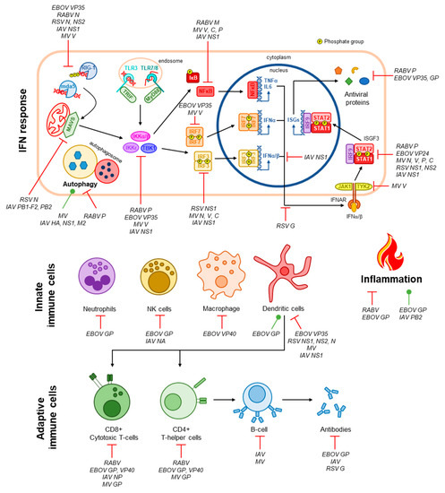
Figure 1. Immune evasion by NSVs. The IFN response is the primary antiviral pathway activated following virus infection. NSVs are detected by intracellular PRRs such as RIG-1 and mda5 in the cytoplasm or TLR3/7/8 in the endosomal compartment via interactions with viral RNA [12]. Signalling cascades involving MAVS (for RIG-I and mda5) or TRIF/MyD88 (for TLRs) result in the activation of the kinases IKKα/β/ε and TBK1 that subsequently activate the latent transcription factors IRF3, IRF7, and NFκB via phosphorylation [12]. These transcription factors promote the expression and secretion of proinflammatory and antiviral cytokines (TNFα, IL-6, and IFNα/β) [12]. IFNs signal in an autocrine and paracrine manner by binding to the IFNα/β receptor (IFNAR), resulting in the activation of the ISGF3 complex (STAT1, STAT2, and IRF9) through phosphorylation of STAT1 and STAT2 by IFNAR-associated kinases JAK1 and TYK2 [12]. ISGF3 then promotes the expression of antiviral genes [12]. Innate and adaptive immune cells, as well as inflammation and autophagy, which additionally contribute to viral clearance, are also depicted. Targeting of the antiviral immune response by NSVs, and the viral proteins involved if known, are indicated [33][34][35][36][37][38][39][40][41][42][43][45][44][46][47][48][49][50][51].

Therefore, the quality and magnitude of adaptive immunity is dependent on the innate immune response [13]. Supporting this, Nakaya et al. found that antibody titers at one-month post vaccination were positively correlated with early expression of type I IFNs associated genes [14]. Early induction of IFN was also reported to be important for the development of antibody responses in LAIV (live attenuated influenza vaccines) and TIV (trivalent influenza vaccines) vaccinated children [15]. TLRs have been shown to play an important role in both cell-mediated and antibody mediated protection in measles vaccine response. Secretion of type-I IFNs promotes the recruitment and activation of specialised immune cells that aid in the clearance of viral pathogens. Of this cell-mediated immune response, dendritic cells are central to bridging innate and adaptive immunity through their functions as antigen-presenting cells, leading to the activation T-helper cells that, in turn, activate humoral immunity via the production of a pathogen-specific antibody by B-cells [16]. Robust activation of T- and B-cells also leads to the generation of immune memory, a key requirement for vaccines to induce long-lasting immunity [17] (Figure 1). The importance of specific innate cell subsets such as natural killer (NK) and T follicular helper cells has been observed as correlates of protection in recipients of Ebola vaccine. Additionally, polymorphisms in dendritic cell-specific intercellular adhesion molecule-3 grabbing nonintegrin (DC-SIGN), a measles-specific receptor, may modulate cytokine responses to the measles component of the MMR vaccine [18]. Although the adaptive immune response has traditionally been the primary focus of vaccine development, these studies have highlighted the importance of innate immune responses after vaccination and infection. Thus, strategies can be developed to trigger specific innate pathways that can lead to stronger adaptive immune responses which are protective against infection.

T-cells and B-cells are critical components in adaptive immunity against IAV infection. CD4+ T-cells target IAV-infected epithelial cells through binding to MHC class II molecules and contribute to B cell activation promoting antibody production [19]. CD8+ T-cells differentiate into cytotoxic T lymphocytes (CTLs) and defend against IAV infection via producing cytokines and effector molecules in addition to direct cytotoxic effects on infected cells mediated by MHC class I [20]. Since cytotoxic T-cells target infected cells, and not the virus directly, T-cell mediated immunity typically provides protection without complete virus neutralization, allowing some degree of virus replication [21]. T-cell recognition of conserved viral antigens presented by antigen presenting cells (APC) can contribute to a more qualitative antibody response. However, current inactivated vaccines do not elicit a strong T-cell response, which may in part be overcome by the use of optimal adjuvants [22]. Polymorphisms in the signaling lymphocyte activation molecule (SLAM) and CD46 have been shown to drastically reduce MV-specific antibodies in individuals’ post-vaccination, which illustrates the importance of genetic variation in our immune cell surface markers on vaccine responses [23]. A correlation in the number of plasmacytoid dendritic cells and MV-specific antibodies has also been observed post-vaccination in infants, indicating another important link between the adaptive and innate immunity [24]. Induction of antibodies targeting conserved IAV antigens is desirable for vaccine responses with the potential to provide broad protection. Virus-neutralizing antibodies usually target viral surface antigens, for example the IAV haemagglutinin (HA) head domain. However, multiple immune mechanisms that do not result in virus neutralization can also contribute to heterosubtypic immunity against different subtypes of influenza viruses [25]. Ways by which the humoral branch of the vaccine response can also contribute to protection other than virus neutralization is through Fc receptor engagement via antibody-dependent cellular phagocytosis (ADCP) [26][27] or antibody-dependent cellular cytotoxicity (ADCC) [28][29][30], as well as antibody-dependent complement mediated-lysis (ADCL) [31]. Additionally, external factors that can modulate the effectiveness of the adaptive immune system, such as post-transplant immunosuppressant medication, can lead to a reduction in B-cells and protective antibody responses to MV over time [32].

3. Immune Evasion by NSVs

As mentioned in Section 2, viruses must overcome robust immune defences in order to establish productive infection. The NSVs discussed in this manuscript expertly evade these responses through a multitude of mechanisms summarised in Figure 1 and reviewed elsewhere [33][34][35][36][52]. Understanding how these viruses evade these immune defenses, especially the host–pathogen interfaces involved, are integral to the development of efficacious vaccine strains both by reducing pathogenicity and ensuring formation of immunological memory. In this section, we highlight a few points of convergence where these NSVs target our immune responses and the implications for vaccine development.

The IFN response is the primary defense against viral infection. Two stages of this response that are commonly targeted by NSVs are proverbial ‘bottlenecks’, viral RNA sensing and the ISGF3 transcription complex (consisting of STAT1, STAT2, and IRF9). Viral RNA is detected by two families of pattern recognition receptors (PRRs), RIG-I-like receptors (RLRs), and toll-like receptors (TLRS). The RLRs RIG-I and mda-5 detect uncapped RNA and double-stranded RNA (dsRNA), respectively, in the cytoplasmic compartment [53]. Toll-like receptors (TLRs) instead sense viral RNA within the endosomal compartment, these include TLR3, which detects dsRNA, and TLR7 and 8, which detected single-stranded RNA (ssRNA) [54]. RLRs are the principal PRRs that detect the NSVs discussed in this entry  [53] and, as such, each has a mechanism evading detection. In some cases, this is believed to be a ‘passive’ mechanism whereby the viral RNA is hidden from RLRs, such as by encapsidation by the RABV N protein and sequestration by EBOV VP35 [55][56]. RSV NS2 and MV V protein actively associate with RIG-I or MDA5, respectively, to inhibit its activity and RSV N sequesters MDA5 in viral inclusion bodies [51][57][58]. MV V also targets the phosphatases PPIα/γ and IAV NS1 targets the ubiquitin ligase TRIM25, both preventing their functions in the activation of RIG-I [59][60].

ISGF3 is responsible for the expression of hundreds of IFN-stimulated genes (ISGs), many of which have antiviral activity [61]. Thus, it is not unexpected that components of this complex, in particular STAT1, are common targets for viral IFN antagonists. The P gene products of both RABV (P1–P5) and MV (P, V, C) variously inhibit STAT1 function. RABV P1 binds to ‘active’, phosphorylated STAT1 (pY-STAT1) and excludes it from the nucleus, while the smaller P3 tethers pY-STAT1 to cellular microtubules to inhibit its nuclear import and additionally blocks its DNA-binding intranuclearly [62][63][64]. MV P associates with unphosphorylated STAT1 (U-STAT) to inhibit its phosphorylation and subsequent nuclear accumulation, and the V protein prevents phosphorylation of Tyk2, in addition to associating with Jak1, STAT1, and STAT2, to contribute to inhibition of ISGF3 activation and nuclear import [65][66][67][68]. The C protein instead targets pY-STAT1, inhibiting correct formation of STAT1 homodimers in response to the type-III IFN, IFNγ [69]. In addition, the MV N protein also contributes to this STAT1 blockade by inhibiting the nuclear accumulation of pY-STAT1 [70]. ISGF3 targeting by RSV, EBOV, and IAV are less multifaceted, RSV NS1 and NS2 target STAT2 for degradation, and IAV upregulates the expression of SOCS1 and SOCS3 that negatively regulated STAT signaling by dephosphorylating/inactivating JAK1 and TYK2 [71][72][73][74]. Finally, EBOV VP24 inhibits pY-STAT1 nuclear accumulation by competitively binding with the nuclear transport proteins importin-α1, α5, and α6 [75][76]; VP24 has also been found to bind to U-STAT1, but the contributions of this interaction to immune evasion are not yet known [77]. Importantly, inhibition of STAT1 contributes to the pathogenicity of all five viruses, evaluated through infection of STAT1 knockout mice or using mutant virus unable to target STAT1 [72][78][79][80][81]. Thus, disruption of the virus-STAT1 interface is central to the production of attenuated vaccines.

The NSVs discussed in this manuscript commonly target T-cells to prevent their antiviral immune functions and their ability to activate humoral immunity. During RABV infection, only transient infiltration of T-cells into the CNS is observed, this is believed to be due to the depletion of T-cell populations via upregulation of the immunosuppressive molecules FasL, B7-H1, and HLA-G on infected neurons [82][83][84]. MV inhibits the proliferation of uninfected T-cells at the cell surface involving the surface exposed GP complex, consisting of the HA and F proteins, though the specific mechanism is yet to be elucidated [85][86][87]. EBOV has similarly been shown to induce apoptosis of T-cells through interactions of its surface GP with TLR4 [88]. EBOV VP40 is also released from infected cells in exosomes, which have also been proposed to induce apoptosis in uninfected T-cells. While not targeting T-cells specifically, RSV inhibits the activation of T-cells by DCs via N protein, which localises to the surface of DCs and impairs formation of the immunological synapse through reduction of MHC clustering [89]. IAV also does not target T-cell directly, but its high mutation rate in immunological epitopes of its N protein have been shown to prevent recognition of infected cells by CD8+ T-cells [45]; this could possibly extend to evasion of CD4+ T-cells as well. In addition, IAV, RSV, and EBOV all suppress DC maturation, inhibiting their capacity to activate T-cells, while MV does not impact maturation, but the capacity for infected DCs to activate T-cells is greatly reduced [90][91][92][93][94].

References

  1. Afrough, B.; Dowall, S.; Hewson, R. Emerging viruses and current strategies for vaccine intervention. Clin. Exp. Immunol. 2019, 196, 157–166, doi:10.1111/cei.13295.
  2. Carrasco-Hernandez, R.; Jacome, R.; Lopez Vidal, Y.; Ponce de Leon, S. Are RNA Viruses Candidate Agents for the Next Global Pandemic? A Review. ILAR J. 2017, 58, 343–358.
  3. Payne, S. Chapter 10—Introduction to RNA Viruses. In Viruses; Payne, S., Ed.; Academic Press: Cambridge, MA, USA, 2017; pp. 97–105, doi:10.1016/B978-0-12-803109-4.00010-6.
  4. Durmuş, S.; Ülgen, K.Ö. Comparative interactomics for virus–human protein–protein interactions: DNA viruses versus RNA viruses. FEBS Open Bio 2017, 7, 96–107, doi:10.1002/2211-5463.12167.
  5. Duffy, S.; Shackelton, L.A.; Holmes, E.C. Rates of evolutionary change in viruses: Patterns and determinants. Nat. Rev. Genet. 2008, 9, 267–276, doi:10.1038/nrg2323.
  6. Holmes, E.C. The Evolutionary Genetics of Emerging Viruses. Annu. Rev. Ecol. Evol. Syst. 2009, 40, 353–372, doi:10.1146/annurev.ecolsys.110308.120248.
  7. Loomis, R.J.; Johnson, P.R. Emerging Vaccine Technologies. Vaccines 2015, 3, 429–447, doi:10.3390/vaccines3020429.
  8. Regules, J.A.; Beigel, J.H.; Paolino, K.M.; Voell, J.; Castellano, A.R.; Hu, Z.; Munoz, P.; Moon, J.E.; Ruck, R.C.; Bennett, J.W.; et al. A Recombinant Vesicular Stomatitis Virus Ebola Vaccine. N. Engl. J. Med. 2017, 376, 330–341, doi:10.1056/NEJMoa1414216.
  9. Sesterhenn, F.; Yang, C.; Bonet, J.; Cramer, J.T.; Wen, X.; Wang, Y.; Chiang, C.I.; Abriata, L.A.; Kucharska, I.; Castoro, G.; et al. De novo protein design enables the precise induction of RSV-neutralizing antibodies. Science 2020, 368, doi:10.1126/science.aay5051.
  10. Hobernik, D.; Bros, M. DNA Vaccines-How Far from Clinical Use? Int. J. Mol. Sci. 2018, 19, 3605, doi:10.3390/ijms19113605.
  11. Iwasaki, A.; Pillai, P.S. Innate immunity to influenza virus infection. Nat. Rev. Immunol. 2014, 14, 315–328.
  12. Bonjardim, C.A.; Ferreira, P.C.; Kroon, E.G. Interferons: Signaling, antiviral and viral evasion. Immunol. Lett. 2009, 122, 1–11, doi:10.1016/j.imlet.2008.11.002.
  13. Iwasaki, A.; Medzhitov, R. Control of adaptive immunity by the innate immune system. Nat. Immunol. 2015, 16, 343.
  14. Nakaya, H.I.; Hagan, T.; Duraisingham, S.S.; Lee, E.K.; Kwissa, M.; Rouphael, N.; Frasca, D.; Gersten, M.; Mehta, A.K.; Gaujoux, R. Systems analysis of immunity to influenza vaccination across multiple years and in diverse populations reveals shared molecular signatures. Immunity 2015, 43, 1186–1198.
  15. Cao, R.G.; Suarez, N.M.; Obermoser, G.; Lopez, S.M.; Flano, E.; Mertz, S.E.; Albrecht, R.A.; García-Sastre, A.; Mejias, A.; Xu, H. Differences in antibody responses between trivalent inactivated influenza vaccine and live attenuated influenza vaccine correlate with the kinetics and magnitude of interferon signaling in children. J. Infect. Dis. 2014, 210, 224–233.
  16. den Haan, J.M.; Arens, R.; van Zelm, M.C. The activation of the adaptive immune system: Cross-talk between anti-gen-presenting cells, T cells and B cells. Immunol. Lett. 2014, 162, 103–112, doi:10.1016/j.imlet.2014.10.011.
  17. Sallusto, F.; Lanzavecchia, A.; Araki, K.; Ahmed, R. From vaccines to memory and back. Immunity 2010, 33, 451–463, doi:10.1016/j.immuni.2010.10.008.
  18. Clifford, H.D.; Richmond, P.; Khoo, S.K.; Zhang, G.; Yerkovich, S.T.; Le Souef, P.N.; Hayden, C.M. SLAM and DC-SIGN mea-sles receptor polymorphisms and their impact on antibody and cytokine responses to measles vaccine. Vaccine 2011, 29, 5407–5413, doi:10.1016/j.vaccine.2011.05.068.
  19. Swain, S.L.; McKinstry, K.K.; Strutt, T.M. Expanding roles for CD4+ T cells in immunity to viruses. Nat. Rev. Immunol. 2012, 12, 136–148.
  20. Duan, S.; Thomas, P.G. Balancing immune protection and immune pathology by CD8+ T-cell responses to influenza infec-tion. Front. Immunol. 2016, 7, 25.
  21. Spitaels, J.; Roose, K.; Saelens, X. Influenza and memory T cells: How to awake the force. Vaccines 2016, 4, 33.
  22. Tetsutani, K.; Ishii, K.J. Adjuvants in influenza vaccines. Vaccine 2012, 30, 7658–7661.
  23. Dhiman, N.; Poland, G.A.; Cunningham, J.M.; Jacobson, R.M.; Ovsyannikova, I.G.; Vierkant, R.A.; Wu, Y.; Pankratz, V.S. Variations in measles vaccine-specific humoral immunity by polymorphisms in SLAM and CD46 measles virus receptors. J. Allergy Clin. Immunol. 2007, 120, 666–672, doi:10.1016/j.jaci.2007.04.036.
  24. Garcia-Leon, M.L.; Bonifaz, L.C.; Espinosa-Torres, B.; Hernandez-Perez, B.; Cardiel-Marmolejo, L.; Santos-Preciado, J.I.; Wong-Chew, R.M. A correlation of measles specific antibodies and the number of plasmacytoid dendritic cells is observed after measles vaccination in 9 month old infants. Hum. Vaccin. Immunother. 2015, 11, 1762–1769, doi:10.1080/21645515.2015.1032488.
  25. Carragher, D.M.; Kaminski, D.A.; Moquin, A.; Hartson, L.; Randall, T.D. A novel role for non-neutralizing antibodies against nucleoprotein in facilitating resistance to influenza virus. J. Immunol. 2008, 181, 4168–4176.
  26. Ana-Sosa-Batiz, F.; Vanderven, H.; Jegaskanda, S.; Johnston, A.; Rockman, S.; Laurie, K.; Barr, I.; Reading, P.; Lichtfuss, M.; Kent, S.J. Influenza-specific antibody-dependent phagocytosis. PLoS ONE 2016, 11, e0154461.
  27. Ana-Sosa-Batiz, F.; Johnston, A.P.; Hogarth, P.M.; Wines, B.D.; Barr, I.; Wheatley, A.K.; Kent, S.J. Antibody-dependent phag-ocytosis (ADP) responses following trivalent inactivated influenza vaccination of younger and older adults. Vaccine 2017, 35, 6451–6458.
  28. Greenberg, S.B.; Criswell, B.S.; Six, H.R.; Couch, R.B. Lymphocyte Cytotoxicity to Influenza Virus-Infected Cells: II. Re-quirement for Antibody and Non-T Lymphocytes. J. Immunol. 1977, 119, 2100–2106.
  29. Moody, M.A.; Von Holle, T.A. Influenza and antibody-dependent cellular cytotoxicity. Front. Immunol. 2019, 10, 1457.
  30. Vanderven, H.A.; Jegaskanda, S.; Wheatley, A.K.; Kent, S.J. Antibody-dependent cellular cytotoxicity and influenza virus. Curr. Opin. Virol. 2017, 22, 89–96.
  31. Kopf, M.; Abel, B.; Gallimore, A.; Carroll, M.; Bachmann, M.F. Complement component C3 promotes T-cell priming and lung migration to control acute influenza virus infection. Nat. Med. 2002, 8, 373–378.
  32. Rocca, S.; Santilli, V.; Cotugno, N.; Concato, C.; Manno, E.C.; Nocentini, G.; Macchiarulo, G.; Cancrini, C.; Finocchi, A.; Guzzo, I.; et al. Waning of vaccine-induced immunity to measles in kidney transplanted children. Medicine 2016, 95, e4738, doi:10.1097/MD.0000000000004738.
  33. Ito, N.; Moseley, G.W.; Sugiyama, M. The importance of immune evasion in the pathogenesis of rabies virus. J. Vet. Med. Sci. 2016, 78, 1089–1098, doi:10.1292/jvms.16-0092.
  34. Openshaw, P.J.M.; Chiu, C.; Culley, F.J.; Johansson, C. Protective and Harmful Immunity to RSV Infection. Annu. Rev. Im-munol. 2017, 35, 501–532, doi:10.1146/annurev-immunol-051116-052206.
  35. Chen, X.; Liu, S.; Goraya, M.U.; Maarouf, M.; Huang, S.; Chen, J.L. Host Immune Response to Influenza A Virus Infection. Front. Immunol. 2018, 9, 320, doi:10.3389/fimmu.2018.00320.
  36. Audet, J.; Kobinger, G.P. Immune evasion in ebolavirus infections. Viral Immunol. 2015, 28, 10–18, doi:10.1089/vim.2014.0066.
  37. Oshansky, C.M.; Zhang, W.; Moore, E.; Tripp, R.A. The host response and molecular pathogenesis associated with respira-tory syncytial virus infection. Future Microbiol. 2009, 4, 279–297, doi:10.2217/fmb.09.1.
  38. Pleet, M.L.; Erickson, J.; DeMarino, C.; Barclay, R.A.; Cowen, M.; Lepene, B.; Liang, J.; Kuhn, J.H.; Prugar, L.; Stonier, S.W.; et al. Ebola Virus VP40 Modulates Cell Cycle and Biogenesis of Extracellular Vesicles. J. Infect. Dis. 2018, 218, S365–S387, doi:10.1093/infdis/jiy472.
  39. Pleet, M.L.; DeMarino, C.; Lepene, B.; Aman, M.J.; Kashanchi, F. The Role of Exosomal VP40 in Ebola Virus Disease. DNA Cell Biol. 2017, 36, 243–248, doi:10.1089/dna.2017.3639.
  40. Edri, A.; Shemesh, A.; Iraqi, M.; Matalon, O.; Brusilovsky, M.; Hadad, U.; Radinsky, O.; Gershoni-Yahalom, O.; Dye, J.M.; Mandelboim, O.; et al. The Ebola-Glycoprotein Modulates the Function of Natural Killer Cells. Front. Immunol. 2018, 9, 1428, doi:10.3389/fimmu.2018.01428.
  41. Ning, Y.J.; Deng, F.; Hu, Z.; Wang, H. The roles of ebolavirus glycoproteins in viral pathogenesis. Virol. Sin. 2017, 32, 3–15, doi:10.1007/s12250-016-3850-1.
  42. Zhirnov, O.P.; Klenk, H.D. Influenza a virus proteins NS1 and hemagglutinin along with M2 are involved in stimulation of autophagy in infected cells. J. Virol. 2013, 87, 13107–13114, doi:10.1128/JVI.02148-13.
  43. Krug, R.M. Functions of the influenza a virus NS1 protein in antiviral defense. Curr. Opin. Virol. 2015, 12, 1–6, doi:10.1016/j.coviro.2015.01.007.
  44. Rimmelzwaan, G.F.; Boon, A.C.; Voeten, J.T.; Berkhoff, E.G.; Fouchier, R.A.; Osterhaus, A.D. Sequence variation in the in-fluenza A virus nucleoprotein associated with escape from cytotoxic T lymphocytes. Virus Res. 2004, 103, 97–100, doi:10.1016/j.virusres.2004.02.020.
  45. Han, T.; Marasco, W.A. Structural basis of influenza virus neutralization. Ann. N. Y. Acad. Sci. 2011, 1217, 178–190, doi:10.1111/j.1749-6632.2010.05829.x.
  46. Dougan, S.K.; Ashour, J.; Karssemeijer, R.A.; Popp, M.W.; Avalos, A.M.; Barisa, M.; Altenburg, A.F.; Ingram, J.R.; Cragnolini, J.J.; Guo, C.; et al. Antigen-specific B-cell receptor sensitizes B cells to infection by influenza virus. Nature 2013, 503, 406–409, doi:10.1038/nature12637.
  47. Griffin, D.E. Measles virus-induced suppression of immune responses. Immunol. Rev. 2010, 236, 176–189, doi:10.1111/j.1600-065X.2010.00925.x.
  48. Richetta, C.; Gregoire, I.P.; Verlhac, P.; Azocar, O.; Baguet, J.; Flacher, M.; Tangy, F.; Rabourdin-Combe, C.; Faure, M. Sus-tained autophagy contributes to measles virus infectivity. PLoS Pathog. 2013, 9, e1003599, doi:10.1371/journal.ppat.1003599.
  49. Liu, J.; Wang, H.; Gu, J.; Deng, T.; Yuan, Z.; Hu, B.; Xu, Y.; Yan, Y.; Zan, J.; Liao, M.; et al. BECN1-dependent CASP2 incom-plete autophagy induction by binding to rabies virus phosphoprotein. Autophagy 2017, 13, 739–753, doi:10.1080/15548627.2017.1280220.
  50. Lifland, A.W.; Jung, J.; Alonas, E.; Zurla, C.; Crowe, J.E., Jr.; Santangelo, P.J. Human respiratory syncytial virus nucleopro-tein and inclusion bodies antagonize the innate immune response mediated by MDA5 and MAVS. J. Virol. 2012, 86, 8245–8258, doi:10.1128/JVI.00215-12.
  51. Forero, A.; Tisoncik-Go, J.; Watanabe, T.; Zhong, G.; Hatta, M.; Tchitchek, N.; Selinger, C.; Chang, J.; Barker, K.; Morrison, J.; et al. The 1918 Influenza Virus PB2 Protein Enhances Virulence through the Disruption of Inflammatory and Wnt-Mediated Signaling in Mice. J. Virol. 2015, 90, 2240–2253, doi:10.1128/JVI.02974-15.
  52. Pfeffermann, K.; Dorr, M.; Zirkel, F.; von Messling, V. Morbillivirus Pathogenesis and Virus-Host Interactions. Adv. Virus Res. 2018, 100, 75–98, doi:10.1016/bs.aivir.2017.12.003.
  53. Loo, Y.M.; Gale, M., Jr. Immune signaling by RIG-I-like receptors. Immunity 2011, 34, 680–692, doi:10.1016/j.immuni.2011.05.003.
  54. Miyake, K.; Shibata, T.; Ohto, U.; Shimizu, T.; Saitoh, S.I.; Fukui, R.; Murakami, Y. Mechanisms controlling nucleic ac-id-sensing Toll-like receptors. Int. Immunol. 2018, 30, 43–51, doi:10.1093/intimm/dxy016.
  55. Cardenas, W.B.; Loo, Y.M.; Gale, M., Jr.; Hartman, A.L.; Kimberlin, C.R.; Martinez-Sobrido, L.; Saphire, E.O.; Basler, C.F. Ebola virus VP35 protein binds double-stranded RNA and inhibits alpha/beta interferon production induced by RIG-I sig-naling. J. Virol. 2006, 80, 5168–5178, doi:10.1128/JVI.02199-05.
  56. Masatani, T.; Ito, N.; Shimizu, K.; Ito, Y.; Nakagawa, K.; Sawaki, Y.; Koyama, H.; Sugiyama, M. Rabies virus nucleoprotein functions to evade activation of the RIG-I-mediated antiviral response. J. Virol. 2010, 84, 4002–4012, doi:10.1128/JVI.02220-09.
  57. Ling, Z.; Tran, K.C.; Teng, M.N. Human respiratory syncytial virus nonstructural protein NS2 antagonizes the activation of beta interferon transcription by interacting with RIG-I. J. Virol. 2009, 83, 3734–3742, doi:10.1128/JVI.02434-08.
  58. Childs, K.; Stock, N.; Ross, C.; Andrejeva, J.; Hilton, L.; Skinner, M.; Randall, R.; Goodbourn, S. mda-5, but not RIG-I, is a common target for paramyxovirus V proteins. Virology 2007, 359, 190–200, doi:10.1016/j.virol.2006.09.023.
  59. Davis, M.E.; Wang, M.K.; Rennick, L.J.; Full, F.; Gableske, S.; Mesman, A.W.; Gringhuis, S.I.; Geijtenbeek, T.B.; Duprex, W.P.; Gack, M.U. Antagonism of the phosphatase PP1 by the measles virus V protein is required for innate immune escape of MDA5. Cell Host Microbe 2014, 16, 19–30, doi:10.1016/j.chom.2014.06.007.
  60. Gack, M.U.; Albrecht, R.A.; Urano, T.; Inn, K.S.; Huang, I.C.; Carnero, E.; Farzan, M.; Inoue, S.; Jung, J.U.; Garcia-Sastre, A. Influenza A virus NS1 targets the ubiquitin ligase TRIM25 to evade recognition by the host viral RNA sensor RIG-I. Cell Host Microbe 2009, 5, 439–449, doi:10.1016/j.chom.2009.04.006.
  61. Schoggins, J.W.; Wilson, S.J.; Panis, M.; Murphy, M.Y.; Jones, C.T.; Bieniasz, P.; Rice, C.M. A diverse range of gene products are effectors of the type I interferon antiviral response. Nature 2011, 472, 481–485, doi:10.1038/nature09907.
  62. Vidy, A.; El Bougrini, J.; Chelbi-Alix, M.K.; Blondel, D. The nucleocytoplasmic rabies virus P protein counteracts interferon signaling by inhibiting both nuclear accumulation and DNA binding of STAT1. J. Virol. 2007, 81, 4255–4263, doi:10.1128/JVI.01930-06.
  63. Moseley, G.W.; Lahaye, X.; Roth, D.M.; Oksayan, S.; Filmer, R.P.; Rowe, C.L.; Blondel, D.; Jans, D.A. Dual modes of rabies P-protein association with microtubules: A novel strategy to suppress the antiviral response. J. Cell Sci. 2009, 122, 3652–3662, doi:10.1242/jcs.045542.
  64. Brzozka, K.; Finke, S.; Conzelmann, K.K. Inhibition of interferon signaling by rabies virus phosphoprotein P: Activa-tion-dependent binding of STAT1 and STAT2. J. Virol. 2006, 80, 2675–2683, doi:10.1128/JVI.80.6.2675-2683.2006.
  65. Chinnakannan, S.K.; Nanda, S.K.; Baron, M.D. Morbillivirus v proteins exhibit multiple mechanisms to block type 1 and type 2 interferon signalling pathways. PLoS ONE 2013, 8, e57063, doi:10.1371/journal.pone.0057063.
  66. Caignard, G.; Bourai, M.; Jacob, Y.; Infection, M.p.I.M.A.P.; Tangy, F.; Vidalain, P.O. Inhibition of IFN-alpha/beta signaling by two discrete peptides within measles virus V protein that specifically bind STAT1 and STAT2. Virology 2009, 383, 112–120, doi:10.1016/j.virol.2008.10.014.
  67. Devaux, P.; von Messling, V.; Songsungthong, W.; Springfeld, C.; Cattaneo, R. Tyrosine 110 in the measles virus phospho-protein is required to block STAT1 phosphorylation. Virology 2007, 360, 72–83, doi:10.1016/j.virol.2006.09.049.
  68. Ramachandran, A.; Parisien, J.P.; Horvath, C.M. STAT2 is a primary target for measles virus V protein-mediated alpha/beta interferon signaling inhibition. J. Virol. 2008, 82, 8330–8338, doi:10.1128/JVI.00831-08.
  69. Yokota, S.; Okabayashi, T.; Fujii, N. Measles virus C protein suppresses gamma-activated factor formation and vi-rus-induced cell growth arrest. Virology 2011, 414, 74–82, doi:10.1016/j.virol.2011.03.010.
  70. Takayama, I.; Sato, H.; Watanabe, A.; Omi-Furutani, M.; Sugai, A.; Kanki, K.; Yoneda, M.; Kai, C. The nucleocapsid protein of measles virus blocks host interferon response. Virology 2012, 424, 45–55, doi:10.1016/j.virol.2011.12.011.
  71. Pauli, E.K.; Schmolke, M.; Wolff, T.; Viemann, D.; Roth, J.; Bode, J.G.; Ludwig, S. Influenza A virus inhibits type I IFN sig-naling via NF-kappaB-dependent induction of SOCS-3 expression. PLoS Pathog. 2008, 4, e1000196, doi:10.1371/journal.ppat.1000196.
  72. Wei, H.; Wang, S.; Chen, Q.; Chen, Y.; Chi, X.; Zhang, L.; Huang, S.; Gao, G.F.; Chen, J.L. Suppression of interferon lambda signaling by SOCS-1 results in their excessive production during influenza virus infection. PLoS Pathog. 2014, 10, e1003845, doi:10.1371/journal.ppat.1003845.
  73. Whelan, J.N.; Tran, K.C.; van Rossum, D.B.; Teng, M.N. Identification of Respiratory Syncytial Virus Nonstructural Protein 2 Residues Essential for Exploitation of the Host Ubiquitin System and Inhibition of Innate Immune Responses. J. Virol. 2016, 90, 6453–6463, doi:10.1128/JVI.00423-16.
  74. Elliott, J.; Lynch, O.T.; Suessmuth, Y.; Qian, P.; Boyd, C.R.; Burrows, J.F.; Buick, R.; Stevenson, N.J.; Touzelet, O.; Gadina, M.; et al. Respiratory syncytial virus NS1 protein degrades STAT2 by using the Elongin-Cullin E3 ligase. J. Virol. 2007, 81, 3428–3436, doi:10.1128/JVI.02303-06.
  75. Reid, S.P.; Valmas, C.; Martinez, O.; Sanchez, F.M.; Basler, C.F. Ebola virus VP24 proteins inhibit the interaction of NPI-1 subfamily karyopherin alpha proteins with activated STAT1. J. Virol. 2007, 81, 13469–13477, doi:10.1128/JVI.01097-07.
  76. Xu, W.; Edwards, M.R.; Borek, D.M.; Feagins, A.R.; Mittal, A.; Alinger, J.B.; Berry, K.N.; Yen, B.; Hamilton, J.; Brett, T.J.; et al. Ebola virus VP24 targets a unique NLS binding site on karyopherin alpha 5 to selectively compete with nuclear import of phosphorylated STAT1. Cell Host Microbe 2014, 16, 187–200, doi:10.1016/j.chom.2014.07.008.
  77. Zhang, A.P.; Bornholdt, Z.A.; Liu, T.; Abelson, D.M.; Lee, D.E.; Li, S.; Woods, V.L., Jr.; Saphire, E.O. The ebola virus interfer-on antagonist VP24 directly binds STAT1 and has a novel, pyramidal fold. PLoS Pathog. 2012, 8, e1002550, doi:10.1371/journal.ppat.1002550.
  78. Wiltzer, L.; Okada, K.; Yamaoka, S.; Larrous, F.; Kuusisto, H.V.; Sugiyama, M.; Blondel, D.; Bourhy, H.; Jans, D.A.; Ito, N.; et al. Interaction of rabies virus P-protein with STAT proteins is critical to lethal rabies disease. J. Infect. Dis. 2014, 209, 1744–1753, doi:10.1093/infdis/jit829.
  79. Raymond, J.; Bradfute, S.; Bray, M. Filovirus infection of STAT-1 knockout mice. J. Infect. Dis. 2011, 204 (Suppl. 3), S986–S990, doi:10.1093/infdis/jir335.
  80. Durbin, J.E.; Johnson, T.R.; Durbin, R.K.; Mertz, S.E.; Morotti, R.A.; Peebles, R.S.; Graham, B.S. The role of IFN in respiratory syncytial virus pathogenesis. J. Immunol. 2002, 168, 2944–2952, doi:10.4049/jimmunol.168.6.2944.
  81. Devaux, P.; Hudacek, A.W.; Hodge, G.; Reyes-Del Valle, J.; McChesney, M.B.; Cattaneo, R. A recombinant measles virus un-able to antagonize STAT1 function cannot control inflammation and is attenuated in rhesus monkeys. J. Virol. 2011, 85, 348–356, doi:10.1128/JVI.00802-10.
  82. Lafon, M.; Megret, F.; Meuth, S.G.; Simon, O.; Velandia Romero, M.L.; Lafage, M.; Chen, L.; Alexopoulou, L.; Flavell, R.A.; Prehaud, C.; et al. Detrimental contribution of the immuno-inhibitor B7-H1 to rabies virus encephalitis. J. Immunol. 2008, 180, 7506–7515, doi:10.4049/jimmunol.180.11.7506.
  83. Megret, F.; Prehaud, C.; Lafage, M.; Moreau, P.; Rouas-Freiss, N.; Carosella, E.D.; Lafon, M. Modulation of HLA-G and HLA-E expression in human neuronal cells after rabies virus or herpes virus simplex type 1 infections. Hum. Immunol. 2007, 68, 294–302, doi:10.1016/j.humimm.2006.12.003.
  84. Baloul, L.; Camelo, S.; Lafon, M. Up-regulation of Fas ligand (FasL) in the central nervous system: A mechanism of immune evasion by rabies virus. J. Neurovirol. 2004, 10, 372–382, doi:10.1080/13550280490521122.
  85. Niewiesk, S.; Ohnimus, H.; Schnorr, J.J.; Gotzelmann, M.; Schneider-Schaulies, S.; Jassoy, C.; Ter Meulen, V. Measles vi-rus-induced immunosuppression in cotton rats is associated with cell cycle retardation in uninfected lymphocytes. J. Gen. Virol. 1999, 80 Pt 8, 2023–2029, doi:10.1099/0022-1317-80-8-2023.
  86. Niewiesk, S.; Gotzelmann, M.; ter Meulen, V. Selective in vivo suppression of T lymphocyte responses in experimental measles virus infection. Proc. Natl. Acad. Sci. USA 2000, 97, 4251–4255, doi:10.1073/pnas.060012097.
  87. Schlender, J.; Schnorr, J.J.; Spielhoffer, P.; Cathomen, T.; Cattaneo, R.; Billeter, M.A.; ter Meulen, V.; Schneider-Schaulies, S. Interaction of measles virus glycoproteins with the surface of uninfected peripheral blood lymphocytes induces immuno-suppression in vitro. Proc. Natl. Acad. Sci. USA 1996, 93, 13194–13199, doi:10.1073/pnas.93.23.13194.
  88. Iampietro, M.; Younan, P.; Nishida, A.; Dutta, M.; Lubaki, N.M.; Santos, R.I.; Koup, R.A.; Katze, M.G.; Bukreyev, A. Ebola virus glycoprotein directly triggers T lymphocyte death despite of the lack of infection. PLoS Pathog. 2017, 13, e1006397, doi:10.1371/journal.ppat.1006397.
  89. Cespedes, P.F.; Bueno, S.M.; Ramirez, B.A.; Gomez, R.S.; Riquelme, S.A.; Palavecino, C.E.; Mackern-Oberti, J.P.; Mora, J.E.; Depoil, D.; Sacristan, C.; et al. Surface expression of the hRSV nucleoprotein impairs immunological synapse formation with T cells. Proc. Natl. Acad. Sci. USA 2014, 111, E3214–E3223, doi:10.1073/pnas.1400760111.
  90. Gonzalez, P.A.; Prado, C.E.; Leiva, E.D.; Carreno, L.J.; Bueno, S.M.; Riedel, C.A.; Kalergis, A.M. Respiratory syncytial virus impairs T cell activation by preventing synapse assembly with dendritic cells. Proc. Natl. Acad. Sci. USA 2008, 105, 14999–15004, doi:10.1073/pnas.0802555105.
  91. Munir, S.; Le Nouen, C.; Luongo, C.; Buchholz, U.J.; Collins, P.L.; Bukreyev, A. Nonstructural proteins 1 and 2 of respiratory syncytial virus suppress maturation of human dendritic cells. J. Virol. 2008, 82, 8780–8796, doi:10.1128/JVI.00630-08.
  92. Fernandez-Sesma, A.; Marukian, S.; Ebersole, B.J.; Kaminski, D.; Park, M.S.; Yuen, T.; Sealfon, S.C.; Garcia-Sastre, A.; Moran, T.M. Influenza virus evades innate and adaptive immunity via the NS1 protein. J. Virol. 2006, 80, 6295–6304, doi:10.1128/JVI.02381-05.
  93. Coughlin, M.M.; Bellini, W.J.; Rota, P.A. Contribution of dendritic cells to measles virus induced immunosuppression. Rev. Med. Virol. 2013, 23, 126–138, doi:10.1002/rmv.1735.
  94. Yen, B.; Mulder, L.C.; Martinez, O.; Basler, C.F. Molecular basis for ebolavirus VP35 suppression of human dendritic cell maturation. J. Virol. 2014, 88, 12500–12510, doi:10.1128/JVI.02163-14.
More
Information
Subjects: Virology
Contributor MDPI registered users' name will be linked to their SciProfiles pages. To register with us, please refer to https://encyclopedia.pub/register :
View Times: 487
Revisions: 3 times (View History)
Update Date: 07 Feb 2021
1000/1000