Submitted Successfully!
To reward your contribution, here is a gift for you: A free trial for our video production service.
Thank you for your contribution! You can also upload a video entry or images related to this topic.
Version Summary Created by Modification Content Size Created at Operation
1 + 3103 word(s) 3103 2021-08-25 09:52:21 |
2 format correct Meta information modification 3103 2021-09-10 08:59:26 |

Video Upload Options

Do you have a full video?

Confirm

Are you sure to Delete?
Cite
If you have any further questions, please contact Encyclopedia Editorial Office.
De Palma, F. Serrated Pathway in Colorectal Cancer. Encyclopedia. Available online: https://encyclopedia.pub/entry/14048 (accessed on 16 April 2024).
De Palma F. Serrated Pathway in Colorectal Cancer. Encyclopedia. Available at: https://encyclopedia.pub/entry/14048. Accessed April 16, 2024.
De Palma, Fatima. "Serrated Pathway in Colorectal Cancer" Encyclopedia, https://encyclopedia.pub/entry/14048 (accessed April 16, 2024).
De Palma, F. (2021, September 09). Serrated Pathway in Colorectal Cancer. In Encyclopedia. https://encyclopedia.pub/entry/14048
De Palma, Fatima. "Serrated Pathway in Colorectal Cancer." Encyclopedia. Web. 09 September, 2021.
Serrated Pathway in Colorectal Cancer
Edit

Colorectal cancer (CRC) is a leading cause of cancer death worldwide. It includes different subtypes that differ in their clinical and prognostic features. In the past decade, in addition to the conventional adenoma-carcinoma model, an alternative multistep mechanism of carcinogenesis, namely the “serrated pathway”, has been described. Approximately, 15 to 30% of all CRCs arise from neoplastic serrated polyps, a heterogeneous group of lesions that are histologically classified into three morphologic categories: hyperplastic polyps, sessile serrated adenomas/polyps, and the traditional serrated adenomas/polyps. Serrated polyps are characterized by genetic (BRAF or KRAS mutations) and epigenetic (CpG island methylator phenotype (CIMP)) alterations that cooperate to initiate and drive malignant transformation from normal colon mucosa to polyps, and then to CRC. The high heterogeneity of the serrated lesions renders their diagnostic and pathological interpretation difficult. 

colorectal cancer serrated pathway serrated lesions serrated polyp CIMP DNA methylation MSI CIN serrated adenocarcinoma gut microbiota

1. Introduction

The Conventional Model of Colorectal Carcinogenesis and the “Serrated” Pathway

Colorectal cancer is a multifactorial and heterogeneous disease [1]. Most CRCs (75%) are sporadic, whereas about 20% of CRC patients report a family history of the disease. Finally, 3–5% of CRCs are hereditary, with subjects bearing highly penetrant germline mutations that are associated with well-defined cancer-predisposing syndromes such as the hereditary nonpolyposis colorectal cancer (HNPCC), best known as Lynch syndrome (1–3%), or the familial adenomatous polyposis (FAP) (<1%), or again the hamartomatous polyposis syndrome, which displays the lowest incidence (<0.1%) [2].
CRC pathogenesis is due to the progressive accumulation of genetic and epigenetic alterations, some of which being responsible for activating oncogenes or inactivating oncosuppressor genes, that are able to drive the malignant evolution from normal epithelium through early neoplastic lesions (aberrant crypt foci, adenomas, and serrated adenomas) to CRC [3][4]. Such malignant transformation requires up to 15 years, depending on the characteristics of the lesion and on other independent risk factors such as gender, body weight, body mass index, physical inactivity [5].
Neoplastic transformation affecting the colon epithelium is characterized by two distinct morphological pathways of carcinogenesis, namely the conventional and the alternative/serrated neoplasia pathways, each one being defined by specific genetic and epigenetic alterations, typical clinical and histological features and leading to different phenotypes [6][7][8].
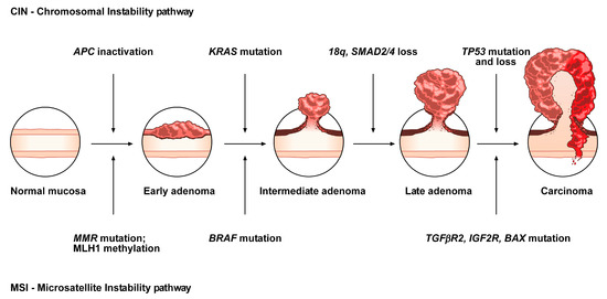
The conventional model, the so-called adenoma-carcinoma sequence, is histologically homogeneous and morphologically characterized the adenoma, including tubular or tubulovillous adenoma, as a precursor lesion [1]. The adenoma-carcinoma sequence is a multistep mutational pathway, in which each histological alteration is the consequence of a molecular dysregulation [9][10][11][12]. At the molecular level, this model recognizes a heterogeneous background, based on two mechanisms of tumorigenesis: (i) chromosomal instability (CIN) or (ii) microsatellite instability (MSI) [13][14][15][16] (Figure 1).
Figure 1. Conventional adenoma-to-carcinoma sequence. The chromosomal instability (CIN) pathway begins with bi-allelic mutations in the tumor suppressor gene APC within the normal colonic mucosa. The latter progressively differentiate into adenocarcinoma upon acquisition of additional mutations in the genes KRASSMAD4, and TP53, with consequent dysregulation of the Wnt/β-catenin, MAPKPI3K and TGF-β signaling pathways. Alternatively, the MSI pathway involves an initial alteration of the Wnt signaling that leads to the formation of an early adenoma. Then, BRAF mutation followed by alterations of the genes TGFBR2IGF2R, and BAX, participate in the progression toward the intermediate and late stages of carcinogenesis.
CIN represents the most prevalent form of genomic instability. It is detected in 85% of sporadic CRCs and is frequently observed in distal, rather than proximal, colon cancer sites [15][16]. CIN consists of a gain or loss of all or part of chromosome(s), and is usually associated with mutations in proto-oncogenes or tumor suppressor genes, such as KRAS or APC, respectively [13][16].
MSI occurs in about 15% of CRCs, predicts a favorable outcome in CRC and can also be detected in the serrated pathway [15][17][18][19]. This genomic instability does not affect chromosomal integrity but consists of an accumulation of insertions/deletions of short nucleotide repeats (microsatellites) that is consecutive to hereditary (5%) or sporadic (10%) alterations in genes involved in DNA mismatch repair (MMR) [14][15].
Although MSI, according to the National Cancer Institute, is frequently determined using a panel of five markers (BAT25, BAT26, D2S123, D5346, and D17S250), a variety of commercially available panels are currently used in most laboratories [20]. Depending on the number of microsatellites associated with these markers, tumors have been subclassified into: (i) high, labeled “MSI”, (ii) low, labeled “MSI-L” or (iii) stable, labeled “MSS” [21]. MSI-L tumors have been regrouped with MSS tumors, due to low differences in their clinicopathological characteristics or in most of their molecular features [22].
Approximately, 3–15% of all CRCs are represented by sporadic forms with MSI [21][23]. Several studies have demonstrated that epigenetic hypermethylation (80% of MSI CRCs), and the consequent silencing and inactivation of the gene MLH1, is the event that triggers malignant transformation and determines a high rate of MSI [21][23]. Moreover, mutations in MMR genes (20% of MSI CRCs) can also determine MSI tumors, associated with HNPCC (3% of CRCs) [21][23]. HNPCC is an autosomal dominant disease due to germline mutations in some MMR genes (e.g., MSH2, MLH1, MSH6, PMS2, and PMS1), causing consequent inactivation of the DNA repair system and the accumulation of mutated microsatellites [24]. In addition, germline deletions in the 3’ end of EPCAM result in epigenetic inactivation of the adjacent gene MSH2 and represent another mutational mechanism responsible for HNPCC (1–3% of HNPCC patients) [25]. HNPCC is not characterized by MLH1 hypermethylation. Thus, MSI analysis, in addition to MLH1 evaluation and BRAF mutation analysis, is currently one of the first steps for the diagnosis of this disease [24][26].
In contrast to the conventional adenoma-carcinoma pathway, an alternative pathway, featured by the presence of serrated adenomas/polyps as precursor lesions, has been documented over the last 10 years [21][27][28][29][30][31]. It has been estimated that 15 to 30% of all CRCs arise from early neoplastic serrated lesions. These lesions, that are histologically characterized by a “serrated” (or saw-toothed) appearance of the epithelial glandular crypts within the precursor polyps, have long been considered innocuous [31][32][33][34]. Nevertheless, serrated lesions are among the main causes of the “interval” CRCs and are associated with synchronous and metachronous advanced colorectal neoplasia [35][36].
At the molecular level, serrated colorectal lesions rarely present truncating APC mutations. The majority of CRCs arising from serrated lesions carry BRAF mutations (whose prevalence varies among the different serrated subtypes), while KRAS mutations remain less frequent. They are also associated with two pathways, namely MSI and the CpG island methylator phenotype (CIMP), which are involved in genomic instability; the latter being considered as the major mechanism that drives the serrated pathway toward CRC [37][38].
Although the role of APC mutations, and the subsequent aberrant activation of the WNT pathway, is fully understood in the conventional adenoma-carcinoma sequence, its role in the serrated pathway remains unclear. To address this issue, the mutational landscape of APC in serrated precursors and BRAF mutant cancers has been recently explored [39]. In the cited study, even if the WNT pathway was notably activated in dysplastic serrated lesions and BRAF mutant cancers, it was not due to truncating APC mutations, suggesting the existence of alternative mechanisms of activation of the WNT signaling. Moreover, the role of missense APC mutations, which are relatively frequent in serrated lesions and BRAF mutant cancers with MSI, should be further investigated in the serrated pathway.
Overall, CRCs have been classified into five molecular subtypes based on their MSI and CIMP status, among which the three following signatures describe serrated lesions [21]:
  • CIMP-H, MLH1 methylated, MSI, BRAF mutated lesions, known as sporadic MSI;
  • CIMP-H, MLH1 partially methylated, MSS, BRAF mutated lesions;
  • CIMP-L, MGMT methylated, MSS, KRAS mutated lesions.

2. Histopathological and Endoscopic Features of Serrated Colorectal Lesions

Serrated neoplasia of the colorectum represents one of the CRC subtypes 
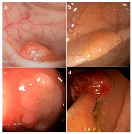
[40]. They are histologically classified by the World Health Organization (WHO) into three morphological categories: (i) hyperplastic polyp (HP), (ii) sessile serrated adenoma/polyp (SSA/P) with or without cytological dysplasia (SSAD), and (iii) the traditional serrated adenoma/polyp (TSA) (Figure 2) (Table 1) [41]. The serrated subtypes, identified by their cytological characteristics and lesion area, have a distinct endoscopic appearance, share some histological features, and are unique at the biological and molecular levels [34][42].
Figure 2. Representative endoscopic appearance of serrated lesions of the colorectum. (a) Hyperplastic polyp; (b) Sessile serrated adenoma/polyp; (c) Sessile serrated adenoma/polyp with dysplasia; (d) Traditional serrated adenoma. (Courtesy of Prof. Dr. Giovanni D. De Palma, University of Naples Federico II, Naples, Italy).
Table 1. Morphologic categories and features of serrated colorectal lesions.
Histological Classification Frequency (%) * Location Shape Mucin Type Size
Hyperplastic polyp (HP) 80–90% Distal Sessile, Flat Variable <5 mm
 Microvesicular HP (MVHP) 60% Distal Sessile Microvesicular <5 mm
 Goblet cell HP (GCHP) 30% Distal Sessile Goblet cells <5 mm
 Mucin poor HP (MPHP) 10% Distal Sessile Poor <5 mm
Sessile serrated adenoma/polyp (SSA/P) 15–20% Proximal Sessile/Flat Microvesicular >5 mm
Traditional serrated polyp (TSA) 1–6% Distal Sessile/Pedunculated Not present >5 mm
* Frequency of all serrated polyps.
HP precursor lesions are the most frequent polyps (80–90%) and mostly remain benign (Figure 2a) (Table 1) [43]. HPs may be flat or sessile, are preferentially located in the distal colon, show a smaller size (< 5mm) than other subtypes, and remain hardly detectable by endoscopic exam. Based on the epithelial mucin content, HP lesions can be histologically subclassified into microvesicular HP (MVHP), goblet cell HP (GCHP), and mucin poor HP (MPHP) polyps (Table 1) [28][30][34]. Although the frequency of each HP subtypes is variable, MVHPs are the most common, while MPHPs are the rarest form of HPs [44][45].
SSA/Ps, which account for 15 to 20% of all serrated polyps, especially develop in the proximal colon, are pale lesions that can be either sessile or flat, with a variable size usually larger than 5 mm in diameter (Figure 2b) (Table 1) [43][46][47]. SSA/Ps can develop either as primary tumors or evolve from hyperplastic polyps. SSA/Ps lesions cannot be easily distinguished from MVHPs, however, MVHPs larger than 10 mm in diameter can be considered clinically equivalent to SSA/Ps. Additionally, SSA/Ps can be subclassified according to the absence or presence of dysplasia (SSAD); the latter being detected in about 0.20% of all serrated lesions (Figure 2c) [46]. Overall, by combining both serrated and dysplastic features, SSADs consist of advanced lesions that usually evolve rapidly toward carcinoma.
TSA lesions are the rarest form of colorectal serrated polyps (1–6%) (Figure 2d) (Table 1) [34][43]. TSAs, that arise either from HPs or SSA/Ps, are precancerous sessile or, more often, pedunculated polypoid lesions, which preferentially develop in the distal colon and rectum, and show a larger size (>5 mm) than HPs [45]. A less aggressive variant form of TSA is the filiform serrated adenoma [48]. TSA can also be subclassified according to the presence of dysplasia that can be of two types: the well-known “adenomatous dysplasia” and the less frequently observed “serrated dysplasia”, which is related to the serrated pathway. The latter can be graded as low- or high-grade dysplasia depending on the absence or presence of cytological and architectural atypia, respectively. Recently, it has also been evidenced that TSA can co-exist with other lesions such as HPs, SSA/Ps and tubulovillous adenomas [49]. Usually, only TSA with serrated dysplasia develops into invasive carcinomas.
However, alternative molecular classifications have been proposed based on recent findings, such as those defined by the CRC Subtyping Consortium (CRCSC) or by Fennell et al. [50][51][52].
The heterogeneity of serrated lesions and the presence of morphological features shared with different subtypes, make difficult the accurate CRC classification during the diagnostic process and also the physiopathological interpretation of the observed lesions. In addition, serrated lesions with a distinctive endoscopic appearance are more difficult to detect compared to conventional lesions. In fact, detection of serrated lesions, particularly those located in the proximal colon, is difficult and endoscopist-dependent [53][54]. Therefore, characterization of molecular markers specific for each CRC subtype may improve the identification of CRCs arising from this alternative pathway, and consequently support the diagnostic process as well as the clinical decision-making.

3. Molecular Features of the Serrated Colorectal Precursor Lesions

3.1. Hyperplastic Polyps

As described above, hyperplastic polyps can be histologically subclassified into MPHP, GCHP and MVHP lesions (Table 1). The endoscopic diagnosis between these subtypes, and furthermore between HPs and SSA/Ps, is difficult and may be supported by the detection of specific biomarkers.
MPHP is the rarest form of HP and is not well described; in fact, to date, it has only been associated with CIMP-H (Table 3). MVHP and GCHP are the most common HP subtypes. At the molecular level, MVHP is particularly characterized by BRAF V600E mutation and CIMP-H (Table 3); for that reason, it is considered a precursor of SSA/Ps [17][55].
Table 3. Molecular profile of serrated colorectal lesions.
Serrated Lesion BRAF/KRAS Status CIMP Rate Gene Methylation MSI Rate
HP BRAF mutated CIMP-H MLH1 not methylated MSS
 MPHP * controversial CIMP-H controversial controversial
 GCHP * KRAS mutated CIMP-L MLH1 not methylated MSS
 MVHP * BRAF mutated CIMP-H MLH1 not methylated MSS
SSA/P BRAF mutated CIMP-H MLH1 not methylated MSS
SSAD BRAF mutated CIMP-H MLH1 hypermethylated MSI
TSA KRAS/BRAF mutated or neither CIMP-L/-H MLH1 not methylated MSS
TSA HGD KRAS mutated CIMP-L MGMT hypermethylated MSS
* MPHP, GCHP, MVHP are HP subtypes.
GCHP is linked to KRAS mutations (often missense substitutions at glycine codons 12 or 13) and CIMP-L (Table 3) [19][56]. Mutations in BRAF or KRAS, that rarely coexist in CRC, constitutively activate the MAPK signaling pathway, which is involved in the regulation of several cellular processes, and inhibit the apoptosis mechanism, thus supporting tumor cell proliferation.
To shed light on other molecular features of HP lesions, early markers of potentially malignant serrated precursor lesions have been identified, such as MUC5AC [57][58]. MVHP and SSA/P lesions present an hypomethylated MUC5AC when compared to GCHPs. Interestingly, this gene hypomethylation occurs early in the serrated pathway, gradually increasing from MVHP to SSA and SSAD, and is particularly related to lesions with BRAF mutations, CIMP-H and MSI. Thus, the epigenetic alteration of MUC5AC could be a potential marker to evaluate the malignant evolution of serrated precursor polyps.

3.2. Sessile Serrated Adenoma/Polyps

At the molecular level, SSA/Ps are mainly characterized by BRAF mutations, MSS, CIMP-H and unmethylated MLH1 (Table 3) [17][59]. Other molecular characteristics of sessile serrated lesions, as well as a subtype-specific gene signature, have been explored and, in some cases, identified based on epigenetic and transcriptomic approaches. An example is the recent molecular characterization of SSA/Ps in a large African American cohort, in which the over-expression of FSCN1 and TRNP1 seemed to segregate with race [60].
The formation of SSA/Ps has been associated with the tumor suppressor gene SLIT2, who is down-expressed in SSA/Ps compared to TSAs/adenomas/normal tissues as a result of promoter hypermethylation and loss of heterozygosity [61]. The high rate of SFRP4 methylation in SSA/P compared to the corresponding adenoma series has also been evidenced [59]CTSETFF1 and ANXA10 were identified as potential clinical markers of SSA/P lesions [62][63][64]. In particular, ANXA10 expression levels significantly increased at the gene and protein levels in SSA/Ps in comparison to MVHPs [63][65]Hes-1, a downstream target of the Notch signaling pathway which involved in intestinal development, has also been described as a SSA/P-specific biomarker due to its immunohistochemical (IHC) loss of expression in SSA/Ps compared to HPs or normal colonic mucosa [66].
In a large-scale study, differentially expressed genes and immunohistochemical markers were also identified when comparing SSA/Ps to controls [67]. In particular, among the 1294 genes identified, VSIG1 and MUC17, were uniquely and significantly increased in SSA/Ps with respect to controls/HPs/adenomas, thus evidencing a molecular signature specific of the polyps and the involvement of different molecular pathways across distinct CRC lesions.
Recently, a platform-independent approach was adopted in order to differentiate SSA/P from HP lesions [68]. SSA/Ps have been characterized by a specific molecular profile of up-/down-regulated genes involved in the inflammatory process, immune response, epithelial–mesenchymal transition (EMT), extracellular matrix (ECM) interaction, cell migration and cell growth. This profile defines the malignant potential of SSA/P lesions and allow to distinguish them from HPs.
As for MVHP polyps, SSA/P immunohistochemically expresses MUC2, MUC5AC and MUC6 [57][69]. The role of MUC6 was evaluated in SSA/Ps lesions and is controversial [57]. Nevertheless, MUC6 was assessed to be a supportive immunohistochemical marker to differentiate SSA/Ps from TSAs [70][71].

3.3. Sessile Serrated Adenoma/Polyps with Dysplasia

The main molecular characteristics found in SSADs are BRAF mutations, a nuclear β-catenin accumulation, CIMP-H and MSI as a consequence of MLH1 gene silencing due to promoter hypermethylation (Table 3) [72]. Although the loss of MLH1 expression is related to the development of dysplasia, MLH1 inactivation can also be detected in SSA/P non-dysplastic crypts, indicating the putative biomarker role of MLH1 in predicting dysplastic progression of these polyps [73].
Moreover, it has been demonstrated that SSAD lesions can be also associated with MSI, harboring different genetic alterations [72]. In particular, MSI lesions are characterized by a high mutational rate of FBXW7 and the loss of MLH1 expression, while MSSs display TP53 mutations without FBXW7 mutations. Thus, FBXW7 alterations can be correlated to the progression of MSI serrated lesions in CRC.
SSAD polyps, and particularly the progression of SSA/P lesions toward dysplasia, can also be characterized by alterations in WNT signaling-associated genes like protein-truncating mutations of RNF43APCZNRF3 and the hypermethylation of AXIN2 and MCC [72][74][75][76]. Furthermore, frameshift mutations in RNF43 are frequently present in patients with MLH1-deficient SSA/P with dysplasia [74].
Genetic variants of DNA-regulatory elements, such as single nucleotide polymorphisms (SNPs), can influence gene expression and be associated with cancer risk. An example is the well-known CRC-associated rs1800734, or MLH1-93G>A, which can be detected in the promoter region of MLH1. This SNP enhances DCLK3 expression which in turn promotes CRC development [77]. MLH1–93 G/a polymorphism has been significantly associated with MLH1 hypermethylation in SSAD lesions compared to TSAs, and represents a putative risk factor for MLH1 promoter methylation [78].

3.4. Traditional Serrated Adenoma/Polyps with and without Dysplasia

TSA is an enigmatic subtype due to its heterogeneous molecular features [49][79]. TSAs can harbor KRAS or BRAF mutations, or neither, and can be CIMP-low or high, and MSS (Table 3) [80]. In contrast with SSA/Ps, TSA lesions do not show MLH1 promoter hypermethylation but can present MGMT hypermethylation (Table 3) [81]. Evaluation of the mutational status of WNT genes in TSA lesions has identified precursor- and TSA-specific alterations that elucidate the mechanism involved in the genetic transition from precursor polyps to TSAs [82]. PTPRK-RSPO3 fusion and mutations in PTENRNF43APC, or CTNNB1 are genetic features of TSAs [83][84].
Several epigenetic biomarkers have also been associated with TSA. An example is SMOC1 which is down-expressed, due to methylations in its promoter region, in TSAs as compared to SSA/Ps [85]. In particular, SMOC1 methylation increases during TSA development and has been correlated with KRAS mutation and CIMP-L. In addition, low expression of the SMOC1 protein in TSA can be verified by IHC assay, supporting its exploitation as a TSA-specific diagnostic biomarker.
The transition of TSAs toward dysplasia is characterized by nuclear β-catenin accumulation, TP53 mutation and p16 inactivation, as detailed below [21]. TSA-HGDs are usually characterized by CIMP-H, BRAF mutations and MSS (Table 3). The high rate of differentially methylated region in the promoter P0 of IGF2 gene (IGF2 DMR0) and hypomethylation of LINE-1 are two epigenetic biomarkers of TSA-HGD [86]LINE-1 hypomethylation in CRC has been associated with early age onset and family history of CRC. In several studies, LINE-1 hypomethylation has been inversely correlated with MSI and CIMP-H phenotypes. Interestingly, the methylation level of LINE-1 has recently been assessed from plasma-circulating cell-free DNA, thus comforting a putative role as a preventive biomarker in CRC [87][88].

References

  1. Kuipers, E.J.; Grady, W.M.; Lieberman, D.; Seufferlein, T.; Sung, J.J.; Boelens, P.G.; van de Velde, C.J.H.; Watanabe, T. Colorectal cancer. Nat. Rev. Dis. Primer 2015, 1, 15065.
  2. Nguyen, H.T.; Nguyen, H.T.; Duong, H.-Q.; Duong, H.-Q. The molecular characteristics of colorectal cancer: Implications for diagnosis and therapy (Review). Oncol. Lett. 2018, 16, 9–18.
  3. Bogaert, J.; Prenen, H. Molecular genetics of colorectal cancer. Ann. Gastroenterol. 2014, 27, 9–14.
  4. Wong, K.; Xie, G. Updates on the Molecular Genetics of Colorectal Cancer. Colorectal Cancer Open Access 2017, 3.
  5. Kuipers, E.J.; Rösch, T.; Bretthauer, M. Colorectal cancer screening--optimizing current strategies and new directions. Nat. Rev. Clin. Oncol. 2013, 10, 130–142.
  6. Bardhan, K.; Liu, K. Epigenetics and colorectal cancer pathogenesis. Cancers 2013, 5, 676–713.
  7. Grady, W.M.; Carethers, J.M. Genomic and epigenetic instability in colorectal cancer pathogenesis. Gastroenterology 2008, 135, 1079–1099.
  8. Pancione, M.; Remo, A.; Colantuoni, V. Genetic and Epigenetic Events Generate Multiple Pathways in Colorectal Cancer Progression. Available online: https://www.hindawi.com/journals/pri/2012/509348/ (accessed on 27 February 2019).
  9. Vogelstein, B.; Fearon, E.R.; Hamilton, S.R.; Kern, S.E.; Preisinger, A.C.; Leppert, M.; Nakamura, Y.; White, R.; Smits, A.M.; Bos, J.L. Genetic alterations during colorectal-tumor development. N. Engl. J. Med. 1988, 319, 525–532.
  10. Fearon, E.R.; Vogelstein, B. A genetic model for colorectal tumorigenesis. Cell 1990, 61, 759–767.
  11. Cho, K.R.; Vogelstein, B. Genetic alterations in the adenoma--carcinoma sequence. Cancer 1992, 70, 1727–1731.
  12. Armaghany, T.; Wilson, J.D.; Chu, Q.; Mills, G. Genetic Alterations in Colorectal Cancer. GCR 2012, 5, 19–27.
  13. Geigl, J.B.; Obenauf, A.C.; Schwarzbraun, T.; Speicher, M.R. Defining “chromosomal instability”. Trends Genet. 2008, 24, 64–69.
  14. Boland, C.R.; Goel, A. Microsatellite instability in colorectal cancer. Gastroenterology 2010, 138, 2073–2087.
  15. Gupta, R.; Sinha, S.; Paul, R.N. The impact of microsatellite stability status in colorectal cancer. Curr. Probl. Cancer 2018, 42, 548–559.
  16. Pino, M.S.; Chung, D.C. The Chromosomal Instability Pathway in Colon Cancer. Gastroenterology 2010, 138, 2059–2072.
  17. O’Brien, M.J.; Yang, S.; Mack, C.; Xu, H.; Huang, C.S.; Mulcahy, E.; Amorosino, M.; Farraye, F.A. Comparison of microsatellite instability, CpG island methylation phenotype, BRAF and KRAS status in serrated polyps and traditional adenomas indicates separate pathways to distinct colorectal carcinoma end points. Am. J. Surg. Pathol. 2006, 30, 1491–1501.
  18. Gryfe, R.; Kim, H.; Hsieh, E.T.; Aronson, M.D.; Holowaty, E.J.; Bull, S.B.; Redston, M.; Gallinger, S. Tumor microsatellite instability and clinical outcome in young patients with colorectal cancer. N. Engl. J. Med. 2000, 342, 69–77.
  19. Murcia, O.; Juárez, M.; Hernández-Illán, E.; Egoavil, C.; Giner-Calabuig, M.; Rodríguez-Soler, M.; Jover, R. Serrated colorectal cancer: Molecular classification, prognosis, and response to chemotherapy. World J. Gastroenterol. 2016, 22, 3516–3530.
  20. Hegde, M.; Ferber, M.; Mao, R.; Samowitz, W.; Ganguly, A.; Working Group of the American College of Medical Genetics and Genomics (ACMG) Laboratory Quality Assurance Committee. ACMG technical standards and guidelines for genetic testing for inherited colorectal cancer (Lynch syndrome, familial adenomatous polyposis, and MYH-associated polyposis). Genet. Med. 2014, 16, 101–116.
  21. Jass, J.R. Classification of colorectal cancer based on correlation of clinical, morphological and molecular features. Histopathology 2007, 50, 113–130.
  22. Ogino, S.; Nosho, K.; Kirkner, G.J.; Kawasaki, T.; Meyerhardt, J.A.; Loda, M.; Giovannucci, E.L.; Fuchs, C.S. CpG island methylator phenotype, microsatellite instability, BRAF mutation and clinical outcome in colon cancer. Gut 2009, 58, 90–96.
  23. Yamagishi, H.; Kuroda, H.; Imai, Y.; Hiraishi, H. Molecular pathogenesis of sporadic colorectal cancers. Chin. J. Cancer 2016, 35.
  24. Lynch, H.T.; Snyder, C.L.; Shaw, T.G.; Heinen, C.D.; Hitchins, M.P. Milestones of Lynch syndrome: 1895–2015. Nat. Rev. Cancer 2015, 15, 181–194.
  25. Tutlewska, K.; Lubinski, J.; Kurzawski, G. Germline deletions in the EPCAM gene as a cause of Lynch syndrome—Literature review. Hered. Cancer Clin. Pract. 2013, 11, 9.
  26. Bessa, X.; Ballesté, B.; Andreu, M.; Castells, A.; Bellosillo, B.; Balaguer, F.; Castellví-Bel, S.; Paya, A.; Jover, R.; Alenda, C.; et al. A prospective, multicenter, population-based study of BRAF mutational analysis for Lynch syndrome screening. Clin. Gastroenterol. Hepatol. 2008, 6, 206–214.
  27. Harvey, N.T.; Ruszkiewicz, A. Serrated neoplasia of the colorectum. World J. Gastroenterol. 2007, 13, 3792–3798.
  28. Rex, D.K.; Ahnen, D.J.; Baron, J.A.; Batts, K.P.; Burke, C.A.; Burt, R.W.; Goldblum, J.R.; Guillem, J.G.; Kahi, C.J.; Kalady, M.F.; et al. Serrated Lesions of the Colorectum: Review and Recommendations from an Expert Panel. Am. J. Gastroenterol. 2012, 107, 1315–1330.
  29. Patai, Á.V.; Molnár, B.; Tulassay, Z.; Sipos, F. Serrated pathway: Alternative route to colorectal cancer. World J. Gastroenterol. 2013, 19, 607–615.
  30. Leggett, B.; Whitehall, V. Role of the serrated pathway in colorectal cancer pathogenesis. Gastroenterology 2010, 138, 2088–2100.
  31. Bettington, M.; Walker, N.; Clouston, A.; Brown, I.; Leggett, B.; Whitehall, V. The serrated pathway to colorectal carcinoma: Current concepts and challenges. Histopathology 2013, 62, 367–386.
  32. Longacre, T.A.; Fenoglio-Preiser, C.M. Mixed hyperplastic adenomatous polyps/serrated adenomas. A distinct form of colorectal neoplasia. Am. J. Surg. Pathol. 1990, 14, 524–537.
  33. East, J.E.; Atkin, W.S.; Bateman, A.C.; Clark, S.K.; Dolwani, S.; Ket, S.N.; Leedham, S.J.; Phull, P.S.; Rutter, M.D.; Shepherd, N.A.; et al. British Society of Gastroenterology position statement on serrated polyps in the colon and rectum. Gut 2017, 66, 1181–1196.
  34. Pai, R.K.; Bettington, M.; Srivastava, A.; Rosty, C. An update on the morphology and molecular pathology of serrated colorectal polyps and associated carcinomas. Mod. Pathol. 2019, 1.
  35. Kahi, C.J. Screening Relevance of Sessile Serrated Polyps. Clin. Endosc. 2019, 52.
  36. Hamoudah, T.; Ma, K.; Esteban, M.; Hayat, W.; Berger, D.; Mahon, B.; Jakate, S.; Melson, J. Patients with small and diminutive proximal hyperplastic polyps have higher rates of synchronous advanced neoplasia compared with patients without serrated lesions. Gastrointest. Endosc. 2018, 87, 1518–1526.
  37. Mojarad, E.N.; Kuppen, P.J.; Aghdaei, H.A.; Zali, M.R. The CpG island methylator phenotype (CIMP) in colorectal cancer. Gastroenterol. Hepatol. Bed Bench 2013, 6, 120–128.
  38. Barault, L.; Charon-Barra, C.; Jooste, V.; de la Vega, M.F.; Martin, L.; Roignot, P.; Rat, P.; Bouvier, A.-M.; Laurent-Puig, P.; Faivre, J.; et al. Hypermethylator phenotype in sporadic colon cancer: Study on a population-based series of 582 cases. Cancer Res. 2008, 68, 8541–8546.
  39. Borowsky, J.; Dumenil, T.; Bettington, M.; Pearson, S.-A.; Bond, C.; Fennell, L.; Liu, C.; McKeone, D.; Rosty, C.; Brown, I.; et al. The role of APC in WNT pathway activation in serrated neoplasia. Mod. Pathol. 2018, 31, 495–504.
  40. Torlakovic, E.; Skovlund, E.; Snover, D.C.; Torlakovic, G.; Nesland, J.M. Morphologic reappraisal of serrated colorectal polyps. Am. J. Surg. Pathol. 2003, 27, 65–81.
  41. Bosman, F.T.; Carneiro, F.; Hruban, R.H.; Theise, N.D. WHO Classification of Tumours of the Digestive System, 4th ed.; World Health Organization, International Agency for Research on Cancer (IARC): Lyon, France, 2010.
  42. Cassese, G.; Amendola, A.; Maione, F.; Giglio, M.C.; Pagano, G.; Milone, M.; Aprea, G.; Luglio, G.; De Palma, G.D. Serrated Lesions of the Colon-Rectum: A Focus on New Diagnostic Tools and Current Management. Gastroenterol. Res. Pract. 2019, 2019, 9179718.
  43. Aust, D.E.; Baretton, G.B.; Members of the Working Group GI-Pathology of the German Society of Pathology. Serrated polyps of the colon and rectum (hyperplastic polyps, sessile serrated adenomas, traditional serrated adenomas, and mixed polyps)-proposal for diagnostic criteria. Virchows Arch. Int. J. Pathol. 2010, 457, 291–297.
  44. Yamane, L.; Scapulatempo-Neto, C.; Reis, R.M.; Guimarães, D.P. Serrated pathway in colorectal carcinogenesis. World J. Gastroenterol. 2014, 20, 2634–2640.
  45. IJspeert, J.E.G.; Vermeulen, L.; Meijer, G.A.; Dekker, E. Serrated neoplasia—role in colorectal carcinogenesis and clinical implications. Nat. Rev. Gastroenterol. Hepatol. 2015, 12, 401–409.
  46. Murakami, T.; Sakamoto, N.; Nagahara, A. Endoscopic diagnosis of sessile serrated adenoma/polyp with and without dysplasia/carcinoma. World J. Gastroenterol. 2018, 24, 3250–3259.
  47. Torlakovic, E.; Snover, D.C. Serrated adenomatous polyposis in humans. Gastroenterology 1996, 110, 748–755.
  48. Yantiss, R.K.; Oh, K.Y.; Chen, Y.-T.; Redston, M.; Odze, R.D. Filiform serrated adenomas: A clinicopathologic and immunophenotypic study of 18 cases. Am. J. Surg. Pathol. 2007, 31, 1238–1245.
  49. Kalimuthu, S.N.; Chelliah, A.; Chetty, R. From traditional serrated adenoma to tubulovillous adenoma and beyond. World J. Gastrointest. Oncol. 2016, 8, 805–809.
  50. Guinney, J.; Dienstmann, R.; Wang, X.; de Reyniès, A.; Schlicker, A.; Soneson, C.; Marisa, L.; Roepman, P.; Nyamundanda, G.; Angelino, P.; et al. The Consensus Molecular Subtypes of Colorectal Cancer. Nat. Med. 2015, 21, 1350–1356.
  51. Bae, J.M.; Kim, J.H.; Kwak, Y.; Lee, D.-W.; Cha, Y.; Wen, X.; Lee, T.H.; Cho, N.-Y.; Jeong, S.-Y.; Park, K.J.; et al. Distinct clinical outcomes of two CIMP-positive colorectal cancer subtypes based on a revised CIMP classification system. Br. J. Cancer 2017, 116, 1012–1020.
  52. Fennell, L.; Dumenil, T.; Wockner, L.; Hartel, G.; Nones, K.; Bond, C.; Borowsky, J.; Liu, C.; McKeone, D.; Bowdler, L.; et al. Integrative Genome-Scale DNA Methylation Analysis of a Large and Unselected Cohort Reveals Five Distinct Subtypes of Colorectal Adenocarcinomas. Cell. Mol. Gastroenterol. Hepatol. 2019.
  53. Kahi, C.J.; Hewett, D.G.; Rex, D.K. Relationship of non-polypoid colorectal neoplasms to quality of colonoscopy. Gastrointest. Endosc. Clin. N. Am. 2010, 20, 407–415.
  54. Kahi, C.J.; Hewett, D.G.; Norton, D.L.; Eckert, G.J.; Rex, D.K. Prevalence and variable detection of proximal colon serrated polyps during screening colonoscopy. Clin. Gastroenterol. Hepatol. 2011, 9, 42–46.
  55. Yang, S.; Farraye, F.A.; Mack, C.; Posnik, O.; O’Brien, M.J. BRAF and KRAS Mutations in hyperplastic polyps and serrated adenomas of the colorectum: Relationship to histology and CpG island methylation status. Am. J. Surg. Pathol. 2004, 28, 1452–1459.
  56. Spring, K.J.; Zhao, Z.Z.; Karamatic, R.; Walsh, M.D.; Whitehall, V.L.J.; Pike, T.; Simms, L.A.; Young, J.; James, M.; Montgomery, G.W.; et al. High Prevalence of Sessile Serrated Adenomas with BRAF Mutations: A Prospective Study of Patients Undergoing Colonoscopy. Gastroenterology 2006, 131, 1400–1407.
  57. Gibson, J.A.; Hahn, H.P.; Shahsafaei, A.; Odze, R.D. MUC expression in hyperplastic and serrated colonic polyps: Lack of specificity of MUC6. Am. J. Surg. Pathol. 2011, 35, 742–749.
  58. Renaud, F.; Mariette, C.; Vincent, A.; Wacrenier, A.; Maunoury, V.; Leclerc, J.; Coppin, L.; Crépin, M.; Seuningen, I.V.; Leteurtre, E.; et al. The serrated neoplasia pathway of colorectal tumors: Identification of MUC5AC hypomethylation as an early marker of polyps with malignant potential. Int. J. Cancer 2016, 138, 1472–1481.
  59. Murakami, T.; Mitomi, H.; Saito, T.; Takahashi, M.; Sakamoto, N.; Fukui, N.; Yao, T.; Watanabe, S. Distinct WNT/β-catenin signaling activation in the serrated neoplasia pathway and the adenoma-carcinoma sequence of the colorectum. Mod. Pathol. 2015, 28, 146–158.
  60. Ashktorab, H.; Delker, D.; Kanth, P.; Goel, A.; Carethers, J.M.; Brim, H. Molecular Characterization of sessile serrated adenoma/polyps from a large African American cohort. Gastroenterology 2019.
  61. Beggs, A.D.; Jones, A.; Shepherd, N.; Arnaout, A.; Finlayson, C.; Abulafi, A.M.; Morton, D.G.; Matthews, G.M.; Hodgson, S.V.; Tomlinson, I.P.M. Loss of Expression and Promoter Methylation of SLIT2 Are Associated with Sessile Serrated Adenoma Formation. PLoS Genet. 2013, 9.
  62. Caruso, M.; Moore, J.; Goodall, G.J.; Thomas, M.; Phillis, S.; Tyskin, A.; Cheetham, G.; Lerda, N.; Takahashi, H.; Ruszkiewicz, A. Over-expression of cathepsin E and trefoil factor 1 in sessile serrated adenomas of the colorectum identified by gene expression analysis. Virchows Arch. Int. J. Pathol. 2009, 454, 291–302.
  63. Gonzalo, D.H.; Lai, K.K.; Shadrach, B.; Goldblum, J.R.; Bennett, A.E.; Downs-Kelly, E.; Liu, X.; Henricks, W.; Patil, D.T.; Carver, P.; et al. Gene expression profiling of serrated polyps identifies annexin A10 as a marker of a sessile serrated adenoma/polyp. J. Pathol. 2013, 230, 420–429.
  64. Kanth, P.; Bronner, M.P.; Boucher, K.M.; Burt, R.W.; Neklason, D.W.; Hagedorn, C.H.; Delker, D.A. Gene Signature in Sessile Serrated Polyps Identifies Colon Cancer Subtype. Cancer Prev. Res. Phila. Pa 2016, 9, 456–465.
  65. Bae, J.M.; Kim, J.H.; Rhee, Y.-Y.; Cho, N.-Y.; Kim, T.-Y.; Kang, G.H. Annexin A10 expression in colorectal cancers with emphasis on the serrated neoplasia pathway. World J. Gastroenterol. 2015, 21, 9749–9757.
  66. Cui, M.; Awadallah, A.; Liu, W.; Zhou, L.; Xin, W. Loss of Hes1 Differentiates Sessile Serrated Adenoma/Polyp from Hyperplastic Polyp. Am. J. Surg. Pathol. 2016, 40, 113–119.
  67. Delker, D.A.; McGettigan, B.M.; Kanth, P.; Pop, S.; Neklason, D.W.; Bronner, M.P.; Burt, R.W.; Hagedorn, C.H. RNA Sequencing of Sessile Serrated Colon Polyps Identifies Differentially Expressed Genes and Immunohistochemical Markers. PLoS ONE 2014, 9.
  68. Rahmatallah, Y.; Khaidakov, M.; Lai, K.K.; Goyne, H.E.; Lamps, L.W.; Hagedorn, C.H.; Glazko, G. Platform-independent gene expression signature differentiates sessile serrated adenomas/polyps and hyperplastic polyps of the colon. BMC Med. Genomics 2017, 10.
  69. Mikhaleva, L.M.; Vandysheva, R.A.; Shakhpazyan, N.K.; Fedorov, E.D.; Biryukov, A.E.; Midiber, K.Y.; Pechnikova, V.V. [Comparative assessment of the expression of Muc 2, Muc 5AC, and Muc 6 in serrated neoplasms of the colon]. Arkh. Patol. 2019, 81, 10–17.
  70. Owens, S.R.; Chiosea, S.I.; Kuan, S.-F. Selective expression of gastric mucin MUC6 in colonic sessile serrated adenoma but not in hyperplastic polyp aids in morphological diagnosis of serrated polyps. Mod. Pathol. 2008, 21, 660–669.
  71. Travaglino, A.; D’Armiento, F.P.; Cassese, G.; Campanino, M.R.; Borrelli, G.; Pignatiello, S.; Luglio, G.; Maione, F.; De Palma, G.D.; D’Armiento, M. Clinico-pathological factors associated with BRAF-V600E mutation in colorectal serrated adenomas. Histopathology 2019.
  72. Murakami, T.; Akazawa, Y.; Yatagai, N.; Hiromoto, T.; Sasahara, N.; Saito, T.; Sakamoto, N.; Nagahara, A.; Yao, T. Molecular characterization of sessile serrated adenoma/polyps with dysplasia/carcinoma based on immunohistochemistry, next-generation sequencing, and microsatellite instability testing: A case series study. Diagn. Pathol. 2018, 13, 88.
  73. Yozu, M.; Kem, M.; Cenaj, O.; Mino-Kenudson, M.; Odze, R.D.; Misdraji, J. Loss of expression of MLH1 in non-dysplastic crypts is a harbinger of neoplastic progression in sessile serrated adenomas/polyps. Histopathology 2019.
  74. Hashimoto, T.; Yamashita, S.; Yoshida, H.; Taniguchi, H.; Ushijima, T.; Yamada, T.; Saito, Y.; Ochiai, A.; Sekine, S.; Hiraoka, N. WNT Pathway Gene Mutations Are Associated with the Presence of Dysplasia in Colorectal Sessile Serrated Adenoma/Polyps. Am. J. Surg. Pathol. 2017, 41, 1188–1197.
  75. Bond, C.E.; McKeone, D.M.; Kalimutho, M.; Bettington, M.L.; Pearson, S.-A.; Dumenil, T.D.; Wockner, L.F.; Burge, M.; Leggett, B.A.; Whitehall, V.L.J. RNF43 and ZNRF3 are commonly altered in serrated pathway colorectal tumorigenesis. Oncotarget 2016, 7, 70589–70600.
  76. Koinuma, K.; Yamashita, Y.; Liu, W.; Hatanaka, H.; Kurashina, K.; Wada, T.; Takada, S.; Kaneda, R.; Choi, Y.L.; Fujiwara, S.-I.; et al. Epigenetic silencing of AXIN2 in colorectal carcinoma with microsatellite instability. Oncogene 2006, 25, 139–146.
  77. Liu, N.Q.; ter Huurne, M.; Nguyen, L.N.; Peng, T.; Wang, S.-Y.; Studd, J.B.; Joshi, O.; Ongen, H.; Bramsen, J.B.; Yan, J.; et al. The non-coding variant rs1800734 enhances DCLK3 expression through long-range interaction and promotes colorectal cancer progression. Nat. Commun. 2017, 8, 14418.
  78. Fennell, L.J.; Jamieson, S.; McKeone, D.; Corish, T.; Rohdmann, M.; Furner, T.; Bettington, M.; Liu, C.; Kawamata, F.; Bond, C.; et al. MLH1-93 G/a polymorphism is associated with MLH1 promoter methylation and protein loss in dysplastic sessile serrated adenomas with BRAFV600E mutation. BMC Cancer 2018, 18, 35.
  79. Torlakovic, E.E.; Gomez, J.D.; Driman, D.K.; Parfitt, J.R.; Wang, C.; Benerjee, T.; Snover, D.C. Sessile serrated adenoma (SSA) vs. traditional serrated adenoma (TSA). Am. J. Surg. Pathol. 2008, 32, 21–29.
  80. Bettington, M.L.; Walker, N.I.; Rosty, C.; Brown, I.S.; Clouston, A.D.; McKeone, D.M.; Pearson, S.-A.; Klein, K.; Leggett, B.A.; Whitehall, V.L.J. A clinicopathological and molecular analysis of 200 traditional serrated adenomas. Mod. Pathol. 2015, 28, 414–427.
  81. Whitehall, V.L.J.; Walsh, M.D.; Young, J.; Leggett, B.A.; Jass, J.R. Methylation of O–6-Methylguanine DNA Methyltransferase Characterizes a Subset of Colorectal Cancer with Low-level DNA Microsatellite Instability. Cancer Res. 2001, 61, 827–830.
  82. Hashimoto, T.; Ogawa, R.; Yoshida, H.; Taniguchi, H.; Kojima, M.; Saito, Y.; Sekine, S. Acquisition of WNT Pathway Gene Alterations Coincides With the Transition From Precursor Polyps to Traditional Serrated Adenomas. Am. J. Surg. Pathol. 2019, 43, 132–139.
  83. Sekine, S.; Yamashita, S.; Tanabe, T.; Hashimoto, T.; Yoshida, H.; Taniguchi, H.; Kojima, M.; Shinmura, K.; Saito, Y.; Hiraoka, N.; et al. Frequent PTPRK-RSPO3 fusions and RNF43 mutations in colorectal traditional serrated adenoma. J. Pathol. 2016, 239, 133–138.
  84. Tsai, J.-H.; Jeng, Y.-M.; Yuan, C.-T.; Lin, Y.-L.; Cheng, M.-L.; Liau, J.-Y. Traditional Serrated Pathway-associated Colorectal Carcinoma: Morphologic Reappraisal of Serrated Morphology, Tumor Budding, and Identification of Frequent PTEN Alterations. Am. J. Surg. Pathol. 2019, 43.
  85. Aoki, H.; Yamamoto, E.; Takasawa, A.; Niinuma, T.; Yamano, H.-O.; Harada, T.; Matsushita, H.-O.; Yoshikawa, K.; Takagi, R.; Harada, E.; et al. Epigenetic silencing of SMOC1 in traditional serrated adenoma and colorectal cancer. Oncotarget 2017, 9, 4707–4721.
  86. Naito, T.; Nosho, K.; Ito, M.; Igarashi, H.; Mitsuhashi, K.; Yoshii, S.; Aoki, H.; Nomura, M.; Sukawa, Y.; Yamamoto, E.; et al. IGF2 differentially methylated region hypomethylation in relation to pathological and molecular features of serrated lesions. World J. Gastroenterol. 2014, 20, 10050–10061.
  87. Kerachian, M.A.; Kerachian, M. Long interspersed nucleotide element-1 (LINE-1) methylation in colorectal cancer. Clin. Chim. Acta Int. J. Clin. Chem. 2019, 488, 209–214.
  88. Nagai, Y.; Sunami, E.; Yamamoto, Y.; Hata, K.; Okada, S.; Murono, K.; Yasuda, K.; Otani, K.; Nishikawa, T.; Tanaka, T.; et al. LINE-1 hypomethylation status of circulating cell-free DNA in plasma as a biomarker for colorectal cancer. Oncotarget 2017, 8, 11906–11916.
More
Information
Subjects: Oncology
Contributor MDPI registered users' name will be linked to their SciProfiles pages. To register with us, please refer to https://encyclopedia.pub/register :
View Times: 406
Entry Collection: Gastrointestinal Disease
Revisions: 2 times (View History)
Update Date: 10 Sep 2021
1000/1000