Submitted Successfully!
To reward your contribution, here is a gift for you: A free trial for our video production service.
Thank you for your contribution! You can also upload a video entry or images related to this topic.
Version Summary Created by Modification Content Size Created at Operation
1 + 1616 word(s) 1616 2021-08-19 11:22:34 |
2 format corrected. + 166 word(s) 1782 2021-08-27 12:17:44 |

Video Upload Options

Do you have a full video?

Confirm

Are you sure to Delete?
Cite
If you have any further questions, please contact Encyclopedia Editorial Office.
Tsirogianni, A. Perinuclear Anti-Neutrophil Cytoplasmic Antibodies (P-ANCA). Encyclopedia. Available online: https://encyclopedia.pub/entry/13637 (accessed on 19 April 2024).
Tsirogianni A. Perinuclear Anti-Neutrophil Cytoplasmic Antibodies (P-ANCA). Encyclopedia. Available at: https://encyclopedia.pub/entry/13637. Accessed April 19, 2024.
Tsirogianni, Alexandra. "Perinuclear Anti-Neutrophil Cytoplasmic Antibodies (P-ANCA)" Encyclopedia, https://encyclopedia.pub/entry/13637 (accessed April 19, 2024).
Tsirogianni, A. (2021, August 27). Perinuclear Anti-Neutrophil Cytoplasmic Antibodies (P-ANCA). In Encyclopedia. https://encyclopedia.pub/entry/13637
Tsirogianni, Alexandra. "Perinuclear Anti-Neutrophil Cytoplasmic Antibodies (P-ANCA)." Encyclopedia. Web. 27 August, 2021.
Perinuclear Anti-Neutrophil Cytoplasmic Antibodies (P-ANCA)
Edit

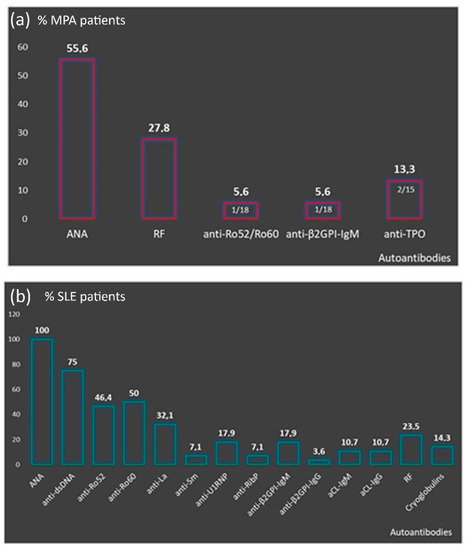
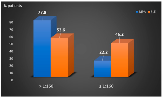
Perinuclear anti-neutrophilic cytoplasmic antibodies (P-ANCA) recognize heterogeneous antigens, including myeloperoxidase (MPO), lactoferrin, elastase, cathepsin-G and bactericidal/permeability-increasing protein. Although P-ANCA have diagnostic utility in vasculitides, they may also be found in patients with various other systemic autoimmune rheumatic diseases (SARDs). Nevertheless, the clinical significance and the targets recognized by P-ANCA in such patients remain unclear. For this purpose, herein we investigated the occurrence of ANCA-related antigenic specificities in 82 P-ANCA-positive sera by multiplex ELISA, as well as their association with other autoantibodies. The P-ANCA-positive sera corresponded to patients with vasculitides (n = 24), systemic lupus erythematosus (n = 28), antiphospholipid syndrome (n = 5), Sjögren’s syndrome (n = 7), rheumatoid arthritis (n = 3), systemic scleroderma (n = 1), sarcoidosis (n = 1) and Hashimoto′s thyroiditis (n = 13). In most P-ANCA-positive patients studied (51/82, 62.3%), these autoantibodies occurred in high titers (>1:160). The analysis of P-ANCA-positive sera revealed reactivity to MPO in only 50% of patients with vasculitides, whereas it was infrequent in the other disease groups studied. Reactivity to other P-ANCA-related autoantigens was also rarely detected. Our findings support that high P-ANCA titers occur in SARD. The P-ANCA-positive staining pattern is associated with MPO specificity in vasculitides, while in other autoimmune diseases, it mostly involves unknown autoantigens. 

P-ANCA autoantibodies myeloperoxidase elastase vasculitis systemic autoimmune rheumatic diseases

1. Introduction

Anti-neutrophil cytoplasmic antibodies (ANCA) are autoantibodies, mainly of IgG isotype, directed against proteins in the cytoplasmic granules of neutrophils and lysosomal proteins of monocytes. Depending on their staining pattern on alcohol-fixed neutrophils, ANCA are classified as diffuse cytoplasmic (C-ANCA), perinuclear (P-ANCA) and atypical (A-ANCA), the first two being highly significant for the diagnosis of ANCA-associated vasculitides. Myeloperoxidase (MPO) represents the major autoantigen recognized by P-ANCA, followed by neutrophil elastase, lactoferrin, cathepsin G, bactericidal/permeability-increasing protein (BPI), catalase and lysozyme, among others [1]. C-ANCA targeting proteinase-3 (PR3) has been associated with granulomatosis with polyangiitis (GPA), whereas P-ANCA targeting MPO is associated with microscopic polyangiitis (MPA). Patients with vasculitis and P-ANCA targeting MPO are most likely suffering from MPA (55–65%), followed by eosinophilic granulomatosis with polyangiitis (EGPA) (30–40%) and GPA (20–30%) [2]. Emerging evidence suggests that ANCA specificity associates with disease activity and may affect the clinical phenotype, as well as response to treatment, risk of relapse and long-term prognosis. To this end, MPA patients with MPO-ANCAs are more likely to develop isolated crescentic glomerulonephritis [3][4], pulmonary fibrosis and peripheral neuropathy [5][6], while MPO+-GPA patients have more frequently limited disease, without severe organ involvement, less need for cyclophosphamide or rituximab therapy and fewer relapses than those with proteinase-3 (PR3)-ANCA [7][8]. Interestingly, reappearance of MPO-ANCAs indicates relapse in more than 75% of patients [9].
Beyond MPA, P-ANCA have been described in a variety of other systemic autoimmune rheumatic diseases (SARDs), as well as chronic infections [10]. Indeed, MPO-ANCAs have been reported in systemic lupus erythematosus (SLE; 9.3%) [11], rheumatoid arthritis (RA; 4–18%) [12], Sjögren’s syndrome (SS; <3%) [13] and systemic sclerosis (SScl; <2.4%) [14]. Their presence has been associated with vasculitic patterns of glomerulonephritis and/or pulmonary involvement, while other P-ANCA-specific autoantigens, such as lactoferrin, neutrophil elastase, cathepsin and lysozyme, have also been described, although without known clinical significance [13][15][16]. In this context, P-ANCA and their distinct targets may have a potential role in distinguishing clinical phenotypes, disease prognosis and/or treatment monitoring. The aim of this study was to investigate the occurrence and the autoantigenic targets recognized by P-ANCA in various SARDs.

2. Analysis on Results

2.1. Patients’ Characteristics

Eighty two of 550 tested patients were found to be positive for P-ANCA autoantibodies (in titer ≥1:20 dilution), 69 (84.2%) of whom fulfilled the criteria of a systemic autoimmune rheumatic disease (SARD) and 13 (15.9%) who presented with Hashimoto thyroiditis (HT). The 69 P-ANCA-positive SARD patients included 28 (40.6%) with systemic lupus erythematosus (SLE), 24 (34.8%) with a form of systemic vasculitis (18 with microscopic polyangiitis [MPA], two each with Behcet’s disease [BD] and Henoch–Schönlein purpura [HSP] and one each with aortitis and cryoglobulinemic vasculitis), 7 (10.2%) with Sjögren’s syndrome (SS), 5 (7.2%) with primary antiphospholipid syndrome (APS), 3 (4.4%) with rheumatoid arthritis (RA), and one each with systemic sclerosis (SSCL) and sarcoidosis.
The majority of SARD patients (57/69, 82.6%), as well as HT patients (11/13, 84.6%) were women. The median age at the time of P-ANCA measurement was 58 years (range: 20–85) for the SARD group and 55 years (range: 34–77) for those with HT patients. A more detailed description of patients’ characteristics per disease group is presented in Supplementary Tables S1 and S2.

2.2. P-ANCA Titers and Serum Autoantigen Specificity Per Autoimmune Disease

As detected by IIF, the titers of P-ANCA autoantibodies in the 82 P-ANCA-positive sera ranged from 1:20 to 1:640 (median: 640, Table 1). The majority of patients with SARD who were studied (50/69, 72.5%) presented with high P-ANCA-titer, namely, ≥1:80 (in 50/69, 72.5%) or ≥1:160 (in 43/69, 62.3%). Microscopic polyangiitis patients had higher P-ANCA titers compared to SLE patients (Figure 1). Among the 18 sera of P-ANCA-positive MPA patients, 11 (61.1%) presented reactivity to MPO (MPA-P-ANCA-MPO-positive), whereas the remaining seven did not exhibit any reactivity against the various autoantigens examined (MPA-P-ANCA-NS). On the other hand, the vast majority of the patients with other SARD were not found to recognize any of the P-ANCA-related antigens under investigation, including 25/28 (89.3%) of SLE patients studied. In fact, monospecific P-ANCA-positive patient cases with anti-MPO reactivity included one each with SLE, APS, RA and systemic sclerosis, whereas one patient with SS reacted with elastase and a SLE patient with lactoferrin. In addition, a SLE patient had double specificity for MPO/lactoferrin (Table 2).
Table 1. Serum P-ANCA titers in the various P-ANCA-positive autoimmune disease patients studied.
Patient Groups P-ANCA Serum Titers (No Positive)
≥1:640 1:320 1:160 1:80 1:40 1:20
Vasculitides MPA (n = 18) 9 5 1 1 1 1
  BD (n = 2) 1 1 0 0 0 0
  Aortitis (n = 1) 1 0 0 0 0 0
  HSP (n = 2) 0 0 1 1 0 0
  CV (n = 1) 0 1 0 0 0 0
SLE (n = 28)   9 6 3 7 23 0
APS (n = 5)   1 1 1 1 0 1
SS (n = 7)   5 0 0 0 0 2
RA (n = 3)   2 0 0 1 0 0
SSCL (n = 1)   0 1 0 0 0 0
Sarcoidosis (n = 1) 0 0 1 0 0 0
Hashimoto (n = 13) 2 1 4 2 1 3
P-ANCA: perinuclear antineutrophil cytoplasmic antibodies, MPA: microscopic polyangiitis, BD: Behcet’s disease, HSP: Henoch–Schonlein purpura, CV: cryoglobulinemic vasculitis, SLE: systemic lupus erythematosus, APS: antiphospholipid syndrome, SS: Sjögren’s syndrome, RA: rheumatoid arthritis, SSCL: systemic sclerosis.
Figure 1. Comparison of the percentages of P-ANCA-positive MPA and SLE patients according to low ≤1/160 or high >1/160 titers. MPA: microscopic polyangiitis, SLE: systemic lupus erythematosus.
Table 2. P-ANCA-related antigenic specificities in the various P-ANCA-positive autoimmune disease patients studied.
Autoimmune Diseases Antigens Recognized by P-ANCA Positive Sera (No Positive)
MPO Elastase Cathepsin G BPI Lactoferrin MPO/Lactoferrin
Vasculitides MPA (n = 18) 11 0 0 0 0 0
  BD (n = 2) 1 0 0 0 0 0
  Aortitis (n = 1) 0 0 0 0 0 0
  HSP (n = 2) 0 0 0 0 0 0
  CV (n = 1) 0 0 0 0 1/1 (100) 0
SLE (n = 28) 1 0 0 0 1/28 (3.6) 1/28 (3.6)
APS (n = 5) 1 0 0 0 0 0
SS (n = 7) 0 1 0 0 0 0
RA (n = 3) 1 0 0 0 0 0
SSCL (n = 2) 1 0 0 0 0 0
Sarcoidosis (n = 1) 0 0 0 0 0 0
Hashimoto (n = 13) 0 0 0 0 0 0
P-ANCA: perinuclear antineutrophil cytoplasmic antibodies, MPO: myeloperoxidase, BPI: bactericidal/permeability-increasing protein, MPA: microscopic polyangiitis, BD: Behcet’s disease, HSP: Henoch–Schonlein purpura, CV: cryoglobulinemic vasculitis, SLE: systemic lupus erythematosus, APS: antiphospholipid syndrome, SS: Sjögren’s syndrome, RA: rheumatoid arthritis, SSCL: systemic sclerosis. Positive values are highlighted by bold type letters.


2.3. Autoantibody Profile of P-ANCA Positive Patients and P-ANCA Related Specificity

Microscopic polyangiitis-P-ANCA patients had increased frequency of ANA with a titer ranging from 1:160 to 1:1280 [1:160: 30%, n = 3/10, 1:320: 50%, n = 5/10, 1:640: 10%, n = 1/10 and 1:1280: 10%, n = 1/10) and RF (27.8%, n = 5/18) (Figure 2A). Compared to MPA-P-ANCA-NS, MPA-P-ANCA-MPO-positive patients had higher prevalence of ANA (63.6%, n = 7/11 vs. 42.9%, n = 3/7) and RF (36.4%, n = 4/11 vs. 14.3%, n = 1/7) and lower prevalence of anti-β2GPI-IgM (0% vs. 14.3%, n = 1/7), anti-Ro52/Ro60 (0% vs. 14.3%, n = 1/7) and anti-TPO (9,1%, n = 1/11 vs. 14.3%, n = 1/7), although these differences did not reach statistical significance (data not shown). The autoantibody profile of P-ANCA-positive SLE patients is presented in Figure 2B. All were ANA positive (28/28), with the vast majority having anti-dsDNA antibodies (in 21/25, 75%) and anti-Ro60/SSA autoantibodies (in 14/28, 50%). The majority of P-ANCA-positive SS patients were ANA positive (5/7, 71.4%) and approximately half of them had also anti-Ro52/Ro60 autoantibodies (3/7, 42.9%). A detailed description of the autoantibody profile of the remaining SARD, as well as HT patients, is presented in Supplementary Table S3.
Figure 2. Autoantibody profile of P-ANCA positive patients. (a) Presence other than P-ANCA autoantibodies in MPA patients; (b) distribution of autoantibodies in the context of SLE. P-ANCA: perinuclear antineutrophil cytoplasmic antibodies, MPA: microscopic polyangiitis, SLE: systemic lupus erythematosus, ANA: antinuclear antibodies, RF: rheumatoid factor, aCL: anti-cardiolipin.

2.4. Disease Features of P-ANCA Positive Patients

The small size of each disease group hampered the reliable statistical analysis of possible associations between P-ANCA-reactivity and clinical phenotypes. However, it seems that the clinical features of P-ANCA-positive patients fall within the typical disease clinical spectrum. More analytically, MPA-P-ANCA patients presented with non-specific clinical manifestations, including fatigue (14/18) and fever (12/18), as well as organ threatening disease, such as interstitial lung disease (ILD) and/or infiltrates (12/18) and glomerulonephritis (11/18). The most frequent clinical manifestations of SLE-P-ANCA patents were skin rash and/or photosensitivity (22/28), arthralgia/arthritis (18/28), anemia of chronic disease (13/28) and renal involvement (9/28). Furthermore, the three SLE patients recognizing P-ANCA-related antigens presented with different clinical phenotype. The anti-MPO-positive patient suffered from fatigue, fever, arthralgia/arthritis, photosensitivity and hematological findings, including anemia, leukopenia and thrombocytopenia; the anti-lactoferrin-positive SLE patient had more severe disease with non-specific manifestations (fatigue, fever, sicca symptoms), inflammatory arthritis, skin rash/photosensitivity, lymphadenopathy, serositis, Libman-Sacks endocarditis, enteritis, anemia and leukopenia, while the SLE patient with the double anti-MPO/lactoferrin specificity presented with fatigue, sicca manifestations, arthralgias, skin rash/photosensitivity and hematological findings (anemia, leukopenia and thrombocytopenia). The SS-P-ANCA+ patients all had sicca symptoms, musculoskeletal manifestations (6/7), Raynaud’s phenomenon (3/7) and anemia of chronic disease (3/7). The anti-elastase-positive SS patient had sicca manifestations, lymphadenopathy and ILD. The P-ANCA-positive APS patients had major vascular events, including pulmonary emboli, and thrombocytopenia, while the anti-MPO/elastase patient had lupus-like phenotype with mouth ulcers, alopecia, Raynaud’s phenomenon, pulmonary emboli, leukopenia and thrombocytopenia. The SSCL-anti-MPO+ patient developed acute renal failure. The clinical and laboratory features of all P-ANCA(+) patients included in the study are summarized in Supplementary Tables S1 and S2.

3. Current Insights

In this report, we investigated the occurrence of P-ANCA autoantibodies and their specificity in various autoimmune diseases. In accordance with previous studies [2], this study further indicates that MPO is the predominant autoantigen targeted by P-ANCA in MPA patients, whereas reactivity to other P-ANCA-related autoantigens, such as lactoferrin, may only sporadically be observed [17]. Nevertheless, our study indicates that the antigenic specificity of P-ANCA autoantibodies remains elusive in a significant proportion of such patients. On the other hand, our results support that high titers of P-ANCA autoantibodies are frequently observed in patients with systemic autoimmune diseases, other than MPA. Interestingly, despite the relatively high P-ANCA titers, the vast majority of these patient groups have unidentified specificities. In line with previous reports [4][11][12][16], the most frequently recognized antigen in P-ANCA-positive patients is MPO, whereas reactivity against other P-ANCA-related autoantigens, such as lactoferrin and elastase, were rarely observed. Importantly, P-ANCA-positive SLE and MPA patients presented with enriched autoantibody profile, implying systemic autoimmune responses against ubiquitous self-antigens. Although the number of SLE P-ANCA positive patients is small, it is noteworthy that P-ANCA positivity among these patients is associated with skin, renal and hematologic manifestations. Interestingly, the anti-MPO positive lupus patient had no renal involvement, while anti-lactoferrin specificity was linked to the most severe clinical phenotype as opposed to double anti-MPO/lactoferrin specificity relayed to a milder clinical picture. These findings imply that P-ANCA specificity may have clinical significance; however, further multicentric studies of large patient cohorts are needed to verify these observations.
 

References

  1. Savige, J.; Pollock, W.; Trevisin, M. What do antineutrophil cytoplasmic antibodies (ANCA) tell us? Best Pract. Res. Clin. Rheumatol. 2005, 19, 263–276.
  2. Kitching, A.R.; Anders, H.J.; Basu, N.; Brouwer, E.; Gordon, J.; Jayne, D.R.; Kullman, J.; Lyons, P.A.; Merkel, P.A.; Savage, C.O.S.; et al. ANCA-associated vasculitis. Nat. Rev. Dis. Primers 2020, 6, 71.
  3. Lionaki, S.; Blyth, E.R.; Hogan, S.L.; Hu, Y.; Senior, B.A.; Jennette, C.E.; Nachman, P.H.; Jennette, J.C.; Falk, R.J. Classification of antineutrophil cytoplasmic autoantibody vasculitides: The role of antineutrophil cytoplasmic autoantibody specificity for myeloperoxidase or proteinase 3 in disease recognition and prognosis. Arthritis Rheum. 2012, 64, 3452–3462.
  4. Falk, R.J.; Jennette, J.C. Anti-neutrophil cytoplasmic autoantibodies with specificity for myeloperoxidase in patients with systemic vasculitis and idiopathic necrotizing and crescentic glomerulonephritis. N. Engl. J. Med. 1988, 318, 1651–1657.
  5. Comarmond, C.; Crestani, B.; Tazi, A.; Hervier, B.; Adam-Marchand, S.; Nunes, H.; Cohen-Aubart, F.; Wislez, M.; Cadranel, J.; Housset, B.; et al. Pulmonary fibrosis in antineutrophil cytoplasmic antibodies (ANCA)-associated vasculitis: A series of 49 patients and review of the literature. Medicine 2014, 93, 340–349.
  6. Sada, K.E.; Yamamura, M.; Harigai, M.; Fujii, T.; Dobashi, H.; Takasaki, Y.; Ito, S.; Yamada, H.; Wada, T.; Hirahashi, J.; et al. Classification and characteristics of Japanese patients with antineutrophil cytoplasmic antibody-associated vasculitis in a nationwide, prospective, inception cohort study. Arthritis Res. Ther. 2014, 16, R101.
  7. Stone, J.H.; Merkel, P.A.; Spiera, R.; Seo, P.; Langford, C.A.; Hoffman, G.S.; Kallenberg, C.G.; St Clair, E.W.; Turkiewicz, A.; Tchao, N.K.; et al. Rituximab versus cyclophosphamide for ANCA-associated vasculitis. N. Engl. J. Med. 2010, 363, 221–232.
  8. Unizony, S.; Villarreal, M.; Miloslavsky, E.M.; Lu, N.; Merkel, P.A.; Spiera, R.; Seo, P.; Langford, C.A.; Hoffman, G.S.; Kallenberg, C.M.; et al. Clinical outcomes of treatment of anti-neutrophil cytoplasmic antibody (ANCA)-associated vasculitis based on ANCA type. Ann. Rheum. Dis. 2016, 75, 1166–1169.
  9. Terrier, B.; Saadoun, D.; Sene, D.; Ghillani, P.; Amoura, Z.; Deray, G.; Fautrel, B.; Piette, J.C.; Cacoub, P. Antimyeloperoxidase antibodies are a useful marker of disease activity in antineutrophil cytoplasmic antibody-associated vasculitides. Ann. Rheum. Dis. 2009, 68, 1564–1571.
  10. Moiseev, S.; Cohen Tervaert, J.W.; Arimura, Y.; Bogdanos, D.P.; Csernok, E.; Damoiseaux, J.; Ferrante, M.; Flores-Suarez, L.F.; Fritzler, M.J.; Invernizzi, P.; et al. 2020 international consensus on ANCA testing beyond systemic vasculitis. Autoimmun. Rev. 2020, 19, 102618.
  11. Galeazzi, M.; Morozzi, G.; Sebastiani, G.D.; Bellisai, F.; Marcolongo, R.; Cervera, R.; De Ramon Garrido, E.; Fernandez-Nebro, A.; Houssiau, F.; Jedryka-Goral, A.; et al. Anti-neutrophil cytoplasmic antibodies in 566 European patients with systemic lupus erythematosus: Prevalence, clinical associations and correlation with other autoantibodies. European Concerted Action on the Immunogenetics of SLE. Clin. Exp. Rheumatol. 1998, 16, 541–546.
  12. Cambridge, G.; Williams, M.; Leaker, B.; Corbett, M.; Smith, C.R. Anti-myeloperoxidase antibodies in patients with rheumatoid arthritis: Prevalence, clinical correlates, and IgG subclass. Ann. Rheum. Dis. 1994, 53, 24–29.
  13. Font, J.; Ramos-Casals, M.; Cervera, R.; Bosch, X.; Mirapeix, E.; Garcia-Carrasco, M.; Morla, R.M.; Ingelmo, M. Antineutrophil cytoplasmic antibodies in primary Sjogren’s syndrome: Prevalence and clinical significance. Br. J. Rheumatol. 1998, 37, 1287–1291.
  14. Moxey, J.; Huq, M.; Proudman, S.; Sahhar, J.; Ngian, G.S.; Walker, J.; Strickland, G.; Wilson, M.; Ross, L.; Major, G.; et al. Significance of anti-neutrophil cytoplasmic antibodies in systemic sclerosis. Arthritis Res. 2019, 21, 57.
  15. Aringer, M.; Costenbader, K.; Daikh, D.; Brinks, R.; Mosca, M.; Ramsey-Goldman, R.; Smolen, J.S.; Wofsy, D.; Boumpas, D.T.; Kamen, D.L.; et al. 2019 European League Against Rheumatism/American College of Rheumatology classification criteria for systemic lupus erythematosus. Ann. Rheum. Dis. 2019, 78, 1151–1159.
  16. Kida, I.; Kobayashi, S.; Takeuchi, K.; Tsuda, H.; Hashimoto, H.; Takasaki, Y. Antineutrophil cytoplasmic antibodies against myeloperoxidase, proteinase 3, elastase, cathepsin G and lactoferrin in Japanese patients with rheumatoid arthritis. Mod. Rheumatol. 2011, 21, 43–50.
  17. Talor, M.V.; Stone, J.H.; Stebbing, J.; Barin, J.; Rose, N.R.; Burek, C.L. Antibodies to selected minor target antigens in patients with anti-neutrophil cytoplasmic antibodies (ANCA). Clin. Exp. Immunol. 2007, 150, 42–48.
More
Information
Subjects: Cell Biology
Contributor MDPI registered users' name will be linked to their SciProfiles pages. To register with us, please refer to https://encyclopedia.pub/register :
View Times: 4.1K
Revisions: 2 times (View History)
Update Date: 27 Aug 2021
1000/1000