The investigation aimed to study the in vitro and in silico antioxidant properties of Melissa officinalis subsp. officinalis essential oil (MOEO). The chemical composition of MOEO was determined using GC–MS analysis. Among 36 compounds identified in MOEO, the main were beta-cubebene (27.66%), beta-caryophyllene (27.41%), alpha-cadinene (4.72%), caryophyllene oxide (4.09%), and alpha-cadinol (4.07%), respectively. In vitro antioxidant properties of MOEO have been studied in 2,2’-azino-bis(3-ethylbenzothiazoline-6-sulfonic acid) (ABTS) and 2,2-diphenyl-1-picrylhydrazyl (DPPH) free-radical scavenging, and inhibition of β-carotene bleaching assays. The half-maximal inhibitory concentration (IC50) for the radical scavenging abilities of ABTS and DPPH were 1.225 ± 0.011 μg/mL and 14.015 ± 0.027 μg/mL, respectively, demonstrating good antioxidant activity. Moreover, MOEO exhibited a strong inhibitory effect (94.031 ± 0.082%) in the β-carotene bleaching assay by neutralizing hydroperoxides, responsible for the oxidation of highly unsaturated β-carotene. Furthermore, molecular docking showed that the MOEO components could exert an in vitro antioxidant activity through xanthine oxidoreductase inhibition. The most active structures are minor MOEO components (approximately 6%), among which the highest affinity for the target protein belongs to carvacrol.
1. Introduction
Lipid oxidation represents a significant concern for the food industry because it can occur throughout processing, storage, and distribution, directly affecting food stability, safety, and quality
[1]. Furthermore, it can increase oxidative rancidity, loss of essential fatty acids, generation of off odors and off flavors, and toxic compounds, crucial for the foodstuff shelf life
[1][2][3]. Consequently, to extend the shelf-life of foodstuffs without any adverse effect on their sensory or nutritional qualities, antioxidants have become an indispensable group of food additives for the food industry, mainly the synthetic ones
[3]. They have been reported to act through single or combined mechanisms; particularly, by neutralizing radicals (as radical scavengers), as singlet oxygen quenchers; through synergism with other antioxidants; through complexing of pro-oxidants that catalyze the generation of radicals; and finally, as inhibition of pro-oxidant enzymes that generate radicals (i.e., lipoxygenase, xanthine oxidase, and NADPH oxidase)
[4][5][6]. However, due to potential health risks (i.e., carcinogenic and teratogenic effects)
[7][8], food consumers have an increasing demand for the development of natural antioxidants, which are generally supposed to be safer
[9][10].
The essential oils (EOs) exert various biological activities, prominently antioxidant, antibacterial, and antifungal activities
[11][12]. Those properties are mainly associated with EOs’ chemical composition, which is determined by pedoclimatic conditions and plant genotype
[13][14][15]. Numerous EOs have been confirmed as natural antioxidants
[11][16][17][18] and are recommended as possible replacements of synthetic antioxidants in the food industry. Moreover, the natural extracts’ biological activities can have applicability to the pharmaceutical industry, by inhibiting lipid peroxidative damage associated with pathological disorders, such as aging processes, coronary atherosclerosis, Alzheimer’s disease, and carcinogenesis
[19][20]. The antioxidant activity of natural extracts may be due to a combination of multiple factors that commonly result in the reduction in cellular oxidative stress. In addition to the ability of some substances to act as molecules capable of reducing free radicals with destructive oxidative potential, they can act as inducers of enzymes with antioxidant effects, inducers of endogenous antioxidant compound biosynthesis, or inhibitors of enzymes whose metabolic action generates reactive oxygen species (ROS) as a byproduct
[21]. Enzymes that can produce ROS are usually involved in metabolic oxidative degradation reactions of endogenous/exogenous compounds. Cytochromes, lipoxygenases, or xanthine oxidoreductase fall into this category. The increased activity of such enzymes can generate a high level of oxidative stress which is usually associated with pathological conditions. Oxidative stress caused by xanthine oxidoreductase hyperreactivity is associated with gout
[22], whereas the oxidizing activity of lipoxygenase plays a significant role in oxidative-stress-triggered apoptosis
[23]. Other enzymes that produce reactive oxygen species are involved in the regeneration of coenzymes (NADH, NADPH) that regulate the mitochondrial electron transport. Disruption of the physiological activity of such enzymes can cause a mitochondrial imbalance with increasing ROS levels which can have serious implications for cell proliferation, viability, or programmed cell death
[24].
Melissa officinalis L. (lemon balm), a member of the
Lamiaceae family, is a perennial subshrub endemic to Europe and Central Asia and extensively cultivated in Romania, Spain, Bulgaria, and Turkey
[25]. All three subspecies of
M. officinalis, subsp.
officinalis, subsp.
Inodona, and subsp.
Altissima, have commercial value, but only subsp.
officinalis has been extensively cultivated for its characteristic lemon-scented oil
[25][26].
M. officinalis leaves contain 0.05–0.15% EO in fresh material and 0.1–0.45% EO in dried material, respectively
[27]. Due to its digestive and antispasmodic properties, the leaves of
M. officinalis are utilized in traditional medicine to treat moderate abdominal disorders and biliary dyskinesia
[28]. The
M. officinalis essential oil mainly contains terpenic aldehydes (citral, geranial, neral, and citronellal) and terpenic alcohols (geraniol, linalool, and octen-3-ol-l)
[29]. Moreover, EOs and extracts of lemon balm possess antibacterial, antiparasitic, and antiviral activity
[18][29][30][31]. Moreover, lemon balm oil and extracts demonstrate good potential for antioxidant activity
[18][26][32][33] that recommend them for being used in lipid-containing foods.
2. Results and Discussion
2.1. MOEO Chemical Composition
A pale-yellow color oil with a lemon-like odor was isolated by steam distillation from
M. officinalis leaves with a 0.41% yield. The determined yield revealed that the plant sample from western Romania is rich in essential oil. Moreover, the results match the scientific literature’s values that report yields ranged between 0.01 and 0.45% (dry material)
[34]. Higher yields have been recorded for
M. officinalis from Brazil (0.97%)
[35], Iran (1%)
[36], and Spain (0.8%)
[37]. According to Kittler et al.
[34], the lemon balm EO content is strongly related to the biotic and abiotic conditions, different harvesting years, and genetic makeup of the genotypes.
The GC–MS analysis identified 36 components, representing 98.79% of the total contents of the MOEO (
Table 1). The main constituents are beta-cubebene (27.66%), beta-caryophyllene (27.41%), alpha-cadinene (4.72%), caryophyllene oxide (4.09%), and alpha-cadinol (4.07%). A high content of sesquiterpenoids, such as beta-cubebene (15.41%), beta-caryophyllene (14.24%), alpha-cadinol (7.19%), has also been reported in a MOEO from Turkey
[38]. According to the scientific data, beta-cubebene is a chemical compound commonly found in lower amounts in subsp.
officinalis [15][28]. Only for the subsp.
altissima was there previously recorded a higher amount of beta-cubebene (39%)
[39]. However, caryophyllene, the second major compound of the analyzed oil, has been recorded in large amounts in subsp.
officinalis from Sardinia (20–39%)
[40] and Germany (1.17–18.64%)
[34]. Another peculiarity of the analyzed oil is the low content of alpha-citral (2.06%), beta-citral (1.15%), and citronellal (0.27%), compared with other subsp.
officinalis. These oxygenated monoterpenes are present in large amounts in subsp.
officinalis EOs
[15][18][28] and are responsible for their lemon-like aroma
[39]. This phytochemical polymorphism is significantly determined by genetic factors
[34][41] and also influenced by ontogenetic
[42] and environmental variations
[43].
Table 1. Chemical composition of M. officinalis subsp. officinalis essential oil analyzed by GC–MS.
| No |
Compounds |
RI 1 |
% |
| 1. |
Hydroperoxide, 1-ethylbutyl |
925 |
0.11 |
| 2. |
Hydroperoxide, 1-methylpentyl |
934 |
0.08 |
| 3. |
p-Cymene |
1005 |
0.07 |
| 4. |
beta-trans-Ocimene |
1017 |
0.09 |
| 5. |
beta-cis-Ocimene |
1029 |
0.51 |
| 6. |
gama-Terpinene |
1042 |
0.09 |
| 7. |
Nonanal |
1092 |
0.17 |
| 8. |
(R)-(+)-Citronellal |
1145 |
0.27 |
| 9. |
Decanal |
1206 |
0.11 |
| 10. |
Octyl acetate |
1211 |
0.08 |
| 11. |
beta-Citral |
1241 |
1.15 |
| 12. |
(S)-(−)-Citronellic acid, methyl ester |
1264 |
0.66 |
| 13. |
alpha-Citral |
1275 |
2.06 |
| 14. |
Carvacrol |
1309 |
0.18 |
| 15. |
Methyl geranate |
1333 |
0.19 |
| 16. |
p-Menthane-3,8-diol |
1352 |
2.14 |
| 17. |
alpha-Copaene |
1394 |
2.78 |
| 18. |
beta-Bourbonene |
1402 |
1.16 |
| 19. |
beta-Elemene |
1408 |
2.73 |
| 20. |
beta-Caryophyllene |
1442 |
27.41 |
| 21. |
alpha-Cubebene |
1450 |
0.41 |
| 22. |
alpha-Caryophyllene |
1476 |
3.37 |
| 23. |
Alloaromadendrene |
1481 |
0.87 |
| 24. |
beta-Cubebene |
1504 |
27.66 |
| 25. |
(Z,E)-alpha-Farnesene |
1512 |
1.37 |
| 26. |
alpha-Muurolene |
1520 |
0.96 |
| 27. |
alpha-Farnesene |
1526 |
0.71 |
| 28. |
gamma-Cadinene |
1534 |
1.36 |
| 29. |
alpha-Cadinene |
1540 |
4.72 |
| 30. |
Germacrene D-4-ol |
1596 |
1.96 |
| 31. |
Caryophyllene oxide |
1601 |
4.09 |
| 32. |
alpha-Cadinol |
1669 |
4.07 |
| 33. |
Isoaromadendrene epoxide |
1819 |
0.98 |
| 34. |
Platambin |
1849 |
2.13 |
| 35. |
Murolan-3,9(11)-diene-10-peroxy |
1884 |
1.18 |
| 36. |
Aromadendrene oxide |
1891 |
0.92 |
| |
|
Total: |
98.79 |
2.2. Assessment of Antioxidant Activity
The antioxidant activity of MOEO was evaluated by three in vitro tests, DPPH, ABTS, and
β-carotene/linoleic acid bleaching assays. Results are displayed as mean ± SD of triplicate tests in
Table 2. In the DPPH assay, the MOEO’s ability to act as the donor for hydrogen atoms or electrons in the transformation of DPPH
• into its reduced form DPPH-H was measured spectrophotometrically. The MOEO was able to reduce the stable radical DPPH to the yellow-colored DPPH-H, reaching a 50% reduction with a IC
50 of 14.015 ± 0.027 μg/mL. A comparison of the DPPH scavenging activity of MOEO to those expressed by BHA pointed out very similar IC
50 values (11.006 ± 0.011 μg/mL) with no significant difference (
p > 0.05) observed by the Tukey test. Furthermore, the scavenging ability of the MOEO was significantly (
p < 0.05) higher than that of ascorbic acid (618.117 ± 0.174 μg/mL). These results are comparable with previous studies that report a strong DPPH
• free radical scavenging capacity for EO
[18][44][45][46] and extract isolated from
M. officinalis [33][44][45][47].
Table 2. Antioxidant activities of M. officinalis subsp. officinalis essential oil.
Samples
Tested |
Parameters |
DPPH,
IC50 (μg/mL) |
ABTS,
IC50 (μg/mL) |
β-Carotene/Linoleic Acid,
(% Inhibition Rate) |
| MOEO |
14.015 ± 0.027 |
1.225 ± 0.011 |
94.031 ± 0.082 |
| BHA 1 |
11.006 ± 0.011 |
0.902 ± 0.003 |
100 |
| Ascorbic acid |
618.117 ± 0.174 |
29.434 ± 0.081 |
N.T. |
The ABTS coloring method is an excellent method for determining the antioxidant activity of a broad diversity of substances, such as hydrogen-donating antioxidants or scavengers of aqueous phase radicals and chain-breaking antioxidants or scavengers of lipid peroxyl radicals
[48]. In the ABTS radical scavenging method, MOEO showed a strong antioxidant activity with a IC
50 of 1.225 ± 0.011 μg/mL (
Table 2), which was significantly (
p < 0.05) more pronounced than that of ascorbic acid, IC
50 value 29.434 ± 0.081 μg/mL. However, BHA has a better ability to scavenge ABTS
•− radicals, displaying a IC
50 value of 0.902 ± 0.003 μg/mL, with no significant difference (
p > 0.05) observed. The obtained results appear to be better than the findings of Dastmalchi et al.
[49] and Ben et al.
[50] for ethanolic extracts and Ehsani et al.
[36] for EO of
M. officinalis.
The
β-carotene/linoleic acid bleaching assay determines the antioxidants’ ability to protect target molecules exposed to a free radical source and antioxidants’ capacity to inhibit or delay lipid oxidation
[51]. The assay employs a model lipid substrate, conceded to be a good model for membrane-based lipid peroxidation
[44]. The antioxidant activity of MOEO expressed as relative antioxidant activity (RAA%) was calculated with the equation: RAA = A
MOEO/A
standard (A
standard is the absorption of BHA, the positive control used, and A
MOEO is the absorption of MOEO). MOEO exhibited strong antioxidant activity (94.031 ± 0.082%) in the
β-carotene-linoleic acid test, but lower than that of BHA (100%) (
Table 2). No significant differences (
p > 0.05) in their efficacy were observed. Similar results were recorded for extracts obtained from subsp.
officinalis and subsp.
altissima [26].
The activity of plant-origin natural extracts is often evaluated for their proposed antioxidant activity using established methods such as those used in our current study. The ability of a natural compound or extracts to scavenge free radicals such as DPPH∙ or ABTS∙ reflects its ability to act similarly in the presence of ROS at the cellular/mitochondrial levels. Numerous studies have shown a clear correlation between the ability of an extract to scavenge free radicals assessed by the DPPH or ABTS method and the ability of the same product to decrease ROS production in vitro. Wettasinghe et al. assessed the ROS and DPPH
• scavenging capacity of a borage and evening primrose crude extracts and several standardized fractions
[52]. Their results clearly showed that the most active tested extracts and fractions inhibited DPPH
• and ROS formations in a dose-dependent manner. A more recent study evaluated the antioxidant activity of several
Solanum sisymbriifolium extracts showing that the tested products also demonstrate the ability to scavenge DPPH
• and ABTS
• and, at the same time, reduce ROS production in a dose-dependent manner
[53]. Although these correlations may be difficult to consider due to the complex composition of a plant extract, they are also reported in cases when the antioxidant activity of a single chemical compound is determined. Bai et al. showed that dimethylglycine sodium salt exerted its free radical scavenging capacity against DPPH, ABTS, and H
2O
2 and reduced ROS production at the same time
[54]. Considering our obtained results, we can conclude that the MOEO has in vitro antioxidant potential through the ability to scavenge free radicals such as cellular/mitochondrial level ROS.
2.3. In Silico Prediction of a Protein Target-Based Antioxidant Mechanism by Molecular Docking Analysis
Terpenoids are secondary metabolites in plants and are often used as natural starting compounds in drug development. Their biological properties are due to their ability to target or regulate the activity of key enzymes involved in proliferation, inflammation, or oxidative stress
[55]. Such possible biological effects can be predicted through state-of-the-art computational techniques with continuously increasing prediction capacity. These computational methods are extensively used in different stages of modern-day drug discovery research, aiding scientists in their ongoing quest for developing potent therapeutical active compounds. Molecular docking is a useful technique that can aid in an advanced understanding of plausible action mechanisms exhibited by in vitro biological active molecules.
Herein we used molecular docking to identify a supplementary possible protein-targeted mechanism of action correlated with the potential in vitro antioxidant effect of the terpene-rich MOEO.
For the present study, we chose to investigate, using an in silico-based approach, the potential of the MOEO components to act as inhibitors against available target proteins involved in intracellular antioxidant mechanisms or reactive oxygen species (ROS) generation. For this purpose, lipoxygenase, CYP2C9, NADPH-oxidase, xanthine oxidase, and type II—NADH dehydrogenase were used as protein targets.
Obtained docking scores are listed as a three-colored scheme (red-yellow-green) heat map table that can easily show a clear tendency of a set of compounds to act as potential inhibitors for a certain protein. For each protein target, the color range was set from red (as the energy value corresponding to the native ligand) to green, spanning a 5 kcal/mol interval (Table 3). This approach is applicable especially for sets of compounds that share a high structural similarity. In our case, out of the 37 EO-tested components, the vast majority is represented by monoterpenes/monoterpene derivatives.
Table 3. Heat map of recorded docking scores (binding free energy—kcal/mol) of the M. officinalis subsp. officinalis essential oil components.
| |
Protein PBD ID |
1N8Q |
1OG5 |
2CDU |
3NRZ |
| Ligand |
|
Binding Free Energy ∆G (kcal/mol) 1 |
| Native co-crystalized ligand |
−5.8 |
−9.8 |
−9.3 |
−6.7 |
| Hydroperoxide, 1-ethylbutyl |
−4.6 |
−6.4 |
−6.1 |
−5.7 |
| Hydroperoxide, 1-methylpentyl |
−3.3 |
−5.8 |
−6 |
−5.8 |
| p-Cymene |
−5.1 |
−4.8 |
−4.4 |
−6.9 |
| beta-trans-Ocimene |
−5 |
−5.1 |
−5.1 |
−6.3 |
| beta-cis-Ocimene |
−6 |
−6.7 |
−6 |
−6.2 |
| gamma-Terpinene |
−5.6 |
−6 |
−5.9 |
−6.8 |
| Nonanal |
−6.5 |
−5.7 |
−5.6 |
−6.4 |
| (R)-(+)-Citronellal |
−0.6 |
−7.3 |
−6.8 |
−6.4 |
| Decanal |
−5.3 |
−5.5 |
−4.9 |
−6.5 |
| Octyl acetate |
−5.1 |
−5.9 |
−5.5 |
−6.5 |
| beta-Citral (Neral) |
−4.4 |
−7.4 |
−6.8 |
−6.3 |
| (S)-(−)-Citronellic acid, methyl ester |
−2.8 |
−5.7 |
−5.3 |
−6.5 |
| alpha-Citral (Geranial) |
−3.8 |
−7.6 |
−7.3 |
−6.4 |
| Carvacrol |
−3.9 |
−5.7 |
−5.6 |
−7.2 |
| Methyl geranate |
−0.4 |
−7.7 |
−7.2 |
−7 |
| p-Menthane-3,8-diol |
−5.8 |
−6.3 |
−6.1 |
−6.7 |
| alpha-Copaene |
−4 |
−5.9 |
−5.9 |
−6.1 |
| beta-Bourbonene |
−5.3 |
−6.5 |
−6.2 |
−6.2 |
| beta-Elemene |
−3.5 |
−6.7 |
−6.4 |
−6.4 |
| beta-Caryophyllene |
−4.9 |
−6.4 |
−6.1 |
−5.2 |
| alpha-Cubebene |
−5.9 |
−6.1 |
−6 |
−5.2 |
| alpha-Caryophyllene |
−4.4 |
−6.4 |
−5.8 |
−5.1 |
| Alloaromadendrene |
−5.3 |
−6.3 |
−5.9 |
−5.1 |
| beta-Cubebene |
−4.7 |
−5.8 |
−5.7 |
−5.1 |
| (Z,E)-alpha-Farnesene |
−3.3 |
−5.5 |
−5.9 |
−7.1 |
| alpha-Muurolene |
−4.4 |
−5.6 |
−5.7 |
−5.9 |
| alpha-Farnesene |
−3.2 |
−6.8 |
−6 |
−7 |
| gamma-Cadinene |
−5.2 |
−6 |
−6.3 |
−6.7 |
| alpha-Cadinene |
−5.6 |
−6.3 |
−5.7 |
−6.5 |
| Germacrene D-4-ol |
−4.8 |
−5.5 |
−5 |
−5 |
| Caryophyllene oxide |
−4.6 |
−6.3 |
−5.6 |
−4.5 |
| alpha-Cadinol |
−4.9 |
−4.9 |
−4.7 |
−5 |
| Isoaromadendrene epoxide |
−6 |
−6.2 |
−5.7 |
−4.5 |
| Platambin |
−5.5 |
−6.3 |
−5.7 |
−3.1 |
| Murolan-3,9(11)-diene-10-peroxy |
−4 |
−6.1 |
−5.8 |
−5.2 |
| Aromadendrene oxide |
−1.2 |
−7.9 |
−7.1 |
−4.2 |
Our results show that the EO compounds have a clear tendency to act as xanthine oxidoreductase (PDB ID: 3NRZ) inhibitors. Out of the 37 tested compounds, 7 structures gave docking scores higher or equal to that of the native ligand hypoxanthine. Xanthine oxidoreductase is the enzyme that catalyzes the oxidation of hypoxanthine to xanthine and the subsequent transformation of xanthine to uric acid. In addition to this biological role, mammalian xanthine oxidoreductase is a physiological source of ROS, such as superoxide ions or hydrogen peroxide, which can trigger the activation of various pathways
[56]. Therefore, the inhibition of this particular enzyme could induce a significant in vitro antioxidative effect. According to the obtained docking scores, among the most active compounds are various structures, including monoterpenes (carvacrol, −7.2 kcal/mol), monoterpenoid esters (methyl geranate, −7 kcal/mol), or sesquiterpenes (alpha-farnesene, −7.1 kcal/mol). Xanthine oxidoreductase inhibitors are usually researched for their potential effect in reducing the oxidative stress present in gout. On the same note, a similar study aimed to determine the inhibitory activity of some commercially available mono- and sesquiterpenes
[57]. Published data showed that the assessed compounds showed superior in silico inhibitory activity of xanthine oxidoreductase as compared to allopurinol. Compounds such as beta-caryophyllene (a MOEO constituent) or alpha-terpinene (an isomer of the corresponding MOEO gamma-terpinene) gave similar docking scores as the ones reported in this study. Moreover, the authors determined that these compounds also show an in vitro xanthine oxidoreductase inhibitory activity. The seven most active compounds also account for approximately 6% of the total EO composition. This could mean that even if a biological antioxidant effect could well be correlated to the synergistic activity of most compounds, the highest in vitro antioxidant effect related to xanthine oxidoreductase inhibition is actually attributed to minor-occurring molecules. Our results are consistent with previous in silico studies reported by our research group, where a monoterpene-rich
Mentha smithiana EO gave similar results in terms of a proposed in vitro antioxidant activity by targeting xanthine oxidoreductase
[58].
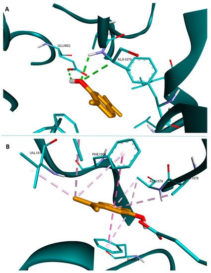
The most active docked compound according to the obtained scores was carvacrol. Our predicted results are in line with a recent study published by Rezaienasab et al. according to which carvacrol inhibits xanthine oxidoreductase in a dose-dependent manner, and its antioxidant activity is related to the decrease om ROS production due to xanthine oxidoreductase inhibition
[59]. Binding interactions analysis of carvacrol reveals the formation of three hydrogen bonds, one with Glu802 and the other two with Ala1079 (
Figure 1A), similar to the binding pattern of the native ligand. The structure is also very well stabilized in the binding pocket through multiple hydrophobic interactions (
Figure 1B). Given the existing above mentioned biological literature data that validate our docking method, we can conclude that MOEO is a strong antioxidant product that could exert its antioxidant activity not only by radical scavenging but also by targeted inhibition of xanthine oxidoreductase.
Figure 1. Structure of xanthine oxidoreductase (3NRZ) in complex with carvacrol (orange); hydrogen bond interactions are depicted as green dotted lines (A), and hydrophobic interactions as purple dotted lines (B); interacting amino acids are shown as light blue sticks.
 +1 point