1000/1000
Hot
Most Recent
 +1 point
Bursera fagaroides is a medicinal tree endemic to México, it belongs to the Burseraceae family and has proven antitumor activity. Modern research, performed principally with the bark extracts, have indicated that lignans are the main active constituents of B. fagaroides, with a high content of aryltetralin, aryldihydronaphtalene, dibenzylbutirolactone, and dibenzylbutane type lignans as the constituents of the active extracts. In general, lignans from B. fagaroides exhibited potent anti-cancer activity, although antitumor, anti-bacterial, anti-protozoal, anti inflammatory, and anti-viral properties have also been described.
Cancer is well known as one of the most important causes of morbidity and mortality worldwide, its effects in both more and less economically developed countries, and its likelihood to rank as the leading cause of death in the 21st century. It is estimated that this public health problem caused 9.6 million deaths worldwide in 2018 [1][2]. Currently, there are various types of therapies for cancer treatment, but chemotherapy is one of the most widely used. However, because of the severe side effects exhibited by commercially available drugs to treat cancer, as well as the drug resistance in tumor cells, it remains a challenge for medicinal chemistry to develop novel agents and treatment strategies to attack this public health problem [3][4]. According to the Food and Drug Administration (FDA), 246 anti-cancer drugs were approved between 1940 and 2014 and around 38% of them are natural products (or derived from them) [5]. In this context, one of the most important sources of anti-cancer secondary metabolites is the Bursera genus because it has been reported that these plants are effective against different types of cancer [6]. The genus Bursera Jacq. ex L. (Burseraceae) consists of about 105 shrubs and trees with a geographical distribution extending from the Southern U.S. to Peru and the Caribbean. In Mexico, the Bursera species grow principally in the tropical dry forests, where about 92 species have been described and most of them (~85%) are endemic [7][8]. Bursera has been divided into two subgenera (subg.), or sections: B. subg. Bursera and B. subg. Elaphrium (previously known as Bullockia) [8]; the bark, among other traits, mainly differentiates between them. The species of section Bursera have colorful trunks and peeling bark, while species of Elaphrium have rough and non-peeling bark [9]. Several Bursera species are recognized because of their characteristic production of an aromatic resin (exuded) known as “copal” that provides a chemical defense against specialized herbivores [10]. Since ancient times, copal resin has been commonly used in México and Central America as incense in religious activities [11][12]. The chemical profile of the species of Bursera includes flavonoids [13][14], triterpenes [15][16], sesquiterpenes [16][17], diterpenes [18], and lignans [19][20]. Most of the Bursera species that produce lignans are widely used in México as traditional, natural medicine due to their pharmacological properties, including analgesic, anti-inflammatory, and antitumoral properties. Also, they can help treat different illnesses, such as colds, polyps, and venereal diseases [6][21]. In general, lignans from the Bursera genus are secondary metabolites, known for their antioxidant, apoptotic, anti-cancer, anti-inflammatory, anti-bacterial, anti-viral, anti-fungal, and anti-protozoal properties. In particular, lignans from B. fagaroides have been reported to have an important anti-cancer effect [6]. This review aims to summarize literature findings on the Mexican B. fagaroides, such as uses in medicinal folk, pharmacological effects of its extracts and chemistry, and the biological activities of its lignans. This review focuses on the biosynthesis, chemical aspects, anti-cancer effects, and molecular mechanisms of lignans from B. fagaroides. The information reported in this work results from a search in ScienceDirect, PubMed, and Scifinder databases.
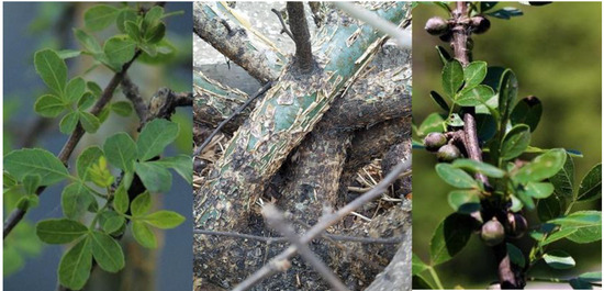
Bursera fagaroides (B. subg. Bursera) (Figure 1), also identified as Elaphrium fagaroides, Amyris fagaroides , and Terebinthus fagaroides, is a Mexican medicinal plant locally known as “copalillo”, “aceitillo”, “copal”, “sarzafrás” “xixote”, “cuajiote amarillo” “jiote”, “palo del diablo”, “papelillo”, and “xicote” [17][22][23][24]. It is an aromatic bush or tree of about 0.5–8 m high, distributed from the Southwestern United States of America to the Isthmus of Tehuantepec in México; it grows mainly at altitudes from 300 to 2200 m [8][17][23]. B. fagaroides, as traditional natural medicine, have been popularly used to treat inflammation, hits, tumors, cancer, and stomach disorders [20][22][25]. These medicinal properties have served as inspiration for various cancer research groups, as described below.
This plant species has been studied principally for its anti-cancer properties, although its antimicrobial and antigiardial effects also have been reported. An overview of the anti-cancer biological studies performed on this plant species shows that the only parts examined have been the bark and the exudate resin from the tree trunk. In vivo and in vitro studies on the extracts of these plant parts have shown important cytotoxic activities.
For instance, in 1969, Bianchi and Cole [26] found that the chloroform extract displayed a 32% reduction in the in vivo Walker carcinoma 256 tumor system (WA16). Further, the ethanol extract from the dried exudates of B. fagaroides showed a concentration-dependent inhibitory effect on cell proliferation against the human colon cell line HT-29, with an IC50 value of 0.41 ± 0.01 μg/mL at 72 h [27].
Another in vivo study by Rojas-Sepulveda [19] reported that the intraperitoneal administration of 100 mg/Kg of the hydroalcoholic extract from the bark on mice, inoculated with L5178Y lymphoma cells, increased the survival time and cured 26% ( p < 0.001) of the treated mice. This extract also significantly inhibited the proliferation of KB (nasopharyngeal, ED50 = 9.6 × 10 −2 μg/mL) , PC-3 (prostate, ED50 = 2.5 × 10 −1 μg/mL), HF-6 (colon, ED50 = 7.1 × 10 −3 μg/mL), and MCF-7 (mama, ED50 = 6.6 μg/mL) tumor cell lines [19]. Later, Acevedo et al. (2015) [28] described the cytotoxicity of the n -hexane and chloroform extracts measured by the sulforhodamine B protein staining assay using KB, HF-6, MCF-7, and PC-3 cancer cell lines, along with a normal skin fibroblast cell line. The results indicated that both extracts displayed an important antiproliferative effect on all the studied cells, including normal cells, corroborating the results obtained previously [28].
In another in vivo study, the hydroalcoholic extract from the bark of B. fagaroides does not affect the number of Histone H3 phosphorylated at serine 10 (H3S10ph)-positive nuclei, with respect to the control without treatment, when measured in whole 24 h post-fertilization (hpf) zebrafish embryos; this indicated that the extract does not induce mitotic cells in the embryos [29]. This result contrasts with the strong in vivo antitumor activity against L5178Y lymphoma in mice and the authors attributed this to the poor bioavailability because of the low concentration of the active compounds present in the studied extract [19]. Further, chromatographic fractionation afforded two rich-lignans fractions that induced a high amount of cells in mitotic arrest in zebrafish embryos [29].
The chemical study of B. fagaroides through the years allowed the characterization of 19 lignan structures named: podophyllotoxin (1), ß-peltatin-A-methylether (2) , 5′desmethoxy-ß-peltatin-A-methylether (3), desoxypodophyllotoxin (4), acetyl podophyllotoxin (5) [[1]], morelensin (6) [[1]], burseranin (7) [[1]],, acetylpicropodophyllotoxin (8) [[2]], desmethoxy-yatein (9), yatein (10) [[1]], hinokinin (11) [40], 7′,8′-dehydropodophyllotoxin (12), 7′,8′-dehydroacethyl podophyllotoxin (13), 7′,8′ dehydro-trans-p-cumaroylpodophyllotoxin (14) [[3]], 9-acetyl-9-pentadecanoildihydroclusin (15), 2,3-demethoxy-secoisolintetralin diacetate (16), dihydroclusin diacetate (17), 2,3 demethoxy-secoisolintetralin monoacetate (18) dihydroclusin mono acetate (19) [[4]]. Eight of these are aryltetralin (1–8), three are dibenzylbutyrolactone (9–11), three are aryldihydronaphtalene (12–14), and five are dibenzylbutane lignans (15–19).
The lignans 1–19 have been isolated from specimens of B. fagaroides, collected in México, principally from the bark (Michoacán state), two reports from Oaxaca state [[4]] and one from Guerrero state [[5]] analyzed the resin. The polarity of the used extracts was chloroform (CHCl3), dichloromethane (CH), ethanol (CH3CH2OH) [27], CH3CH2OH 80% (previously treated with hexane) [[5]], and methanol (CHOH) 70% [19,29,39]. In general, the purification of these extracts was carried out by bioassay-guided chromatographic methods. The extracts were fractionated and the components were separated by repeated column chromatography, eluting with gradients or isocratic mixtures [[1], [6]] of organic solvents through preparative thin layer chromatography (TLC), semi-preparative reverse phase HPLC with a diode array detection system, flash chromatography, or preparative reverse phase TLC, as required. The yields and purity of isolated compounds were based on the peak areas of the HPLC chromatograms[2].
In addition, their 13C NMR (CDCl3) spectra show the occurrence of carbon resonances, ascribable to carboxyl ester groups ( δC ~ 167.7–173.7); signals at δ ~ 171.1 and δ ~ 21.0 also confirm the presence of acetyl groups; carbons at δC7′ ~ 147.2–147.9 and δC8′ ~ 118.7 in lignans 12, 13, and 14 were characteristics of a tetrasubstitued double bound. Other signals also were assigned: methylenedioxy group in ring A [δ C ~ 102.0–103.1], methoxyl groups [δ C ~58.6 - 61.2], aliphatic methylene group [δ C ~ 69.6–70.1], aliphatic methine groups (C7) δ ~74.2–75.2 for lignans 1–8 and 12–14, aliphatic methine groups [δC8 ~ 41.7–43.9], characteristic signals of carbon atoms bearing oxygen at δ ~ 63.95 - 64.3, aromatic carbons for ring B [δC1 ~ 128.2–130.6, δC2 ~ 129.1–132.0, δC3 ~ 110.0–110.2, δC4 ~ 150–153, δC5 ~ 147.5–148.4, and δC6 ~ 104.8–105.8] and those corresponding to substituted aromatic carbons for ring E [δC1′ ~ 129.1–131.2, δC2′ ~ 109.6–110.0, δC3′ ~ 153.0–153.7, δC4′ ~ 135.7–139.5, δC5′ ~ 153.0–154.0, and δC6′ ~ 109.6–110.3], respectively [19][20][25][26][27][29][30]. On the other hand, the absolute configurations of lignans 4, 5, 7, 11, and 12 were determined using vibrational circular dichroism [27], while that of lignan 1 was determined by chemical correlation with D-phenylalanine [31] and by X-ray diffraction analysis of 2′-bromopodophyllotoxin [32].
The molecular studies of lignans isolated from B. fagaroides are diverse and all head to anti-cancer activity. Cancer is a worldwide health problem with 9.6 million cancer deaths in 2018 [2]; due to this, lignans are eye-catching secondary metabolites from medicinal plant research implicated in cancer. Figure 5 summarizes all the assays performed for aryl tetralin and aryldihydronaphtalene lignans isolated from B. fagaroides , from the cytotoxic in vitro results, in vivo assays in mice and zebrafish models, and to the molecular recognition by NMR. Lignans 8, 10, 11, 15–19 do not report any activity.
In vivo studies with Walker carcinoma 256 (intramuscular) were performed for 2 and 3, they exhibited an important antitumoral activity at a level of 10% T/C at 12.5 mg/kg and 20% T/C at 100 mg/kg, respectively [26]. Also, the in vivo model using zebrafish embryos was performed to observe disruption of cell behavior; the results showed a delay cell migration in actin filaments for 1, 2, 5, and 9. Also the same compounds presented a microtubule depolymerization in the same model by α-tubulin immunofluorescence [29].
The cytotoxicity (IC50) of 14 lignans were reported in 9 cancer cell lines: KB, PC-3, MCF-7, MDA-MB-231, BT-549, HF-6, A549, A2780, and SiHa. According to the results, PC-3 and KB cell lines were the most sensitive with IC50 values between 2.29 and 4.43 × 10 −6 µM, respectively [19][20][33][30]. β-peltatin-A-methylether (2) and podophyllotoxin (1) were the most active compounds in KB cells with an IC50 of 4.43 × 10 −6 and 4.61 × 10 −6 µM, respectively [19]. It should be highlighted that the importance of these results is due to prostate cancer occupying the first place in mortality cancer in males [34].
In general, lignans obtained from B. fagaroides are important secondary metabolites with promising pharmacological anti-cancer effects and it could be interesting to explore them as antivirals. These compounds can act by the same mechanism of action of podophyllotoxin and can be considered in clinical trials for cancer.


