Submitted Successfully!
To reward your contribution, here is a gift for you: A free trial for our video production service.
Thank you for your contribution! You can also upload a video entry or images related to this topic.
Version Summary Created by Modification Content Size Created at Operation
1 + 2147 word(s) 2147 2021-05-25 11:42:50 |
2 format change -8 word(s) 2139 2021-06-04 03:03:50 |

Video Upload Options

Do you have a full video?

Confirm

Are you sure to Delete?
Cite
If you have any further questions, please contact Encyclopedia Editorial Office.
Di Giancamillo, A. Gut Development and Piglets. Encyclopedia. Available online: https://encyclopedia.pub/entry/10481 (accessed on 16 April 2024).
Di Giancamillo A. Gut Development and Piglets. Encyclopedia. Available at: https://encyclopedia.pub/entry/10481. Accessed April 16, 2024.
Di Giancamillo, Alessia. "Gut Development and Piglets" Encyclopedia, https://encyclopedia.pub/entry/10481 (accessed April 16, 2024).
Di Giancamillo, A. (2021, June 03). Gut Development and Piglets. In Encyclopedia. https://encyclopedia.pub/entry/10481
Di Giancamillo, Alessia. "Gut Development and Piglets." Encyclopedia. Web. 03 June, 2021.
Gut Development and Piglets
Edit

During the prenatal, neonatal, and weaning periods, the porcine gastrointestinal tract undergoes several morpho-functional, changes together with substantial modification of the microbial ecosystem. Modifications of the overall structure of the small intestine also occur, as well as a rapid increase of the volume, mainly in the last period of gestation: intestinal villi, starting from jejunum, appears shortly before the sixth week of gestation, and towards the end of the third month, epithelial cells diversify into enterocytes, goblet cells, endocrine, and Paneth cells. Moreover, in the neonatal period, colostrum induces an increase in intestinal weight, absorptive area, and brush border enzyme activities: intestine doubles its weight and increases the length by 30% within three days of birth. 

gut development cell turnover morpho-functional activity piglets

1. Introduction

There are many stressful events that affect the health and growth of piglets from birth to weaning: among these, the development of the gastrointestinal tract (GIT) is a complex and delicate process, which begins in the prenatal phase and then continues after birth.
The most important factor that can influence the structure and functions of GIT is diet: in the first postnatal period, the bioactive substances present in the colostrum and in the sow’s milk are essential for proper development and growth; their properties modulate cell physiology, such as the turnover of apoptosis and mitosis [1]. In that way, they also influence the development of GIT, allowing the adaptation of animals to solid feed (Figure 1). On the contrary, the weaning phase is related not only to the diet, but also to the proper development of the intestinal microflora.
Figure 1. Timing of cellular turnover during gut development: apoptosis and mitosis.
Appropriate changes of the anatomical structure of the intestinal mucosa and its enzymatic activities are indispensable to allow to GIT to adapt to diet, as already previously described in pig during weaning [1][2].
This review will focus on the gut morphological development and growth in a wider period, from preterm to weaning. The aim is to provide useful tools to evaluate (predict) new strategies to improve gut homeostasis at the physiological, immunological, and microbial level in the long-term, and to prevent weaning gut alterations; thus, reducing antimicrobial use.

2. Gut Anatomy in Brief

Focus on the Small Intestine

The small intestine is anatomically divided into three regions whose proportions are as follows: duodenum, which in adult pig represents about 4–4.5%, jejunum, about 88–91% and finally ileum, about 4–5% [3]. The newborn has similar proportions to those of the adult, even if the differentiation between jejunum and ileum is not well defined. Although in adults, these three traits have morphological characteristics that clearly distinguish them, they also share many common features as they are all organized in four basic layers: the tunica serosa, the tunica muscularis, the tunica submucosa, and the tunica mucosa. The serosa is the outer layer and morphologically consists of an epithelium lining on connective tissue, which contains blood vessels and nerves, and which eventually forms the mesentery. The tunica muscularis is organized with two distinct layers of muscle fibers; an outer layer of longitudinal fibers and an inner layer of circular fibers, which both are involved in GIT motility. The submucosa is made up of a layer of connective tissue that contains blood and lymphatic vessels and nerves.
A special focus should be done on the submucosa of the duodenum, because it contains the Brunner’s glands, which are specialized to produce alkaline secretion containing bicarbonate, in order to protect the duodenal mucosa from the acidic content of the chyme coming from the stomach; in addition, this secretion stimulates the activation of intestinal enzymes by creating an alkaline environment and lubricating its walls. Nutrition, nervous and/or hormonal stimuli and reflexes increase the secretory activity of Brunner’s glands [4].
The inner layer, the mucosa, is the one that characterizes the morpho-functional activity of each individual trait of the intestine and it is made up of three sublayers: the muscularis mucosa, the lamina propria and the epithelium. The muscularis mucosa is a thin layer of muscle fibers that divides the submucosa from the mucosa and that contributes to form the transient intestinal folds. The lamina propria is made up of connective tissue that contains blood vessels, neurons, lymphocytes and, in the ileum, it also contains lymph nodes called Peyer’s patches; the lamina propria has the duty of sustaining the avascular epithelial layer. This latter consists of a single layer of epithelial cells, which covers the luminal surface of the intestine.
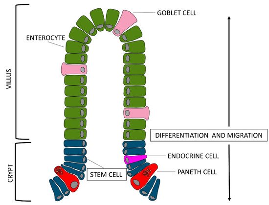
The mucosa as a whole, folds back to form finger-like projections called villi, and at the base of these ones, the Crypts of Lieberkühn, also known as intestinal glands, which are moat-like invaginations.
Villi increase the work surface by at least five times compared to a flat surface of the same size. Furthermore, the luminal surface of the enterocytes has got microvilli at the level of the apical membrane (also called brush border), which further increase the absorptive surface by 15–40 times, simulating the observed system of the villi folds. Microvilli are placed in a gelatinous layer of glycoproteins, the glycocalyx, and have digestive enzymes. The villi morphology changes according to the intestinal tracts and this trend reflects their different functions: the length increases from the duodenum to the mid-jejunum and then decreases again towards the distal ileum. Similarly, the crypts also change in size and composition along the intestine: they are deeper in the duodenum and jejunum and less deep in the ileum. [5].
If we look at the epithelial layer more closely, we notice that there are three types of cells lining on the villus surface: enterocytes, goblet cells, and enteroendocrine cells (piglet gut barrier reviewed by [6]). Enterocytes are the most abundant group of villi cells, about 94%, goblet cells are about 5% and endocrine cells about 1%. All of these cell types originate from stem cells located at the base of the crypts [7]. Enterocytes get mature while they migrate from the base to the tip of the villi; their enzymatic activity, which reflects the digestive function, begins when the enterocytes reach the basal third of the villi axis, while their absorption function begins when enterocytes reach the medium-high level and spreads its maximum when the cells reach the top of the villi. Obviously, the enterocytes present on the surface of the villi are continuously renewed.
The goblet cells are secreting cells: specifically, they secrete viscous mucus and are located between the enterocytes. They increase in number from the proximal portion of the jejunum to the distal one of the ileum. Goblet cells secrete relentlessly viscous mucus and their basal activity increases when the cells come in contact with a secretagogue substance. Mucus production can be altered by several agents: cholinergic agents, neuropeptides, hormones, and toxins for example. Furthermore, nutrients are capable of influencing the thickness and production of mucus (e.g., milk peptides). In addition, the enterocytes that organize the mucosa can also have secreting activity contributing to produce, with the mucus, the intestinal juice: a young pig of about 70 kg, produces approximately 6 L per day [3]. The third cell type, the enteroendocrine cells, produce hormones to regulate the functionality of the gastrointestinal tract [8].
Interesting, recent works suggest the presence in the GIT of pig of another type of cells, the Paneth cells, which have been described extensively in other species [9]. Reference [10] observed these cells, located adjacent to stem cells at the bottom of the crypt of the ileum, and [11] observed Paneth cell-like cells in the gut of piglet from newborn to weaned stages in the same localization. The exact function is unknown, but due to the presence of substances, such as lysozymes and defensins inside microgranules of the cytoplasm, they most likely contribute to maintenance of the gastrointestinal barrier. About the differentiation of Paneth cells, it would be possible to hypothesize that, as described for other species, they move back to the stem cell compartment to be interspersed with stem cells [12].

3. The Development of the Small Intestine during the Preterm Period

The rapid and massive growth of the small intestine in the pig, begins in the prenatal phase, just a few weeks before farrowing and it is by far the fastest growth of the whole organism [13], and there are tissue-specific activities of brush-border enzymes with low maltase and high lactase activities [14]. This may partly be due to increased ingestion of amniotic fluid, as suggested by [15].
The small intestine shows the presence of intestinal villi starting from jejunum, the site of maximum absorption, shortly before the sixth week of gestation and, towards the end of the third month, it is possible to observe the differentiation of the epithelial cells into the three cell types previously described. The morphological peculiarity of fetal enterocytes lies in the presence of large cytoplasmic vacuoles (endocytic activity) that, in the adult, will be replaced with cells without vacuoles. There are two types of vacuoles: (1) transport vacuoles, necessary for the transport of macromolecules from the intestinal lumen to the enterocytes; (2) digestive or lysosomal vacuoles, involved in the intracellular digestion of nutrients and pH control in the intestinal lumen [16]. The transport vacuoles are present throughout the small intestine in the first two days of birth and allow the bioactive compounds of the colostrum to cross the enterocytes without being modified, providing passive immunity; subsequently, there is a reduction of this transport capacity [17]. The specific transport of macromolecules comes cross the epithelium through M cells, which are specialized cells, active in the transcytosis process, a cellular specialization for transport of molecules across the mucosal barrier to the submucosal tissues. [18]. At the same time, tissue-specific activities of brush-border enzymes appear, with a low maltase and high lactase activities [14].This may partly be due to increased ingestion of amniotic fluid, as suggested by [15].
The changes of the epithelium are accompanied by modifications of the overall structure of the small intestine as well. Towards the end of the third month, intestinal crypts are formed and the two layers of muscular tunic differentiate [19][20]. Near birth, the small intestine grows very rapidly and in the last three weeks of gestation the volume relative to the weight of the animal increases by more than 70% [21].
To understand exactly what happens in the last period of gestation in the small intestine, we should focus on cellular turnover pathway.
After stem cell mitosis, the fate of epithelial cells depends on the way they migrate: most of the cells go towards the apex of the villi and differentiate into enterocytes, mucous cells, or endocrine cells; some other cells migrate down the crypts and differentiate into Paneth cells (Figure 2). Enterocytes have a rapid turnover of about 48–72 h that alternates mitosis and apoptosis: the latter, a programmed physiological death, enables the removal of cells when these are old, damaged or mutated during different physiological stages, such as organogenesis, development, regeneration, and involution [22]. The dynamic balance between mitosis and apoptosis allows to maintain the correct physiology of the tissues and, if necessary, the rapid reconstruction of damaged tissues in case of pathologies [23].
Figure 2. After stem cell mitosis (blue cells), some epithelial cells go towards the apex of the villi and differentiate into enterocytes (green cells) or mucous cells (light pink cells); some others migrate down the crypts and differentiate into Paneth cells (red cells) or endocrine cells (pink cell).
Considering this, we observe that the cellular turnover in structural pattern of the gut follows a pretty precise pattern: proliferation in the crypts and migration of enterocytes and goblet cells towards the apex of the villi, where the turnover will occur with cell loss in the intestinal lumen due to apoptosis, while the endocrine cells will follow the path of the depth of the crypts. Such organized turnover allows to maintain a constant number of cells despite the speed with which this process occurs [24].
Starting from the last 3 weeks of gestation, there is a significant increase in the number of mitoses and, at the same time, a reduction in apoptosis which leads to a considerable increase in visceral mass [25][26]. Interestingly, the apoptotic cells are not localized only at the apex of the villi, but are also found along its entire axis and death occurs with detachment of groups of cells and not of single elements. In references [23][27], the authors hypothesized an important role of autophagy starting from the intrauterine pathway for the development of the intestine. Autophagy is an important cellular mechanism for the renewal of cellular components damaged following stress. It is a catabolic process targeted at recycling cellular parts or damaged organelles through a lysosome-dependent degradation pathway [28].
Basal autophagy is necessary for normal cell turnover and it is important for maintaining homeostasis, while excessive or uncontrolled autophagy promotes cell death and morbidity [29]. The mechanism of autophagy in the various intestinal cell compartments has been extensively reviewed by [30]: it is regulated by cathepsins and acts through the formation of autophagolysosomes for the degradation of cellular organelles without any change at the nuclear level [31][32].

References

  1. Domeneghini, C.; di Giancamillo, A.; Arrighi, S.; Bosi, G. Gut-trophic feed additives and their effects upon the gut structure and intestinal metabolism: State of the art in the pig, and perspectives towards humans. Histol. Histopathol. Cell. Mol. Biol. 2006, 21, 273–283.
  2. Pluske, J.R.; Turpin, D.L.; Kim, J.-C. Gastrointestinal tract (gut) health in the young pig. Anim. Nutr. 2018, 4, 187–196.
  3. Lærke, H.N.; Hedemann, M.S. The Digestive System of The Pig (Chapter 5). In Nutritional Physiology of Pigs—Online Publication; Videncenter for Svineproduktion: Foulum, Denmark, 2012.
  4. Moore, B.A.; Morris, G.P.; Vanner, S. A novel in vitro model of Brunner’s gland secretion in the guinea pig duodenum. Am. J. Physiol. Gastrointest. Liver Physiol. 2000, 278, G477–G485.
  5. Sumigray, K.D.; Terwilliger, M.; Lechler, T. Morphogenesis and Compartmentalization of the Intestinal Crypt. Dev. Cell 2018, 45, 183–197.e5.
  6. Modina, S.C.; Polito, U.; Rossi, R.; Corino, C.; di Giancamillo, A. Nutritional Regulation of Gut Barrier Integrity in Weaning Piglets. Anim. Open Access J. MDPI 2019, 9, 1045.
  7. Zhu, G.; Hu, J.; Xi, R. The cellular niche for intestinal stem cells: A team effort. Cell Regen. 2021, 10, 1.
  8. Gribble, F.M.; Reimann, F. Function and mechanisms of enteroendocrine cells and gut hormones in metabolism. Nat. Rev. Endocrinol. 2019, 15, 226–237.
  9. Ouellette, A.J. Paneth Cells. In Physiology of the Gastrointestinal Tract; Elsevier: Amsterdam, The Netherlands, 2012; Volume 2, pp. 1211–1228. ISBN 978-0-12-382026-6.
  10. van der Hee, B.; Loonen, L.M.P.; Taverne, N.; Taverne-Thiele, J.J.; Smidt, H.; Wells, J.M. Optimized procedures for generating an enhanced, near physiological 2D culture system from porcine intestinal organoids. Stem Cell Res. 2018, 28, 165–171.
  11. Verdile, N.; Mirmahmoudi, R.; Brevini, T.A.L.; Gandolfi, F. Evolution of pig intestinal stem cells from birth to weaning. Animal 2019, 13, 2830–2839.
  12. van der Flier, L.G.; Clevers, H. Stem Cells, Self-Renewal, and Differentiation in the Intestinal Epithelium. Annu. Rev. Physiol. 2009, 71, 241–260.
  13. Sangild, P.T.; Fowden, A.L.; Trahair, J.F. How does the foetal gastrointestinal tract develop in preparation for enteral nutrition after birth? Livest. Prod. Sci. 2000, 66, 141–150.
  14. Bjornvad, C.R.; Schmidt, M.; Petersen, Y.M.; Jensen, S.K.; Offenberg, H.; Elnif, J.; Sangild, P.T. Preterm birth makes the immature intestine sensitive to feeding-induced intestinal atrophy. Am. J. Physiol. Regul. Integr. Comp. Physiol. 2005, 289, R1212–R1222.
  15. Trahair, J.F.; Sangild, P.T. Fetal organ growth in response to oesophageal infusion of amniotic fluid, colostrum, milk or gastrin-releasing peptide: A study in fetal sheep. Reprod. Fertil. Dev. 2000, 12, 87–95.
  16. Zabielski, R.; Godlewski, M.M.; Guilloteau, P. Control of development of gastrointestinal system in neonates. J. Physiol. Pharmacol. Off. J. Pol. Physiol. Soc. 2008, 59 (Suppl. 1), 35–54.
  17. Pácha, J. Development of Intestinal Transport Function in Mammals. Physiol. Rev. 2000, 80, 1633–1667.
  18. Kulkarni, D.H.; Newberry, R.D. Intestinal Macromolecular Transport Supporting Adaptive Immunity. Cell. Mol. Gastroenterol. Hepatol. 2019, 7, 729–737.
  19. van Ginneken, C.; Weyns, A.; van Meir, E. Stereologic evaluation of the pig gastric wall and of somatostatinergic and serotoninergic immunoreactive mucosal cells during perinatal development. Eur. J. Morphol. 2001, 39, 113–120.
  20. Willemen, S.A.; De Vos, M.; Huygelen, V.; Fransen, E.; Tambuyzer, B.R.; Casteleyn, C.; Van Cruchten, S.; Van Ginneken, C. Ghrelin in the gastrointestinal tract and blood circulation of perinatal low and normal weight piglets. Animal 2013, 7, 1978–1984.
  21. McPherson, R.L.; Ji, F.; Wu, G.; Blanton, J.R.; Kim, S.W. Growth and compositional changes of fetal tissues in pigs. J. Anim. Sci. 2004, 82, 2534–2540.
  22. Darwich, A.S.; Aslam, U.; Ashcroft, D.M.; Rostami-Hodjegan, A. Meta-Analysis of the Turnover of Intestinal Epithelia in Preclinical Animal Species and Humans. Drug Metab. Dispos. 2014, 42, 2016–2022.
  23. Godlewski, M.M.; Słupecka, M.; Woliński, J.; Skrzypek, T.; Skrzypek, H.; Motyl, T.; Zabielski, R. Into the unknown--the death pathways in the neonatal gut epithelium. J. Physiol. Pharmacol. Off. J. Pol. Physiol. Soc. 2005, 56 (Suppl. 3), 7–24.
  24. Yen, T.-H.; Wright, N.A. The gastrointestinal tract stem cell niche. Stem Cell Rev. 2006, 2, 203–212.
  25. Sangild, P.T.; Thymann, T.; Schmidt, M.; Stoll, B.; Burrin, D.G.; Buddington, R.K. Invited review: The preterm pig as a model in pediatric gastroenterology. J. Anim. Sci. 2013, 91, 4713–4729.
  26. Buddington, R.K.; Sangild, P.T.; Hance, B.; Huang, E.Y.; Black, D.D. Prenatal gastrointestinal development in the pig and responses after preterm birth. J. Anim. Sci. 2012, 90 (Suppl. 4), 290–298.
  27. Biernat, M.; Woliński, J.; Godlewski, M.M.; Motyl, T.; Morisset, J.; Zabielski, R. Apoptosis in the Gut of Neonatal Piglets; University of Alberta: Edmonton, AB, Canada, 2003; pp. 46–48.
  28. Levine, B.; Klionsky, D.J. Development by Self-Digestion: Molecular Mechanisms and Biological Functions of Autophagy. Dev. Cell 2004, 6, 463–477.
  29. Mizushima, N.; Levine, B.; Cuervo, A.M.; Klionsky, D.J. Autophagy fights disease through cellular self-digestion. Nature 2008, 451, 1069–1075.
  30. Haq, S.; Grondin, J.; Banskota, S.; Khan, W.I. Autophagy: Roles in intestinal mucosal homeostasis and inflammation. J. Biomed. Sci. 2019, 26, 19.
  31. Lamparska-Przybysz, M.; Gajkowska, B.; Motyl, T. Cathepsins and BID are involved in the molecular switch between apoptosis and autophagy in breast cancer MCF-7 cells exposed to camptothecin. J. Physiol. Pharmacol. Off. J. Pol. Physiol. Soc. 2005, 56 (Suppl. 3), 159–179.
  32. Motyl, T.; Gajkowska, B.; Zarzyńska, J.; Gajewska, M.; Lamparska-Przybysz, M. Apoptosis and autophagy in mammary gland remodeling and breast cancer chemotherapy. J. Physiol. Pharmacol. Off. J. Pol. Physiol. Soc. 2006, 57 (Suppl. 7), 17–32.
More
Information
Contributor MDPI registered users' name will be linked to their SciProfiles pages. To register with us, please refer to https://encyclopedia.pub/register :
View Times: 687
Revisions: 2 times (View History)
Update Date: 04 Jun 2021
1000/1000