Submitted Successfully!
To reward your contribution, here is a gift for you: A free trial for our video production service.
Thank you for your contribution! You can also upload a video entry or images related to this topic.
Version Summary Created by Modification Content Size Created at Operation
1 + 2111 word(s) 2111 2021-06-23 13:06:20 |
2 Reference formatted Meta information modification 2111 2021-06-24 03:11:28 |

Video Upload Options

Do you have a full video?

Confirm

Are you sure to Delete?
Cite
If you have any further questions, please contact Encyclopedia Editorial Office.
Nolte, M.A. Bone Marrow as memory organ. Encyclopedia. Available online: https://encyclopedia.pub/entry/11204 (accessed on 19 April 2024).
Nolte MA. Bone Marrow as memory organ. Encyclopedia. Available at: https://encyclopedia.pub/entry/11204. Accessed April 19, 2024.
Nolte, Martijn A.. "Bone Marrow as memory organ" Encyclopedia, https://encyclopedia.pub/entry/11204 (accessed April 19, 2024).
Nolte, M.A. (2021, June 23). Bone Marrow as memory organ. In Encyclopedia. https://encyclopedia.pub/entry/11204
Nolte, Martijn A.. "Bone Marrow as memory organ." Encyclopedia. Web. 23 June, 2021.
Bone Marrow as memory organ
Edit

The bone marrow (BM) is key to protective immunological memory because it harbors a major fraction of the body’s plasma cells, memory CD4+ and memory CD8+ T-cells. Despite its paramount significance for the human immune system, many aspects of how the BM enables decade-long immunity against pathogens are still poorly understood. In this review, we discuss the relationship between BM survival niches and long-lasting humoral immunity, how intrinsic and extrinsic factors define memory cell longevity and show that the BM is also capable of adopting many responsibilities of a secondary lymphoid organ. Moreover, we discuss what factors determine the establishment of long-lasting immunological memory in the BM and what we can learn for vaccination technologies and antigen design. Finally, we touch on how a more holistic understanding of the BM is necessary for the development of modern and efficient vaccines against the pandemic SARS-CoV-2.

bone marrow memory T-cells B-cells plasma cells infection residency vaccination COVID-19 SARS-CoV-2

1. Introduction

The organizational challenges of maintaining a healthy and protective equilibrium of an immune system as versatile and complex as the humans are immense. Similarly, the physiological challenges for each individual memory cell are enormous: They need to be able to self-renew, persist long-term and give rise to highly proliferative progeny while staying capable of quickly mounting a recall response upon reinfection [1][2]. At the same time, while in a steady state, terminal differentiation has to be prevented. Possible pitfalls include the exhaustion of proliferative potential, telomere shortening, DNA replication stress as well as the accumulation of mutations. Additionally, epigenetic modifications have to be precisely controlled to facilitate the right amount of flexibility and phenotype plasticity [3][4]. Furthermore, the kinetics of cell migration are also an important factor that needs precise adjustment [5][6].
The BM is essential in enabling many of the functions of the immune system beyond the well-known generation of all blood cells—hematopoiesis [7][8]. In this review, we take a look at how the BM plays a pivotal role in establishing long-lasting immune memory and sustaining protection despite the aforementioned obstacles. (See Table 1 for a list of all of the important molecular factors described in this review.) Furthermore, we hypothesize that the BM is the central site where the threads of maintaining memory cells, inducing primary immune responses to systemic infections as well as secondary immune responses converge.

2. Plasma Cells: The Hidden Treasures of Humoral Immunity

Although their importance for long-lasting immunity to pathogens was originally dismissed, plasma cells have now been the focus of immunological research for a long period of time due to their essential role in the humoral arm of immune defense [9]. Derived from B-cells with the help from T-cells, plasma cells are able to produce antibodies against specific antigens supporting neutralization, agglutination, complement activation and activation of effector cells. It was discovered that plasma cells reside in the BM, where they produce the majority of total and antigen-specific serum IgG [10][11]. These BM plasma cells can be sustained independently of memory B-cells and for varying amounts of time, sometimes a lifetime [12][13][14], which makes them the ideal cell type to engage for successful vaccination strategies. During an immune response, around 10% of the plasmablasts, the plasma cell precursors, are typically selected and become plasma cells [13]. The molecular markers for long-lived plasma cells are still highly debated, but CD38high, CD19 and CD138+ are frequently used to define this group of cells [15][16][17][18][19][20].

3. Memory T-Cells: The Wanderers of the Adaptive Immune System

Both CD4+ and CD8+ T-cells are characterized by their ability to continuously recirculate through the body, thereby increasing the chance of finding their cognate antigen. Whereas naïve and central memory T-cells (TCM) travel between SLOs, and effector T-cells and effector memory T-cells (TEM) through peripheral tissues [21][22], the BM is unique in that it harbors all T-cell subsets, irrespective of their activation or memory status [23]. Although they form only a minor fraction of all BM cells, the absolute number of all memory T-cells present in the BM of the entire body is substantial [24]. Together with the notion that many antigen-specific memory T-cells home to the BM after an infection, this has further raised awareness of the BM as an important immunological memory organ [25][26][27].

4. The BM as a Secondary Lymphoid Organ

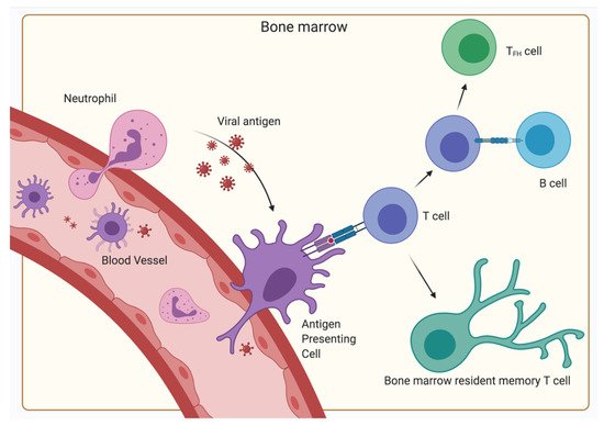
With the BM being such a central organ accommodating a multitude of different kinds of cells, the presentation of antigens and initiation of primary responses—functions typically restricted to SLO—seems to be a possible scenario. Indeed, the initiation of primary T-cell responses of CD4+ as well as CD8+ cells to blood-borne antigens have been observed in the BM, indicating an additional function of the BM as a SLO [25][28]. However, in contrast to classical SLOs, no organized B- and T-cell areas have been described, but instead clusters of dendritic cells and T lymphocytes have been shown [29][30]. These dendritic cells capture, process and present blood-borne antigen to naïve CD4+ and CD8+ T lymphocytes, thereby generating a primary immune response in the BM in the absence of secondary lymphoid organs (Figure 3).
Figure 3. Primary immune response in the BM. Antigen is transported via the blood vessels, antigen-presenting cells (typically dendritic cells) or neutrophils to the BM. There, the antigen-presenting cells display the antigen on their MHC receptors and interact with naïve T-cells inducing their differentiation into BM resident memory T-cells. Some CD4+ T-cells are stimulated to differentiate into TFH by bystander B-cells after the initial antigen-MHC II—TCR interaction. (Created with BioRender.com).
As the BM is not connected to the lymph circulatory system but only the blood circulatory system, the BM might be an important factor for controlling systemic infections. Besides CD11c+ dendritic cells, neutrophils have also been described as a source of antigen transport to the BM [31]. Specifically, virus from the dermis is carried to the BM and induces CD8+ T-cell responses. Along with these primary immune responses, secondary immune responses where memory CD4+ T-cells are reactivated by antigen have been observed to cause aggregation of immune clusters between MHC II expressing cells and antigen-specific T-cells in the BM [32]. The MHC II expressing cells were mostly defined as B lymphocytes. This process amplified the T-cell memory and following the termination of the immune reaction, the CD4+ memory T-cells remained in the BM. These reactions were autonomous to the BM, ergo independent of immigrating T-cells. Even though B-cells were involved, no humoral memory adaptation or GC formation was detected. Furthermore, the expression of signature genes of follicular helper T-cells was significantly lower than in the spleen, indicating a non-follicular reactivation. However, there is some evidence, that dendritic cells may activate CD4+ T-cells and license them to differentiate into resting memory cells in the BM during primary immune responses, while some activated CD4+ T-cells interact with bystander B-cells as a follow up to the initial antigen presentation, leading to their differentiation into TFH (Figure 3) [33]. Furthermore, some studies suggest that BM memory CD4+ T-cells can differentiate into TFH cells during a recall response, indicating that some are committed to the T follicular helper lineage [34]. TFH cells are important for many processes typically associated with SLOs. Memory TFH cells are most likely sustained by a persistence of antigens, potentially via CD11c+ or B-cell presentation [35]. On the other hand, BM resting memory CD4+ T-cells are typically independent from antigen signals; hence, the ratio of TFH cells and BM resting memory cells might be affected by antigen persistence. Interestingly, while TFH cells play an important role in promoting plasma cell survival in SLO via production of IL-21, the plasma cell maintenance in the BM is independent of TFH support as BM plasma cells do not express IL-21R [36]. Overall, while the BM has some competences of a SLO, it is not capable of fulfilling the complete role of a SLO. However, it is unique in the sheer amount of functions it has to implement, being capable of performing primary and secondary immune functions and hemato- and lymphopoiesis. Particularly, the systemic immune control of blood-borne antigen heavily relies on the BM.

5. The Relevance of the BM for Vaccinology

5.1. Disparity between Memory Established by Natural Infections and Vaccination

Considering the importance of the BM in long-lasting immunity, it is very interesting to take a close look at its role in vaccination. While some vaccines are able to induce life-lasting immunity, others have to be refreshed every year. Additionally, comparing a vaccine to its natural infection often reveals big differences in the quality of the immune reaction. This disparity is especially pronounced in current influenza vaccines, as they are especially bad at eliciting a long-lasting immune response, sometimes not even protecting for the whole flu season. Rafi Ahmed’s group elucidated this phenomenon by collecting blood and BM samples at multiple points in time of individuals receiving the inactivated influenza vaccine [37]. They showed that BM plasma cells elicited by the influenza vaccine were only short lived, typically lost within a year. Interestingly, the initial BM plasma cell induction was good, indicating that the quantity of plasma cells induced was not the issue at hand. As it appears that the intrinsic potential of plasma cells and the quality of the survival niche received in the BM determine the longevity of plasma cells (discussed earlier in this review), one or even both factors are not sufficiently achieved with current influenza vaccines. An inadequate CD4+ T-cell response, whose support is needed for the induction of long- lasting immunity, could also play a role.
On the other side of the spectrum are live-attenuated vaccines such as the ones for MMR (measles, mumps, rubella) or smallpox, which elicit strong cellular and humoral immune responses often lasting for several decades [38][39]. What makes this type of vaccine advantageous when it comes to longevity and protective capability and how to transfer these properties to other vaccine technologies is intriguing to investigate, as other types of vaccines are often preferred for safety and manufacturing reasons. One of the advantages of live-attenuated vaccines is that they signal through many different pattern recognition receptors (PRRs), resulting in strong immunogenic capabilities [40]. As full virus particles are able to initiate a bigger variety of PRRs, vaccines that preserve the full virus particle tend to be more immunogenic. For example, virus-vector vaccines, such as the ones based on adenoviruses, appear to be very potent when it comes to the induction of CD8+ T-cell response [40][41][42]. Considering the aforementioned connection between BM memory CD8+ T-cells and neutrophils, this might partly be achieved by means of activating neutrophils via stimulation of their PRRs, promoting the efficient transportation of antigen toward the BM where a potent systemic immune response can be mounted [31]. Adjuvants are often able to make up for the lack of PRR engagement and are therefore hugely important for an efficient vaccine formulation, especially for non-live-attenuated vaccines [43][44].

5.2. Possible Indications for a SARS-CoV-2 Vaccine

Despite worldwide efforts, thousands of lives are still lost every day to the coronavirus disease 2019 (COVID-19) pandemic, with no end in sight [45]. The development and deployment of a vaccine is essential to stop suffering and return to a normal way of living, and the scientific community has reacted accordingly, with currently more than 180 vaccines at various stages of development [46]. The induction of protective immune memory could prove difficult to achieve as the antibody response toward the virus’ spike protein is very varied [47][48][49]. However, similar to other respiratory viruses, severe acute respiratory syndrome coronavirus 2 (SARS-CoV-2) appears to induce an initial surge in virus-specific plasmablasts leading to an increase in the SARS-CoV-2 targeting antibody levels, followed by a decline and stabilization at a baseline. These stabilized antibody serum levels are maintained by long-lived plasma cells and will decide if the individual is protected against re-infection [49][50]. Indeed, studies in non-human primates (NHPs) demonstrated that neutralizing antibodies, but not T-cell responses, correlated with protection [51]. Furthermore, investigating an outbreak of SARS-CoV-2 on a fishing vessel provided evidence that neutralizing antibodies protect humans from SARS-CoV-2 infection [52]. While mucosal antibodies are induced by the virus [53], mucosal immunity typically does not last long, whereas systemic memory can be maintained for extensive periods of time [54]. All of this indicates that the induction of BM resident long-lived plasma cells is key for an effective SARS-CoV-2 vaccine. Looking at the current frontrunners for a successful SARS-CoV-2 vaccine race, two doses of a vaccine will most likely be required in order to elevate the antibody serum levels above the needed threshold [46]. Additionally, booster doses might become necessary at later time points to keep up protective antibody levels. This shows that even with the enormous budgets for COVID-19 research and the modern vaccine technology applied, the induction of long-lived plasma cells can be tricky. More in-depth knowledge about their recruitment is required in order to accelerate the development of vaccines against this and subsequent pandemics.

References

  1. Di Rosa, F. Two Niches in the Bone Marrow: A Hypothesis on Life-long T Cell Memory. Trends Immunol. 2016, 37, 503–512.
  2. Chang, H.-D.; Tokoyoda, K.; Radbruch, A. Immunological memories of the bone marrow. Immunol. Rev. 2018, 283, 86–98.
  3. Youngblood, B.; Hale, J.S.; Ahmed, R. T-cell memory differentiation: Insights from transcriptional signatures and epigenetics. Immunology 2013, 139, 277–284.
  4. Youngblood, B.; Hale, J.S.; Akondy, R. Using epigenetics to define vaccine-induced memory T cells. Curr. Opin. Virol. 2013, 3, 371–376.
  5. Hampton, H.R.; Chtanova, T. Lymphatic Migration of Immune Cells. Front. Immunol. 2019, 10, 19–23.
  6. Griffith, J.W.; Sokol, C.L.; Luster, A.D. Chemokines and Chemokine Receptors: Positioning Cells for Host Defense and Immunity. Annu. Rev. Immunol. 2014, 32, 659–702.
  7. Murphy, D.T.; Moynagh, M.R.; Eustace, S.J.; Kavanagh, E.C. Bone marrow. Magn. Reson. Imaging Clin. N. Am. 2010, 18, 727–735.
  8. García-García, A.; de Castillejo, C.L.F.; Méndez-Ferrer, S. BMSCs and hematopoiesis. Immunol. Lett. 2015, 168, 129–135.
  9. Yoshida, T.; Mei, H.; Dörner, T.; Hiepe, F.; Radbruch, A.; Fillatreau, S.; Hoyer, B.F. Memory B and memory plasma cells. Immunol. Rev. 2010, 237, 117–139.
  10. McMillan, R.; Longmire, R.L.; Yelenosky, R.; Lang, J.E.; Heath, V.; Craddock, C.G. Immunoglobulin synthesis by human lymphoid tissues: Normal bone marrow as a major site of IgG production. J. Immunol. 1972, 109, 1386–1394.
  11. Hibi, T.; Dosch, H.-M. Limiting dilution analysis of the B cell compartment in human bone marrow. Eur. J. Immunol. 1986, 16, 139–145.
  12. Slifka, M.K.; Matloubian, M.; Ahmed, R. Bone marrow is a major site of long-term antibody production after acute viral infection. J. Virol. 1995, 69, 1895–1902.
  13. Manz, R.A.; Thiel, A.; Radbruch, A. Lifetime of plasma cells in the bone marrow. Nature 1997, 388, 133–134.
  14. Hammarlund, E.; Thomas, A.; Amanna, I.J.; Holden, L.A.; Slayden, O.D.; Park, B.; Gao, L.; Slifka, M.K. Plasma cell survival in the absence of B cell memory. Nat. Commun. 2017, 8, 1781.
  15. Nguyen, D.C.; Joyner, C.J.; Sanz, I.; Lee, F.E.-H. Factors Affecting Early Antibody Secreting Cell Maturation Into Long-Lived Plasma Cells. Front. Immunol. 2019, 10, 8–10.
  16. Halliley, J.L.; Tipton, C.M.; Liesveld, J.; Rosenberg, A.F.; Darce, J.; Gregoretti, I.V.; Popova, L.; Kaminiski, D.; Fucile, C.F.; Albizua, I.; et al. Long-Lived Plasma Cells Are Contained within the CD19-CD38hiCD138+ Subset in Human Bone Marrow. Immunity 2015, 43, 132–145.
  17. Tellier, J.; Nutt, S.L. Standing out from the crowd: How to identify plasma cells. Eur. J. Immunol. 2017, 47, 1276–1279.
  18. Kawano, M.; Mihara, K.; Huang, N.; Tsujimoto, T.; Kuramoto, A. Differentiation of early plasma cells on bone marrow stromal cells requires interleukin-6 for escaping from apoptosis. Blood 1995, 85, 487–494.
  19. Medina, F.; Segundo, C.; Campos-Caro, A.; González-García, I.; Brieva, J.A. The heterogeneity shown by human plasma cells from tonsil, blood, and bone marrow reveals graded stages of increasing maturity, but local profiles of adhesion molecule expression. Blood 2002, 99, 2154–2161.
  20. McCarron, M.J.; Park, P.W.; Fooksman, D.R. CD138 mediates selection of mature plasma cells by regulating their survival. Blood 2017, 129, 2749–2759.
  21. Sallusto, F.; Lenig, D.; Förster, R.; Lipp, M.; Lanzavecchia, A. Two subsets of memory T lymphocytes with distinct homing potentials and effector functions. Nature 1999, 401, 708–712.
  22. Gasper, D.J.; Tejera, M.M.; Suresh, M. CD4 T-Cell Memory Generation and Maintenance. Crit. Rev. Immunol. 2014, 34, 121–146.
  23. Schürch, C.M.; Caraccio, C.; Nolte, M.A. Diversity, Localization and (Patho)Physiology of Mature Lymphocyte Populations in the Bone Marrow; American Society of Hematology: Washington, DC, USA, 2021; ISBN 2020007592.
  24. Geerman, S.; Hickson, S.; Brasser, G.; Pascutti, M.F.; Nolte, M.A. Quantitative and Qualitative Analysis of Bone Marrow CD8+ T Cells from Different Bones Uncovers a Major Contribution of the Bone Marrow in the Vertebrae. Front. Immunol. 2016, 6, 1–11.
  25. Di Rosa, F.; Pabst, R. The bone marrow: A nest for migratory memory T cells. Trends Immunol. 2005, 26, 360–366.
  26. Di Rosa, F.; Watts, T.H. Editorial: Bone Marrow T Cells at the Center Stage in Immunological Memory. Front. Immunol. 2016, 7, 24–35.
  27. Okhrimenko, A.; Grun, J.R.; Westendorf, K.; Fang, Z.; Reinke, S.; von Roth, P.; Wassilew, G.; Kuhl, A.A.; Kudernatsch, R.; Demski, S.; et al. Human memory T cells from the bone marrow are resting and maintain long-lasting systemic memory. Proc. Natl. Acad. Sci. USA 2014, 111, 9229–9234.
  28. Tripp, R.A.; Topham, D.J.; Watson, S.R.; Doherty, P.C. Bone marrow can function as a lymphoid organ during a primary immune response under conditions of disrupted lymphocyte trafficking. J. Immunol. 1997, 158, 3716–3720.
  29. Feuerer, M.; Beckhove, P.; Mahnke, Y.; Hommel, M.; Kyewski, B.; Hamann, A.; Umansky, V.; Schirrmacher, V. Bone marrow microenvironment facilitating dendritic cell: CD4 T cell interactions and maintenance of CD4 memory. Int. J. Oncol. 2004, 25, 867–876.
  30. Feuerer, M.; Beckhove, P.; Garbi, N.; Mahnke, Y.; Limmer, A.; Hommel, M.; Hämmerling, G.J.; Kyewski, B.; Hamann, A.; Umansky, V.; et al. Bone marrow as a priming site for T-cell responses to blood-borne antigen. Nat. Med. 2003, 9, 1151–1157.
  31. Duffy, D.; Perrin, H.; Abadie, V.; Benhabiles, N.; Boissonnas, A.; Liard, C.; Descours, B.; Reboulleau, D.; Bonduelle, O.; Verrier, B.; et al. Neutrophils Transport Antigen from the Dermis to the Bone Marrow, Initiating a Source of Memory CD8+ T Cells. Immunity 2012, 37, 917–929.
  32. Siracusa, F.; McGrath, M.A.; Maschmeyer, P.; Bardua, M.; Lehmann, K.; Heinz, G.; Durek, P.; Heinrich, F.F.; Mashreghi, M.-F.; Chang, H.-D.; et al. Nonfollicular reactivation of bone marrow resident memory CD4 T cells in immune clusters of the bone marrow. Proc. Natl. Acad. Sci. USA 2018, 115, 1334–1339.
  33. Hojyo, S.; Sarkander, J.; Männe, C.; Mursell, M.; Hanazawa, A.; Zimmel, D.; Zhu, J.; Paul, W.E.; Fillatreau, S.; Löhning, M.; et al. B Cells Negatively Regulate the Establishment of CD49b+T-bet+ Resting Memory T Helper Cells in the Bone Marrow. Front. Immunol. 2016, 7, 1–8.
  34. Hale, J.S.; Youngblood, B.; Latner, D.R.; Mohammed, A.U.R.; Ye, L.; Akondy, R.S.; Wu, T.; Iyer, S.S.; Ahmed, R. Distinct Memory CD4+ T Cells with Commitment to T Follicular Helper- and T Helper 1-Cell Lineages Are Generated after Acute Viral Infection. Immunity 2013, 38, 805–817.
  35. Baumjohann, D.; Preite, S.; Reboldi, A.; Ronchi, F.; Ansel, K.M.; Lanzavecchia, A.; Sallusto, F. Persistent Antigen and Germinal Center B Cells Sustain T Follicular Helper Cell Responses and Phenotype. Immunity 2013, 38, 596–605.
  36. Rodríguez-Bayona, B.; Ramos-Amaya, A.; Bernal, J.; Campos-Caro, A.; Brieva, J.A. Cutting Edge: IL-21 Derived from Human Follicular Helper T Cells Acts as a Survival Factor for Secondary Lymphoid Organ, but Not for Bone Marrow, Plasma Cells. J. Immunol. 2012, 188, 1578–1581.
  37. Davis, C.W.; Jackson, K.J.L.; McCausland, M.M.; Darce, J.; Chang, C.; Linderman, S.L.; Chennareddy, C.; Gerkin, R.; Brown, S.J.; Wrammert, J.; et al. Influenza vaccine–induced human bone marrow plasma cells decline within a year after vaccination. Science (80-) 2020, 5, eaaz8432.
  38. Depelsenaire, A.C.I.; Kendall, M.A.F.; Young, P.R.; Muller, D.A. Introduction to Vaccines and Vaccination. In Micro- and Nanotechnology in Vaccine Development; Elsevier: Amsterdam, The Netherlands, 2017; ISBN 9780323400299.
  39. Hajj Hussein, I.; Chams, N.; Chams, S.; El Sayegh, S.; Badran, R.; Raad, M.; Gerges-Geagea, A.; Leone, A.; Jurjus, A. Vaccines Through Centuries: Major Cornerstones of Global Health. Front. Public Health 2015, 3, 1–16.
  40. Pulendran, B.; Ahmed, R. Immunological mechanisms of vaccination. Nat. Immunol. 2011, 12, 509–517.
  41. Vetter, V.; Denizer, G.; Friedland, L.R.; Krishnan, J.; Shapiro, M. Understanding modern-day vaccines: What you need to know. Ann. Med. 2018, 50, 110–120.
  42. Ewer, K.J.; Lambe, T.; Rollier, C.S.; Spencer, A.J.; Hill, A.V.S.; Dorrell, L. Viral vectors as vaccine platforms: From immunogenicity to impact. Curr. Opin. Immunol. 2016, 41, 47–54.
  43. Di Pasquale, A.; Preiss, S.; Da Silva, F.T.; Garçon, N. Vaccine adjuvants: From 1920 to 2015 and beyond. Vaccines 2015, 3, 320–343.
  44. Bonam, S.R.; Partidos, C.D.; Halmuthur, S.K.M.; Muller, S. An Overview of Novel Adjuvants Designed for Improving Vaccine Efficacy. Trends Pharmacol. Sci. 2017, 38, 771–793.
  45. Dong, E.; Du, H.; Gardner, L. An interactive web-based dashboard to track COVID-19 in real time. Lancet Infect. Dis. 2020, 20, 533–534.
  46. Krammer, F. SARS-CoV-2 vaccines in development. Nature 2020, 586, 516–527.
  47. Liu, L.; Wang, P.; Nair, M.S.; Yu, J.; Rapp, M.; Wang, Q.; Luo, Y.; Chan, J.F.W.; Sahi, V.; Figueroa, A.; et al. Potent neutralizing antibodies against multiple epitopes on SARS-CoV-2 spike. Nature 2020, 584, 450–456.
  48. Okba, N.M.A.; Müller, M.A.; Li, W.; Wang, C.; GeurtsvanKessel, C.H.; Corman, V.M.; Lamers, M.M.; Sikkema, R.S.; de Bruin, E.; Chandler, F.D.; et al. Severe Acute Respiratory Syndrome Coronavirus 2−Specific Antibody Responses in Coronavirus Disease Patients. Emerg. Infect. Dis. 2020, 26, 1478–1488.
  49. Wajnberg, A.; Amanat, F.; Firpo, A.; Altman, D.R.; Bailey, M.J.; Mansour, M.; McMahon, M.; Meade, P.; Mendu, D.R.; Muellers, K.; et al. Robust neutralizing antibodies to SARS-CoV-2 infection persist for months. Science 2020, 370, 1227–1230.
  50. Iyer, A.S.; Jones, F.K.; Nodoushani, A.; Kelly, M.; Becker, M.; Slater, D.; Mills, R.; Teng, E.; Kamruzzaman, M.; Garcia-Beltran, W.F.; et al. Persistence and decay of human antibody responses to the receptor binding domain of SARS-CoV-2 spike protein in COVID-19 patients. Sci. Immunol. 2020, 5, eabe0367.
  51. Yu, J.; Tostanosk, L.H.; Peter, L.; Mercad, N.B.; McMahan, K.; Mahrokhia, S.H.; Nkolol, J.P.; Liu, J.; Li, Z.; Chandrashekar, A.; et al. DNA vaccine protection against SARS-CoV-2 in rhesus macaques. Science (80-) 2020, 369, 806–811.
  52. Addetia, A.; Crawford, K.H.D.; Dingens, A.; Zhu, H.; Roychoudhury, P.; Huang, M.L.; Jerome, K.R.; Bloom, J.D.; Greninger, A.L. Neutralizing Antibodies Correlate with Protection from SARS-CoV-2 in Humans during a Fishery Vessel Outbreak with a High Attack Rate. J. Clin. Microbiol. 2020, 58, 1–11.
  53. Isho, B.; Abe, K.T.; Zuo, M.; Jamal, A.J.; Rathod, B.; Wang, J.H.; Li, Z.; Chao, G.; Rojas, O.L.; Bang, Y.M.; et al. Persistence of serum and saliva antibody responses to SARS-CoV-2 spike antigens in COVID-19 patients. Sci. Immunol. 2020, 5.
  54. Sallusto, F.; Lanzavecchia, A.; Araki, K.; Ahmed, R. From Vaccines to Memory and Back. Immunity 2010, 33, 451–463.
More
Information
Subjects: Immunology; Biology
Contributor MDPI registered users' name will be linked to their SciProfiles pages. To register with us, please refer to https://encyclopedia.pub/register :
View Times: 438
Revisions: 2 times (View History)
Update Date: 24 Jun 2021
1000/1000