Submitted Successfully!
To reward your contribution, here is a gift for you: A free trial for our video production service.
Thank you for your contribution! You can also upload a video entry or images related to this topic.
Version Summary Created by Modification Content Size Created at Operation
1 + 2457 word(s) 2457 2021-07-26 10:00:03 |
2 update references and layout Meta information modification 2457 2021-07-26 11:00:31 | |
3 update references and layout Meta information modification 2457 2021-07-26 11:01:34 |

Video Upload Options

Do you have a full video?

Confirm

Are you sure to Delete?
Cite
If you have any further questions, please contact Encyclopedia Editorial Office.
Fukuda, T. BMP Signaling in Endometrial Cancer. Encyclopedia. Available online: https://encyclopedia.pub/entry/12422 (accessed on 23 April 2024).
Fukuda T. BMP Signaling in Endometrial Cancer. Encyclopedia. Available at: https://encyclopedia.pub/entry/12422. Accessed April 23, 2024.
Fukuda, Tomohiko. "BMP Signaling in Endometrial Cancer" Encyclopedia, https://encyclopedia.pub/entry/12422 (accessed April 23, 2024).
Fukuda, T. (2021, July 26). BMP Signaling in Endometrial Cancer. In Encyclopedia. https://encyclopedia.pub/entry/12422
Fukuda, Tomohiko. "BMP Signaling in Endometrial Cancer." Encyclopedia. Web. 26 July, 2021.
BMP Signaling in Endometrial Cancer
Edit

The effects of bone morphogenetic proteins (BMPs), members of the transforming growth factor-β (TGF-β) family, in endometrial cancer (EC) have yet to be determined. In this study, we analyzed the TCGA and MSK-IMPACT datasets and investigated the effects of BMP2 and of TWSG1, a BMP antagonist, on Ishikawa EC cells. Frequent ACVR1 mutations and high mRNA expressions of BMP ligands and receptors were observed in EC patients of the TCGA and MSK-IMPACT datasets. Ishikawa cells secreted higher amounts of BMP2 compared with ovarian cancer cell lines. Exogenous BMP2 stimulation enhanced EC cell sphere formation via c-KIT induction. BMP2 also induced EMT of EC cells, and promoted migration by induction of SLUG. The BMP receptor kinase inhibitor LDN193189 augmented the growth inhibitory effects of carboplatin. Analyses of mRNAs of several BMP antagonists revealed that TWSG1 mRNA was abundantly expressed in Ishikawa cells. TWSG1 suppressed BMP7-induced, but not BMP2-induced, EC cell sphere formation and migration. Our results suggest that BMP signaling promotes EC tumorigenesis, and that TWSG1 antagonizes BMP7 in EC. BMP signaling inhibitors, in combination with chemotherapy, might be useful in the treatment of EC patients. 

endometrial cancer BMP ACVR1 EMT cancer stem cells

1. Introduction

Endometrial cancer (EC) arises from uterine endometrial epithelium and invades into uterine myometrium. EC is the sixth most common cancer in women, and shows a rising incidence partly due to increasing obesity and longer life-span. EC patients have relatively good prognosis because they are often diagnosed at early stages with symptoms such as abnormal bleeding and lower abdominal pain, but still about 90,000 patients world-wide die from EC per year [1]. EC is divided into endometrioid carcinoma, which is the most common histological subtype with relatively good prognosis, and non-endometrioid carcinoma with worse prognosis [2]. Endometrial carcinosarcoma (ECS) is a rare histologic subtype of EC, which contains both carcinomatous and sarcomatous components, and causes around 16% of deaths due to malignancies of uterine corpus [3][4]. The sarcomatous component is considered to be derived from the carcinomatous component in most cases [4].

2. Tumor Promoting Effect of BMP Signaling in Endometrial Cancer

2.1. BMP Signaling Is Activated in EC

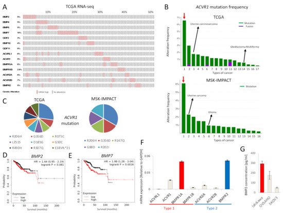
The expression of mRNA for BMP ligands and receptors was found to be frequently increased in EC, as revealed by analysis of the TCGA EC database (Figure 1A). In addition, ACVR1 mutations were more frequently observed in EC compared to other cancers, in both the TCGA and MSK-IMPACT datasets (Figure 1B). Around half of ACVR1 mutations were R206H and G356D (Figure 1C), gain-of-function mutations commonly found in fibrodysplasia ossificans progressiva (FOP) and diffuse intrinsic pontine gliomas (DIPGs) [5]. Moreover, high expression of BMP7 mRNA correlated with significantly lower survival of EC patients; the expression of BMP2 mRNA also showed a correlation, albeit not significant, with poor EC patient survival (Figure 1D,E). We also performed survival analyses of other BMP ligands and receptors (Figure S1). However, there was no correlation between ACVR1 mRNA expression and EC patient survival. To investigate the tumor promoting effect of BMP signaling in EC, we further performed in vitro experiments using Ishikawa EC cells, revealing expression of mRNA for all type I and type II BMP receptors, except ACVRL1 (Figure 1F). BMPR1A mRNA was most abundantly expressed among the type I receptors, whereas BMPR2 mRNA was most abundant among the type II receptors (Figure 1F). In addition, we found that Ishikawa cells secreted BMP2 at a higher level than OVSAHO and SKOV3 ovarian cancer cells, as determined by an ELISA (Figure 1G).
Figure 1. BMP signaling is activated in EC. (A) mRNAs for BMP ligands or receptors are over-expressed in EC. RNA-seq of the TCGA endometrial cancer dataset containing 177 EC tumors was analyzed via cBioPortal. RNA expression cutoff Z score was adjusted to 2.0. The results of 90 tumors are shown. (B) ACVR1 is more frequently mutated in EC compared to other cancers. ACVR1 mutation frequency of the TCGA pancancer atlas studies and of the MSK-IMPACT clinical sequencing cohort was assessed by cBioPortal. Red arrows point to EC. Lanes 1 to 17 (in the top panel); endometrial carcinoma, skin cutaneous melanoma, uterine carcinosarcoma, colorectal adenocarcinoma, bladder urothelial carcinoma, lung adenocarcinoma, mesothelioma, stomach adenocarcinoma, adrenocortical carcinoma, head and neck squamous cell carcinoma, ovarian serous cystadenocarcinoma, lung squamous cell carcinoma, liver hepatocellular carcinoma, glioblastoma multiforme, kidney renal clear cell carcinoma, brain lower grade glioma and breast invasive carcinoma, Lanes 1 to 16 (in the bottom panel); endometrial cancer, uterine sarcoma, melanoma, mesothelioma, cancer of unknown primary, glioma, esophagogastric cancer, colorectal cancer, hepatobiliary cancer, head and neck cancer, mature B-cell neoplasms, germ cell tumor, non-small cell lung cancer, renal cell carcinoma, bladder cancer and breast cancer. (C) Details of ACVR1 mutations found in EC are shown. Eleven cases of the TCGA dataset (out of 244 cases) and six cases of the MSK-IMPACT dataset (out of 113 cases) had the ACVR1 mutations indicated. (D,E) Overall survival was analyzed using RNA-Seq data of KM plotter, which contained 542 EC patients. Patients were divided into two groups, i.e., above or below median mRNA expression. The effects of expression of BMP2 (D) and BMP7 (E) on the survival of EC patients, are shown. (F) mRNA expression levels of BMP receptors in Ishikawa EC cells, as determined by qRT-PCR and normalized relative to GAPDH. (G) BMP2 secretion by Ishikawa cells, and by OVSAHO and SKOV3 ovarian cancer cells for comparison. Confluent cell cultures were incubated in serum-free medium for 24 h; thereafter, the conditioned medium was analyzed for BMP2 by an ELISA. BMP2 concentration was normalized to 1 mg total protein in lysates. The results in panel F and G are shown as the mean ± SE.

2.2. BMP2 Promotes EC Cell Stemness by c-KIT Induction

To determine whether Ishikawa cells responded to BMP stimulation, cells were stimulated by exogenous BMP2, and treated with or without LDN193189, a BMP type I receptor kinase inhibitor. BMP2 induced SMAD1/5/8 phosphorylation, which was inhibited in the presence of LDN193189 (Figure 2A). BMP2 also enhanced stemness of Ishikawa cells, as determined by sphere formation; LDN193189 inhibited the effect (Figure 2B). In accordance with this result, BMP2 increased the expressions of mRNA for the cancer stem cell markers CD44 and c-KIT (Figure 2C). To determine whether c-KIT directly modulated stemness, it was overexpressed in Ishikawa cells (Figure 2D). c-KIT overexpression promoted sphere formation (Figure 2E), thus c-KIT enhanced stemness in Ishikawa cells. Moreover, the importance of c-KIT for the BMP2-induced stemness was investigated by knocking down c-KIT by two different siRNAs in Ishikawa cells; the knock-down efficiencies of the siRNAs were determined by qPCR (Figure S2A). c-KIT knockdown neutralized BMP2-induced sphere formation (Figure 2F). In addition, inhibition of the tyrosine kinase activity of c-KIT by imatinib attenuated BMP2-induced sphere formation in Ishikawa cells (Figure 2G). These results suggest that BMP2 promotes EC stemness via c-KIT induction. To explore whether LDN193189 augments the growth inhibitory effects of carboplatin (CBDCA), a standard chemotherapeutic agent for EC patients, Ishikawa cells were treated with LDN193189 and CBDCA, alone and in combination. LDN193189 significantly potentiated the growth inhibitory effect of CBDCA, as determined by an MTS assay (Figure 2H). LDN193189 also augmented the inhibitory effect of CBDCA on sphere formation, in the absence or presence of BMP2 stimulation (Figure 2I, Figure S3).
Figure 2. BMP2 promotes EC cell stemness by c-KIT induction. (A) BMP2 stimulation induces SMAD1/5/8 phosphorylation in Ishikawa cells. Cells were treated in the absence (CT) or presence of 20 ng/mL BMP2 and 200 nM LDN193189 (LDN) for 24 h. α-tubulin was used as an internal control. (B) BMP2 induces stemness of Ishikawa cells, as determined by a sphere formation assay. Cells were cultured with stem cell medium containing 20 ng/mL BMP2 and 200 nM LDN in 96-well ultra-low attachment plates for eight days; thereafter, sphere numbers per well were counted using a microscope. Images of spheres are shown at the bottom of the graphs. Scale bar = 200 µm. (C) BMP2 induces expression of CD44 and c-KIT mRNA in Ishikawa cells. Cells were treated with PBS (CT), BMP2 (20 ng/mL) or LDN, alone or in combination, for 72 h. mRNA expression was determined by RT-PCR and is shown as fold change relative to control (CT). (D) c-KIT expression was quantified by immunoblots in Ishikawa cells 72 h after transfection with empty vector (CT) or c-KIT (KIT) plasmids. α-tubulin was used as an internal control. (E) Overexpression of c-KIT by transfection induces stemness of Ishikawa cells, as determined by a sphere formation assay. (F,G) BMP2-induced stemness of Ishikawa cells is dependent on c-Kit. Cells were transfected with siNC, siKIT-1, or siKIT-2 for 48 h; thereafter, cells were cultured for an additional eight days in the presence and absence of 20 ng/mL BMP2 (F), or incubated in the presence and absence of 20 ng/mL BMP2 and 10 µM imatinib (G). Cancer stemness was determined by the formation of spheres. (H,I) Ishikawa cells were incubated in the absence (CT) or presence of 200 nM LDN and 500 µM carboplatin (CBDCA). Cell proliferation was determined after 72 h by an MTS assay, and is expressed relative to CT (H), and stemness by a sphere formation assay after eight days (I). The results in panels B, C, E–I are shown as the mean ± SE. * p-value < 0.05, ** p-value < 0.01.

2.3. BMP2 Induces EMT of EC Cells

We investigated the effect of BMP signaling on downstream genes in Ishikawa cells, and found that BMP2 induced ID1, SNAIL and SLUG mRNA in a time-dependent manner (Figure 3A). ID1 induction was sustained till 72 h, whereas SNAIL and SLUG induction peaked rapidly at 2 h after BMP2 stimulation (Figure 3A). Since SNAIL and SLUG are EMT transcription factors, we investigated the expression of EMT markers by immunoblotting. The epithelial marker E-cadherin was suppressed by BMP2, whereas the mesenchymal markers N-cadherin and vimentin were enhanced in a time-dependent manner (Figure 3B); these effects were neutralized by treatment with LDN193189 (Figure 3C). E-cadherin attenuation and vimentin induction were also confirmed by immunofluorescent staining (Figure 3D). These results support the notion that BMP2 induces EMT in Ishikawa cells.
Figure 3. BMP2 induces EMT of EC cells. (A,B) Ishikawa cells were cultured in serum-free medium overnight and treated with 20 ng/mL BMP2 for the indicated time periods. Expression of SNAIL, SLUG, and ID1 mRNA was analyzed by qRT-PCR and normalized relative to 0 h (A), and expression E-cadherin, N-cadherin, vimentin and phospho-SMAD1/5/8 was analyzed by immunoblots, using α-tubulin as a loading control (B). (C,D) Ishikawa cells were incubated in the absence (CT) or presence of 20 ng/mL BMP2 and 200 nM LDN, in 1% FBS-containing medium for 48 h, and then subjected to immunoblotting (C) and immunofluorescent staining (D) for EMT markers. Scale bar = 10 µm. The results in panel A are shown as the mean ± SE.

2.4. BMP2 Enhances EC Cell Migration via SLUG Induction

As BMP2 induced EMT in Ishikawa cells (Figure 3), we investigated whether BMP2 also enhanced cell migration. Using a scratch assay, we observed that BMP2 enhanced Ishikawa cell migration, and that LDN193189 reversed the effect (Figure 4A). Since SNAIL and SLUG were induced after BMP2 stimulation of cells (Figure 3A), we knocked down SNAIL or SLUG by siRNAs. SNAIL knockdown had no effect on BMP2-induced cell migration (Figure 3B), whereas SLUG knockdown inhibited BMP2-induced cell migration (Figure 3C). The induction of SLUG by BMP2 and its suppression by siRNAs was confirmed by qPCR (Figure S2B). Moreover, SLUG knockdown reversed the suppression of E-cadherin induced by BMP2 and slightly attenuated the expressions of the mesenchymal markers N-cadherin and vimentin (Figure 3D). These results suggest that BMP2 enhances migration and EMT of EC cells in a SLUG-dependent manner.
Figure 4. BMP2 enhances EC cell migration via SLUG induction. (A) Migration of Ishikawa cells was evaluated by a scratch assay. Confluent cell cultures were scratched by a 10 µL pipette tip and incubated in the absence (CT) or presence of 20 ng/mL BMP2 and 200 nM LDN193189 (LDN) in 3% FBS-containing medium for 48 h. Cell motility was determined by measuring the gaps between the cell sheets at 0 and 48 h. Scale bar = 100 µm. (BD) BMP2-induced EC cell migration is dependent on SLUG, but not on SNAIL. Ishikawa cells transfected with siNC, siSNAIL (B), or siSLUG-1 or siSLUG-2 (C) for 48 h, were incubated in the absence (CT) and presence of 20 ng/mL BMP2 in 3% FBS-containing medium for an additional 48 h. Cell migration was analyzed by scratch assays (B,C), and expression of EMT markers was determined by immunoblotting (D). The results in panels A, B, and C are shown as the mean ± SE. * p-value < 0.05, ** p-value < 0.01.

2.5. TWSG1 Antagonizes BMP7 in EC Cells

We explored the possibility that BMP antagonists affected BMP signaling in Ishikawa cells. First, we assessed mRNA expressions of ten BMP antagonists in Ishikawa cells; TWSG1 mRNA was most abundantly expressed (Figure 5A). However, according to TNM plot, TWSG1 mRNA expression was significantly decreased in EC compared with normal endometrium (Figure 5B). Expression of the TWSG1 protein was detected both in cell lysates and cultured medium (Figure 5C). Exogenous TWSG1 suppressed BMP7-induced, but not BMP2-induced, SMAD1/5/8 phosphorylation (Figure 5D), and expressions of ID1 (Figure 5E) and SLUG (Figure 5F) mRNA. Consistent with these results, TWSG1 inhibited BMP7-induced, but not BMP2-induced, sphere formation (Figure 5G) and cell migration (Figure 5H) of Ishikawa cells. These results show that TWSG1 antagonizes BMP7 in EC cells.
Figure 5. TWSG1 antagonizes BMP7 in EC cells. (A) TWSG1 mRNA is abundantly expressed in Ishikawa cells. mRNA expression of ten BMP antagonists was determined by qRT-PCR and normalized relative to GAPDH. (B) TWSG1 mRNA expressions of EC and non-cancerous endometrium were analyzed by submitting a query to TNM plot with selection of RNA Seq data. (C) Ishikawa cells secrete TWSG1. TWSG1 protein was detected by immunoblotting in both total cell lysates (TCL) and cultured medium (Medium) of Ishikawa cells. (D) TWSG1 inhibits BMP7-induced SMAD1/5/8 phosphorylation. Ishikawa cells were cultured overnight in serum-free medium, and then treated with or without 20 ng/mL BMP2, 50 ng/mL BMP7, and 1000 ng/mL TWSG1 for 3 h. Cell lysates were subjected to immunoblotting for P-SMAD1/5/8 and SMAD1. (E,F) TWSG1 suppresses BMP7-induced ID1 and SLUG expression. RNA was extracted from Ishikawa cells cultured under the same conditions as (D). ID1 (E) and SLUG (F) mRNA expression was evaluated with qRT-PCR. mRNA expression was normalized relative to no stimulation. (G) TWSG1 decreases BMP7-enhanced sphere formation. Ishikawa cells were incubated with or without 20 ng/mL BMP2, 50 ng/mL BMP7 and 1000 ng/mL TWSG1 for eight days, where spheres were later counted using a microscope. (H) TWSG1 suppresses BMP7-induced EC cell migration. Ishikawa cell cultures were subjected to a scratch, after which cells were incubated in the absence or presence of 20 ng/mL BMP2, 50 ng/mL BMP7 and 1000 ng/mL TWSG1 in 3% FBS-containing medium; after 48 h, the widths of the scratches were determined. The results in panels A, E–H are shown as the mean ± SE. * p-value < 0.05, ** p-value < 0.01.

References

  1. Bray, F.; Ferlay, J.; Soerjomataram, I.; Siegel, R.L.; Torre, L.A.; Jemal, A. Global cancer statistics 2018: GLOBOCAN estimates of incidence and mortality worldwide for 36 cancers in 185 countries. CA Cancer J. Clin. 2018, 68, 394–424.
  2. Johnson, A.L.; Medina, H.N.; Schlumbrecht, M.P.; Reis, I.; Kobetz, E.N.; Pinheiro, P.S. The role of histology on endometrial cancer survival disparities in diverse Florida. PLoS ONE 2020, 15, e0236402.
  3. Gonzalez Bosquet, J.; Terstriep, S.A.; Cliby, W.A.; Brown-Jones, M.; Kaur, J.S.; Podratz, K.C.; Keeney, G.L. The impact of multi-modal therapy on survival for uterine carcinosarcomas. Gynecol. Oncol. 2010, 116, 419–423.
  4. de Jong, R.A.; Nijman, H.W.; Wijbrandi, T.F.; Reyners, A.K.; Boezen, H.M.; Hollema, H. Molecular markers and clinical behavior of uterine carcinosarcomas: Focus on the epithelial tumor component. Mod. Pathol. 2011, 24, 1368–1379.
  5. Han, H.J.; Jain, P.; Resnick, A.C. Shared ACVR1 mutations in FOP and DIPG: Opportunities and challenges in extending biological and clinical implications across rare diseases. Bone 2018, 109, 91–100.
More
Information
Subjects: Oncology
Contributor MDPI registered users' name will be linked to their SciProfiles pages. To register with us, please refer to https://encyclopedia.pub/register :
View Times: 363
Revisions: 3 times (View History)
Update Date: 26 Jul 2021
1000/1000