Pathophysiology of Hemorrhagic Shock: History
Subjects: Emergency Medicine
Contributors:

The pathophysiology of hemorrhagic shock involves a decrease in systemic oxygen delivery to a level less than what is required to maintain cellular function.

  • polytrauma
  • hemorrhage
  • Hemorrhagic Shock
  • Pathophysiology

1. Introduction

For the polytrauma patient, brain injury is the most common cause of early death followed by acute blood loss as the second most common cause of early death [1][2]. In the U.S., 150,000 people die each year due to injury and many of these deaths occur in relatively younger individuals, which causes an aggregate loss of productive life of over 3.3 million years [3]. This results in an annual cost to society of USD 2.34 billion in today’s dollars from lost wages and medical costs. In prospective studies that examine resuscitation after trauma the median time to hemorrhagic death is 2.0 to 2.6 h [4][5][6][7]. Hemorrhage is the most common cause of shock in the injured, and a substantial number of trauma patients will arrive at hospital with profound physiologic disturbances due to acute circulatory failure. Dr. Samuel D Gross, regarded as one of the most innovative and influential surgeons of the 19th century described shock simply as, “… a rude unhinging of the machinery of life”. Indeed, this remarkable characterization of hemorrhagic shock remains as informative today as certainly it was over 175 years ago [8].

The polytrauma victim with significant hemorrhage suffers a life-threatening acute reduction in oxygen delivery (DO 2) to tissue. DO 2 depends on both an adequate circulating blood volume representing sufficient oxygen carrying capacity, and effective cardiovascular function to maintain the circulation of blood to capillary beds in the periphery.

Furthermore, between 25% to 35% of hemorrhaging patients will develop a biochemically evident coagulopathy (trauma-induced coagulopathy; TIC) before arrival to the emergency department, which can manifest clinically as either hypercoagulable or hypocoagulable states, or both. In the polytrauma patient the presence of TIC is associated with higher transfusion requirements, increased I.C.U. and hospital length of stay (LOS), prolonged requirement for mechanical ventilation, an increase in the incidence of multiorgan dysfunction, and, most concerning of all, a threefold to fourfold higher rate of mortality [9][10][11][12][13]. TIC has deleterious effects independent of injury severity, level of shock, degree of acidosis or depth of hypothermia [14].

2. Pathophysiology of Hemorrhagic Sock

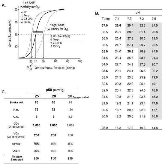
Oxygen Delivery/Utilization Imbalance

The pathophysiology of hemorrhagic shock involves a decrease in systemic DO2 to a level less than what is needed to maintain cellular function (VO2). DO2 equals the rate of blood flow, which is cardiac output (Q; normal = 5–6 L/min) multiplied by the content of oxygen bound to hemoglobin (Hgb) in a volume of blood (normal: male = 20.7 mL O2/dL; female = 18.4 mL O2/dL). A normal DO2 is approximately 1000 to 1250 mL O2/min in males, and approximately 925 to 1100 mL O2/min for females. If oxygen delivery is insufficient, tissue hypoxia develops resulting in anaerobic metabolism and production of lactate.
An important variable in oxygen transport physiology not often considered because it is seldom measured is the oxygen binding affinity of Hgb, expressed as p50 and depicted by oxy-hemoglobin dissociation (OHD) curves (Figure 1A–C). This property of Hgb determines the extent of peripheral oxygen offloading and therefore the quantity of oxygen available for tissue oxygenation. Acidosis shifts the OHD curve to the right (referred to as the Bohr effect) and increases the offloading of oxygen. Conversely, hypothermia shifts the curve to the left tends to decrease offloading of oxygen in the periphery. Acidosis and hypothermia are frequent homeostatic disturbances that complicate resuscitation. Depending on the magnitude of either one at any one moment during resuscitation, offloading of oxygen from Hgb may be enhanced or impeded [15]. These considerations may explain in part variability of responses to resuscitation of different patients. Additionally, of interest is the possibility of enhancing end-organ oxygen availability in patients with compromised oxygen transport by a pharmacological increase in p50 [16].
Figure 1. (A) OHD curve which relates the saturation of Hgb (y-axis) to the degree of partial pressure of oxygen to which Hgb is exposed (x-axis). The pO2 that saturates ½ of Hgb is referred to as p50, which in this example p50 = 27 mmHg. The p50 is the conventional measure of affinity of Hgb for oxygen. The lower the p50 the higher the affinity of Hgb for oxygen. The ‘steep’ portion of the oxyHgb dissociation curve is in the range of pO2 that exists in systemic capillaries (thus a small decrease in systemic capillary pO2 can result in the release of large amounts of oxygen for diffusion to, and uptake by cells). As shown in the figure, several factors increase the affinity of Hgb for oxygen (leftward shift; ↓p50) or decrease affinity (rightward shift; ↑p50). Biochemically, H+ is a heterotropic allosteric inhibitor of Hgb, whereas O2 is a homeotropic allosteric activator of Hgb. (B) Hypothermia and acidosis have opposing effects on p50. Lower temperature shifts the curve to the left increasing Hgb affinity for oxygen and decreasing offloading in capillaries; low pH (increase in H+) decreases the affinity of Hgb for oxygen (Bohr effect) increasing oxygen availability to reverse anaerobic metabolism. A trauma patient may be, and often is hypothermic and acidotic (and coagulopathic). Whether there is a significant change in p50 can be calculated using the Hill–Langmuir equation. (C): Hypothetical oxygen transport variables of a normal subject (Temp = 37 °C; p50 = 25 mmHg) and a subject with hypothermia (Temp = 31 °C; p50 = 20 mmHg), before and after compensation. The p50 at 31 °C and pH = 7.4 is calculated using the Hill–Langmuir equation. A venous blood gas is obtained through a Swan Catheter introducer (7.5Fr) with the tip in the superior vena cava reveals in the hypothermic subject, central venous oxygen saturation (ScvO2) = 85%. This reflects the fact that hypothermia increases the affinity of Hgb for oxygen, shifting the Hgb dissociation curve to the left. A ScvO2 of 85% would imply only 15% of the delivered 1000 mL of oxygen (DO2) prior to compensation is being offloaded, which is approximately 150 mL/min, well below VO2 (250 mL/min). The hypothermic patient can compensate by increasing cardiac output and hence DO2. Assume that stroke volume is unchanged (although a well-known consequence of tachycardia is a reduction in stroke volume), and cardiac output increases by an increase in heart rate (HR) from 72 beats/min to 120 beats/min (a 40% increase in HR causing a substantial increase in myocardial oxygen demand).
Systemic oxygen utilization (VO2), approximately 250 mL O2/min, is the amount of oxygen consumed each minute by all metabolic processes in the body. The physiologic relationship of VO2 to DO2 is expressed as the oxygen extraction ratio (O2ER),
O2DO2
VO2 and thus O2ER differ significantly among different organ systems. For example, extraction ratios measured in the in the heart, liver, and kidney, are 60%, 45% and 15% respectively. Predictably, a higher O2ER is associated with greater DO2 dependency.
O2ER provides an important compensatory mechanism offsetting reductions in DO2 due to acute blood loss and a decrease in cardiac output. An initial reduction in DO2 is offset by an increase in O2ER that maintains VO2 constant. In this hemodynamic state, the value of VO2 is flow-independent. As a compensatory mechanism for blood volume loss, O2ER-mediated flow-independence of VO2 may result in a deceptive clinical presentation of hemodynamic stability (compensated hemorrhagic shock), although as much as 30 percent of blood volume may have been lost. As cardiac output and thus DO2 continue to decline with ongoing hemorrhage, O2ER will increase until eventually the amount of oxygen that can be extracted plateaus (O2ER = 60–70% for most tissues). From this point, any further decrease in DO2 will cause VO2 to decline such that the value of VO2 is now flow-dependent. The value of DO2 that represents the boundary between flow-independent VO2 and flow-dependent VO2 is designated DO2 CRIT. Any DO2 < DO2 CRIT is associated with a decrease in VO2 and impaired oxygen-dependent cellular processes as metabolism shifts from aerobic to anaerobic pathways.
DO2 CRITmarks the onset of lactic acidosis and the beginning of an accumulating oxygen debt [17] (Figure 2). Without effective resuscitation, ongoing hemorrhage progresses to decompensated shock, characterized by hemodynamic instability and diminished blood flow that cannot maintain life-sustaining physiologic processes; and then to refractory shock, representing exhaustion of physiological reserves, hemodynamic collapse, vital organ dysfunction and subsequent failure, and ultimately, death.
Figure 2. DO2 CRIT defines shock. As DO2 (solid black line) decreases secondary to a fall in cardiac output, drop in Hgb concentration, or both, O2ER (solid grey line) increases to maintain VO2 constant until extraction is maximized. At this point, designated as DO2 CRIT (also referred to as the anaerobic threshold), VO2 begins to decrease with further decreases in DO2. When DO2 > DO2 CRIT t, VO2 is flow-independent; when DO2 < DO2 CRIT, VO2 becomes flow-dependent. In addition, DO2 CRIT is associated with the onset of lactate formation and accumulation. Thus, shock can be defined conceptually as the presence of DO2 less than DO2 CRIT, producing a reduction in VO2. Normal DO2 = 800 mL O2/min/m2; normal VO2 = 200 mL O2/min/m2; normal O2ER = 25%.
Therefore, a principal objective of care for the polytrauma patient in shock is to restore DO2 to a level (DO2 =350–450 mL O2/min/m2) such that, to a first approximation, DO2 > DO2 CRIT. However, targeting even higher, supranormal values for DO2 (DO2 > 600 mL O2/min/m2) with aggressive fluid administration predisposes to secondary complications of volume overload. Higher values of DO2 likely will not improve survival and, in fact, are associated with detrimental patient outcomes [18].
DO2 can be determined from the Hgb concentration, SaO2 and stroke volume (hence, cardiac output). Stroke volume can be obtained non-invasively, expeditiously, and to a reasonable degree of accuracy [19] by transthoracic echocardiographic measurement of blood flow velocity at the left ventricular outflow track [20][21][22]. VO2 can be estimated as 125 mL/min/m2 × BSA (BSA m2 = 0.007184 × (W)0.425 kg × (H)0.725 cm), determined by indirect calorimetry, or calculated using the Fick equation [23]. However, DO2 CRIT is not an exact transition point from flow-independent to flow-dependent VO2 [24] and varies considerably from one organ system to another. Moreover, direct point-of-care measurement of many critical parameters of oxygen transport generally are neither practical, nor feasible during resuscitation. Nevertheless, we believe familiarity with the physiology of oxygen delivery/utilization balance, and an appreciation for the meaning of O2ER and DO2 CRIT, establishes an important conceptual foundation that informs critical decisions typically required during resuscitation.

This entry is adapted from the peer-reviewed paper 10.3390/jcm10204793

References

  1. Demetriades, D.; Murray, J.; Martin, M.; Velmahos, G.; Salim, A.; Alo, K.; Rhee, P. Pedestrians injured by automobiles: Relationship of age to injury type and severity. J. Am. Coll. Surg. 2004, 199, 382–387.
  2. Moore, F.A.; Nelson, T.; McKinley, B.A.; Moore, E.E.; Nathens, A.B.; Rhee, P.; Puyana, J.C.; Beilman, G.J.; Cohn, S.M.; StO2 Study Group. Is there a role for aggressive use of fresh frozen plasma in massive transfusion of civilian trauma patients? Am. J. Surg. 2008, 196, 948–958; discussion 958–960.
  3. Cannon, J.W. Hemorrhagic Shock. N. Engl. J. Med. 2018, 378, 370–379.
  4. Tisherman, S.A.; Schmicker, R.H.; Brasel, K.J.; Bulger, E.M.; Kerby, J.D.; Minei, J.P.; Powell, J.L.; Reiff, D.A.; Rizoli, S.B.; Schreiber, M.A. Detailed description of all deaths in both the shock and traumatic brain injury hypertonic saline trials of the Resuscitation Outcomes Consortium. Ann. Surg. 2015, 261, 586–590.
  5. Hauser, C.J.; Boffard, K.; Dutton, R.; Bernard, G.R.; Croce, M.A.; Holcomb, J.B.; Leppaniemi, A.; Parr, M.; Vincent, J.L.; Tortella, B.J.; et al. Results of the CONTROL trial: Efficacy and safety of recombinant activated Factor VII in the management of refractory traumatic hemorrhage. J. Trauma 2010, 69, 489–500.
  6. Holcomb, J.B.; del Junco, D.J.; Fox, E.E.; Wade, C.E.; Cohen, M.J.; Schreiber, M.A.; Alarcon, L.H.; Bai, Y.; Brasel, K.J.; Bulger, E.M.; et al. The prospective, observational, multicenter, major trauma transfusion (PROMMTT) study: Comparative effectiveness of a time-varying treatment with competing risks. JAMA Surg. 2013, 148, 127–136.
  7. Fox, E.E.; Holcomb, J.B.; Wade, C.E.; Bulger, E.M.; Tilley, B.C.; Group, P.S. Earlier Endpoints are Required for Hemorrhagic Shock Trials Among Severely Injured Patients. Shock 2017, 47, 567–573.
  8. Mullins, R.J.; Trunkey, D.D. Samuel, D. Gross: Pioneer academic trauma surgeon of 19th century America. J. Trauma 1990, 30, 528–538.
  9. Maegele, M.; Lefering, R.; Yucel, N.; Tjardes, T.; Rixen, D.; Paffrath, T.; Simanski, C.; Neugebauer, E.; Bouillon, B.; AG Polytrauma of the German Trauma Society (DGU). Early coagulopathy in multiple injury: An analysis from the German Trauma Registry on 8724 patients. Injury 2007, 38, 298–304.
  10. MacLeod, J.B.; Lynn, M.; McKenney, M.G.; Cohn, S.M.; Murtha, M. Early coagulopathy predicts mortality in trauma. J. Trauma 2003, 55, 39–44.
  11. Niles, S.E.; McLaughlin, D.F.; Perkins, J.G.; Wade, C.E.; Li, Y.; Spinella, P.C.; Holcomb, J.B. Increased mortality associated with the early coagulopathy of trauma in combat casualties. J. Trauma 2008, 64, 1459–1463; discussion 1463–1465.
  12. Brohi, K.; Cohen, M.J.; Ganter, M.T.; Matthay, M.A.; Mackersie, R.C.; Pittet, J.F. Acute traumatic coagulopathy: Initiated by hypoperfusion: Modulated through the protein C pathway? Ann. Surg. 2007, 245, 812–818.
  13. Brohi, K.; Singh, J.; Heron, M.; Coats, T. Acute traumatic coagulopathy. J. Trauma 2003, 54, 1127–1130.
  14. Kutcher, M.E.; Howard, B.M.; Sperry, J.L.; Hubbard, A.E.; Decker, A.L.; Cuschieri, J.; Minei, J.P.; Moore, E.E.; Brownstein, B.H.; Maier, R.V.; et al. Evolving beyond the vicious triad: Differential mediation of traumatic coagulopathy by injury, shock, and resuscitation. J. Trauma Acute Care Surg. 2015, 78, 516–523.
  15. Ekeloef, N.P.; Eriksen, J.; Kancir, C.B. Evaluation of two methods to calculate p50 from a single blood sample. Acta Anaesthesiol. Scand. 2001, 45, 550–552.
  16. Srinivasan, A.J.; Morkane, C.; Martin, D.S.; Welsby, I.J. Should modulation of p50 be a therapeutic target in the critically ill? Expert Rev. Hematol. 2017, 10, 449–458.
  17. Leach, R.M.; Treacher, D.F. The pulmonary physician in critical care * 2: Oxygen delivery and consumption in the critically ill. Thorax 2002, 57, 170–177.
  18. Abdelsalam, M.; Cheifetz, I.M. Goal-directed therapy for severely hypoxic patients with acute respiratory distress syndrome: Permissive hypoxemia. Respir. Care 2010, 55, 1483–1490.
  19. Mercado, P.; Maizel, J.; Beyls, C.; Titeca-Beauport, D.; Joris, M.; Kontar, L.; Riviere, A.; Bonef, O.; Soupison, T.; Tribouilloy, C.; et al. Transthoracic echocardiography: An accurate and precise method for estimating cardiac output in the critically ill patient. Crit. Care 2017, 21, 136.
  20. Jozwiak, M.; Monnet, X.; Teboul, J.L. Monitoring: From cardiac output monitoring to echocardiography. Curr. Opin. Crit. Care 2015, 21, 395–401.
  21. Cecconi, M.; De Backer, D.; Antonelli, M.; Beale, R.; Bakker, J.; Hofer, C.; Jaeschke, R.; Mebazaa, A.; Pinsky, M.R.; Teboul, J.L.; et al. Consensus on circulatory shock and hemodynamic monitoring. Task force of the European Society of Intensive Care Medicine. Intensive Care Med. 2014, 40, 1795–1815.
  22. Antonelli, M.; Levy, M.; Andrews, P.J.; Chastre, J.; Hudson, L.D.; Manthous, C.; Meduri, G.U.; Moreno, R.P.; Putensen, C.; Stewart, T.; et al. Hemodynamic monitoring in shock and implications for management. International Consensus Conference, Paris, France, 27–28 April 2006. Intensive Care Med. 2007, 33, 575–590.
  23. Narang, N.; Thibodeau, J.T.; Levine, B.D.; Gore, M.O.; Ayers, C.R.; Lange, R.A.; Cigarroa, J.E.; Turer, A.T.; de Lemos, J.A.; McGuire, D.K. Inaccuracy of estimated resting oxygen uptake in the clinical setting. Circulation 2014, 129, 203–210.
  24. Lubarsky, D.A.; Smith, L.R.; Sladen, R.N.; Mault, J.R.; Reed, R.L., 2nd. Defining the relationship of oxygen delivery and consumption: Use of biologic system models. J. Surg. Res. 1995, 58, 503–508.
More