Subjects:
Virology
1. Introduction
Almost 120 years have passed since Walter Reed, James Carroll, Aristides Agramonte, and Jesse Lazear established that yellow fever is caused by a filterable infectious agent which is transmitted by the bite of a mosquito, then known as Stegomyia fasciata (Aedes aegypti). Lazear, who like his colleagues, had been stationed by the US Army in Cuba to study the disease, died of yellow fever in September 1900 after being exposed experimentally to mosquitos that had fed on sick patients. At about the same time in South Africa, James Spreull and Sir Arnold Theiler demonstrated that bluetongue disease of sheep is caused by an “ultravisible” agent that could be transmitted by the injection of an infected serum. Epidemiological evidence suggested that the agent was vector-borne, and it was subsequently shown by R.M. du Toit that the disease occurred in sheep inoculated experimentally with suspensions of wild-caught biting midges (Culicoides imicola). These and other seminal discoveries precipitated a century of research into vector-borne and zoonotic viral diseases, resulting in the discovery and isolation of many hundreds of novel viruses from insects or vertebrate hosts. Some were identified as important human or veterinary pathogens. Many other viruses were archived in reference collections, with only basic characterization of their biological or molecular properties. In recent years, the advent of next generation sequencing (NGS) has transformed this situation. Complete genome sequences are now available for many of the archived isolates, allowing more accurate taxonomic assignments, analysis of their phylogenetic and evolutionary relationships with other viruses, and evaluation of the potential risks they may present to humans and wild or domestic animal populations. NGS has also opened the door to viral metagenomics, which has greatly increased the pace of new virus discovery from a wide range of hosts, usually with complete or near-complete viral coding sequences, but no virus isolate and minimal biological data. This has presented both opportunities and challenges for virologists and epidemiologists, as well as viral taxonomists, evolutionary biologists, and bioinfomaticians. Sadly, this technological revolution has been accompanied by a period of progressive disinvestment in training in classical virology. In this review, we recall the rich history of the discovery of arboviruses and other zoonotic viruses in various settings around the world and the many outstanding scientists who have contributed to the endeavour. We also consider the impacts of NGS and metagenomic analysis, and the implications of these new technologies for the future of this important field of research.
2. History of Arbovirus Discovery
2.1. Rockefeller Foundation and Yale Arbovirus Research Unit (YARU)
The Rockefeller Foundation (RF) was organized in 1913 for “the well-being of mankind throughout the world” [1]. At the time, yellow fever was still epidemic in many tropical and subtropical regions of Africa and the Americas, so in 1916, the RF established the Yellow Fever Commission, with the lofty goal of eradicating yellow fever from the world. During the next 25 years, the RF supported an international group of scientists working on yellow fever in New York City in the United States (U.S.); Rio de Janeiro and Salvador in Brazil; Bogota, Colombia; Yabba, Nigeria; and Entebbe, Uganda [1,2]. Much of the work and accomplishments of RF-funded personnel during this period was described in Strode’s classic book, entitled “Yellow Fever” [3]. The major accomplishments included:
-
Confirmation of earlier work by the Reed Commission in Cuba, demonstrating that yellow fever was caused by a filterable agent, yellow fever virus (YFV), that was transmitted by the bite of infected mosquitoes, Aedes aegypti;
-
Discovery that the rhesus monkey and the white mouse are susceptible to infection with YFV, providing models for subsequent studies on the pathogenesis, transmission, epidemiology, and control of the disease;
-
Demonstration that convalescent sera of humans and animals infected with YFV neutralize the virus. This discovery led to the development of the mouse neutralization test, which allowed investigators to map the geographic distribution of the virus;
-
Discovery of the forest or sylvan cycle of YFV and the importance of mosquitoes other than Ae. aegypti in the transmission and maintenance of the virus;
-
Development of the 17D vaccine strain of YFV and its first human trials. Max Theiler, son of South African bluetongue researcher Sir Arnold Theiler, received a Nobel Prize in 1951 for this work.
As a by-product of the overseas yellow fever investigations, RF-funded researchers also isolated a number of other previously unknown arboviruses, including West Nile, Zika, Semliki Forest, Bunyamwera, Bwamba, Uganda S, Ilheus, and Anopheles A and B viruses [2].
The threat and onset of World War II changed the priorities of the RF, and most of its overseas YF research activities ceased during this period. Many of the former American RF staff became involved in studies of diseases of military importance, such as typhus, malaria, sandfly fever, and dengue, some as civilians and others as members of the U.S. Armed Forces.
In 1950, after the end of the war, the RF decided to develop a major new program to study arthropod-borne viruses and to discover “what might be out there”, as well as their disease associations, life cycles, and vectors. This led to the development of the Rockefeller Foundation Virus Program, a world-wide virus discovery program that was funded from 1951 to 1970 [1]. Over the next several years, field laboratories staffed by both RF and local scientists were established with foreign governments in Pune, India; Port of Spain, Trinidad; Belem, Brazil; Johannesburg, South Africa; Ibadan, Nigeria; Cali, Colombia; and Cairo, Egypt [2]. In Egypt, the Cairo laboratory was associated with the U.S. Naval Medical Research Unit No. 3. Scientists in these field laboratories were involved in the detection and investigation of human diseases in their respective geographic regions, surveying human and animal populations for serologic evidence of past viral infection, and searching for viruses in a wide variety of arthropods, mammals, birds, reptiles, and amphibians [2]. All virus isolations were done on site, using the classic technique of intracranial inoculation of newborn white mice, but later, as vertebrate cell cultures became available, inoculation of cell cultures was also used. In addition, sentinel animals, such as non-human primates, mice, and hamsters, were also used in the field to detect virus activity. Viruses isolated in the overseas laboratories were initially characterized locally, lyophilized for preservation and storage, and then aliquots of each new virus were shipped back to the RF central virus laboratory in New York for further characterization and study. It was during this period that Jordi Casals, Delphine Clarke, Loring Whitman, and others developed the sucrose-acetone method for preparation of viral antigens and began to adapt the hemagglutination inhibition (HI) and complement fixation (CF) tests to identify and group arboviruses [4,5,6].
The RF Virus Program was extremely productive and many novel arboviruses, as well as non-arthropod-borne viruses (i.e., hantaviruses and arenaviruses), were discovered by RF-funded investigators during this period. The productivity of this search strategy in detecting novel viruses affecting humans was recently reviewed by Rosenberg et al. [7]. The virus discovery rate was highest in the period 1950–1969, which coincided with the RF Virus Program. A similar approach was also practiced by the Institut Pasteur and other international groups involved in virus discovery during this period, as described in this article and in other publications [8,9,10,11,12,13], resulting in the detection of a high proportion of pathogenic arboviruses. The second advantage of the strategy was that it yielded actual virus isolates, whose pathogenesis could be studied experimentally in vivo and in vitro in vertebrate and arthropod models. In contrast, current search methods for new viruses, which generally use metagenomics and other sophisticated genetic techniques to detect novel viral agents, do not usually yield live viruses, only their nucleotide sequences. A complete or partial genetic sequence alone rarely provides insight into the epidemiology and ecology, host range, pathogenesis and disease potential, or transmission modes of the new viruses.
In 1960, the RF made a decision to phase-out its Virus Program and to devote more of its efforts and resources to other projects, such as population control and increasing food production (“the green revolution”). Over the next 10 years, the RF-funded investigators working in overseas laboratories were withdrawn and the respective laboratories were turned over to local institutions or governments. In 1964, the RF made arrangements with Yale University to transfer its Arbovirus Group to New Haven. Accordingly, the Virus Laboratory in New York City was closed, and most of the remaining investigators (Casals, Clarke, Downs, Theiler, Buckley, Shope, Aitken, etc.), their equipment, virus stocks, and reagents were moved to New Haven, with a small endowment to temporarily support them. The newly established group was designated the Yale Arbovirus Research Unit (YARU) and was housed in the newly constructed Laboratory of Epidemiology and Public Health building on the grounds of the Yale University School of Medicine. Upon arrival, the former RF staff members in turn became members of the Yale faculty [1,2]. Wilbur Downs was designated as the first Director of YARU, and in 1974 when Downs retired, Robert E. Shope became the Director.
The YARU was subsequently designated by WHO as an International Reference Center for Arboviruses, and for a number of years, it received virus samples from investigators worldwide for identification and confirmation [14]. Under Shope’s leadership, the World Reference Center for Arboviruses (WRCA) was established. Initially, it consisted of the original viruses (known and unknown) that came along with the staff from the former RF Virus Laboratory in New York, but over the years, as new viruses were submitted for study or were isolated by YARU investigators, they were also added to the WRCA collection [14]. As the former RF personnel retired, new researchers were recruited to YARU as full Yale University faculty [1]. Because of its prestige and university affiliation, many visiting scientists, graduate students, and other trainees from the U.S. and a variety of other countries came to YARU during this period to learn arboviral techniques, to do original research, and to identify samples collected by them during outbreaks or field studies. These samples were also added to the WRCA collection. Many of these visitors, as well as some of the younger YARU faculty, eventually moved on to assume prominent positions in academia, government service, or the corporate world. Shope [14] and Downs [1] have described some of the significant activities, accomplishments, and international collaborations during the early years of YARU.
Without continued support from the RF or from Yale University, the YARU staff, like other university scientists in the U.S., had to compete for grants and contracts to support their research. This eventually changed the nature of YARU’s research activities from international outbreak investigation, diagnostic support, and training to investigator-initiated, hypothesis-driven grant research or contract activities. Meanwhile, Shope and other YARU staff members continued to serve as consultants to U.S. Government agencies [National Institutes of Health (NIH), National Science Foundation (NSF), United States Department of Agriculture (USDA), Department of Defense (DOD), United States Agency for International Development (USAID), and the Centers for Disease Control and Prevention (CDC)], international organizations [World Health Organization (WHO), Pan American Health Organization (PAHO), and Food and Agriculture Organization of the United Nations (FAO)], and foreign governments. In 1992, Shope was a co-editor of the Institute of Medicine’s seminal publication, Emerging Infections: Microbial Threats to Health in the United States, which highlighted the threat that emerging infections pose for the U.S. and global public health [15]. Shope was also an early proponent of the effect of ecological and global climate changes on arboviral and other vector-borne diseases [15,16], and he often served on national and international committees addressing this problem.
Consultancies, service on government committees and free diagnostic laboratory support (the old RF model) could not provide sufficient funds to maintain and recruit new personnel, upgrade equipment, or support a major research program at a university in the U.S. In turn, leaders at Yale Medical School decided that arbovirology was passé and that other areas, such as HIV/AIDS, environmental health, and molecular biology, offered better funding and research opportunities for the 1980s. Thus, YARU gradually lost university support, faculty positions, and space. In 1995, after 30 years at Yale, Shope retired. Along with his colleague Robert Tesh, he moved to the newly established Center for Tropical Diseases at the University of Texas Medical Branch in Galveston [17]. The extensive WRCA virus and reagent collections went with them. Their departure and the loss of the WRCA collections was soon followed by the departure of other, younger, faculty associates, resulting in the eventual demise of YARU as a center for arbovirus research and discovery.
2.2. Pasteur Institutes and the French Biomedical Research Network in West Africa
Since the establishment of the Institut Pasteur in Paris in 1888, Louis Pasteur sent collaborators to various countries, mainly in the French colonies of Indochina and Africa. In that early time, Pasteur wanted to set up rabies centers where the disease was highly and dramatically prevalent; naturally, the research potential of these centers was rapidly extended to tropical infectious diseases [18]. Currently, spread among 25 countries on five continents, there are 22 institutions, 19 of which bear the name of “Institut Pasteur” (IP). Altogether, these institutions constitute a structure long called the "Institut Pasteur d’Outre-mer", which in 1988 became the International Network of Pasteur Institutes (Réseau International de l’Institut Pasteur, RIIP) and Associated Institutes. From this emerging network, the first African laboratory for microbiology was created in Saint Louis, Senegal in 1896, and then transferred to Dakar to become the Pasteur Institute of Dakar (Institut Pasteur de Dakar, IPD).
Another French multidisciplinary worldwide institution, the French Institute of Research for Development (Office de la Recherche Scientifique et Technique Outre-Mer, ORSTOM) joined the RIIP’s efforts in the intertropical zone of Africa in its fight against infectious diseases, by bringing expertise on medical entomology and vector transmitted diseases. ORSTOM was created in 1946 from a previously existing Office of Colonial Research (Office de la Recherche Scientifique Coloniale, ORSC), established by Charles de Gaulle in 1943.
Yellow fever was to play a key role in the research at IPD. In the 1930s, IPD had developed a vaccine against yellow fever (strain FNV) and produced it commercially. The re-emergence of yellow fever in Africa in the 1960s occurred mainly in the savannah zone and revealed the lack of knowledge about the natural maintenance of the virus during the inter-epidemic period. A large study to understand the emergence and re-emergence of sylvatic yellow fever was established between the IPD, Institute Pasteur Abidjan (IPA) in Côte d’Ivoire, and Institute Pasteur Bangui (IPB) in the Central African Republic, in order to detect the circulation of the YFV and map its emergence in the different ecozones. Permanent research stations were developed in rain forests and savannas to detect virus circulation in vectors and hosts from the tree canopies to the savanna ground. Year-round mosquito and monkey sampling over a period of more than 10 years made possible the detection of YFV in various monkey and mosquito species (e.g., Aedes africanus, Aedes opok, Aedes furcifer-taylori, and Aedes luteocephalus), thus elucidating the mechanism of YFV maintenance in nature. Epidemiological observations during this period allowed Max Germain to formulate the concept of a “yellow fever zone of emergence” in West and Central Africa [19]. These ecological transition zones constitute ecotones adjacent to sylvatic environments, where prevailing ecological conditions, such as vector abundance, presence of non-human primates, and closeness for human contact, enable the cross-species transmission of viruses into humans—a “zone of emergence”, which clearly appeared as the main source of epidemics in West Africa [20]. Thus, this specific ecosystem constitutes an ideal transition ecotype, where vaccination campaigns for the containment of epizootics and ultimately eradication ought to concentrate [21]. Lastly, virus isolation in male mosquitoes (Ae. furcifer-taylori) allowed the documentation of vertical transmission, allowing for virus maintenance in the inter-epidemic periods [22].
In the early 1960s, research laboratories focusing broadly on arboviruses were established under the umbrella of the Collaborating Center for Reference and Research on Arboviruses (CRORA), led by Paul Brés. CRORA laboratories were created at various IPs throughout Africa, including IPD, IPA, and IPB, as well as IP Yaoundé (Cameroon), and IP Tananarive (Madagascar). One of the major activities was to establish an inventory of arboviruses circulating in various ecosystems. Although virus isolations were made at various IPs, virus characterization and identification were carried out at the CRORA reference center at IPD, followed by confirmation at YARU, with final registration in the International Arbovirus Catalogue [23].
At the end of the 1970s, following an outbreak of the Ebola epidemic in Zaire in 1976, IPB set up a research program on viral hemorrhagic fevers that lasted until 1983 [24]. This research program was the result of an important collaboration between the various Pasteur Institutes in Africa and researchers of ORSTOM. In 1977, ORSTOM extended its field of research and expertise to tropical medical virology, thus supporting teams from the RIIP. The partnership between RIIP and OSTROM was instrumental in expanding the scope of field research in Africa, with seminal studies on the vertical transmission of arboviruses in mosquitoes [25] or the role of environmental factors, such as climate and latitude, in arbovirus transmission from arthropod to vertebrate hosts, using YFV [26,27] and dengue virus (DENV) [28,29,30] as models.
Research at IPB was also critical in elucidating the etiology of exanthematous fevers, coined “Congolese red fevers”, that have long been attributed to rickettsia. A total of 16 arboviruses (chikungunya, Igbo-Ora, O’Nyong Nyong, Sindbis, Bouboui, yellow fever, Wesselsbron, West Nile, Zika, Ilesha, Bwamba, Dugbe, Tataguine, Nyando, Bangui, and Rift Valley fever viruses) were associated with these syndromes. Symptoms, consisting of fever, diffuse pain, and exanthem, were present in more than 60% of the cases, with etiology dominated by four viruses—Chikungunya, Ilesha, Bwamba, and Tataguine [31,32]. Between 1976 and 1986, two field research stations were established in the Central African Republic, one located adjacent to the forest (Bouboui) and the other in the wooded savanna (Bozo) (Figure 1). During this period, more than 420,000 mosquitoes of identified species in 14,591 pools were preserved for virus isolation. The most common anthropophilic mosquitoes caught were Aedes (stegomiya) africanus and Ae. (st.) opok, inside the forest gallery, Aedes vittatus, in the savannah, and Anopheles gambiae and An. Funestus, in the houses of the village of Bozo. A total of 321 viruses were isolated and assigned to 24 different species. These included chikungunya, Bagaza, Bouboui, Bozo, Bwamba, Ilesha, Kamese, Kedougou, Middleburg, Mossuril, M’Poko, Nyando, Orungo, Pata, Pongola, Simbu, Sindbis, Tataguine, Wesselsbron, West Nile, yellow fever, and Zika viruses. Altogether, six arboviruses were found in the forest gallery, including Bouboui, Bozo, chikungunya, Orungo, yellow fever, and Zika viruses, vectored primarily by Ae. africanus.
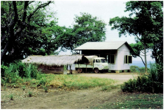
Figure 1. The field station in Bozo, Central African Republic.
Research at IPD and IPB was also instrumental in demonstrating the expanded range of the Crimean–Congo hemorrhagic fever virus (CCHV) in West and Central Africa [33,34,35], followed by repeated isolation of the virus. This allowed a clear understanding of its eco-epidemiology in the region, including north–south migration of the infected ticks through the cattle traffic migration patterns in the Sahel [34,36]. Likewise, active circulation of the Rift Valley fever virus (RVFV) was also demonstrated in Senegal [37], Mauritania [35], Upper Volta (present day Burkina Faso) [35,38], and the Central African Republic [39].
Support for RIIP laboratories located in Africa was provided by the IPP and OSTROM teams, as well as through collaboration with various research centers in the U.S., such as Harvard University, supporting the initial study on yellow fever and FNV vaccine; YARU, as a partner for new arbovirus classification; the Centers for Diseases control (CDC) at Fort Collins Colorado, for the study of arboviruses; the CDC in Atlanta and the U.S. Army Medical Research Institute of Infectious Diseases (USAMRIID), for the initiation of viral hemorrhagic fever research in Central and West Africa.
2.3. Australia
The era of virus discovery in Australia can be traced to the summer of 1950–1951, when a major epidemic of encephalitis swept through southeastern Australia. There were clinical cases reported in Victoria, New South Wales, and South Australia, of which 19 (42%) were fatal [40]. A similar epidemic of unknown etiology (named Australian X disease) had occurred in eastern Australia from 1916 until 1925, with almost 300 reported cases and an average case/fatality rate of 68% [40,41]. Surprisingly, no further cases were reported in the intervening 25 years. Amongst those investigating the 1950–1951 epidemic were John A.R. Miles and colleagues at the Institute of Medical and Veterinary Science in Adelaide, and Eric L. French of the Walter and Elisa Hall Institute of Medical Research in Melbourne who, almost simultaneously, reported the isolation of a virus from the brain tissue of clinical cases [42,43]. The virus, named Murray Valley encephalitis virus (MVEV), was shown to be closely antigenically related to Japanese encephalitis virus (JEV), a flavivirus (then designated group B arbovirus) known to cause fatal encephalitis in East and Southeast Asia [43].
The subsequent development of arbovirology and the pathway to virus discovery in Australia were linked intimately with the 1947 establishment of the Queensland Institute of Medical Research (now the QIMR-Berghofer Institute) at Herston in Brisbane (Figure 2). Ralph L. Doherty joined the staff and, in 1957, established a program of virus isolation from mosquitoes, based at a field station at Innisfail in the far north Queensland. In 1959, Doherty isolated the Ross River virus (RRV) from Aedes vigilax mosquitoes collected in Townsville [44]. He subsequently showed that RRV neutralizing antibodies occurred commonly in human sera in eastern Australia [45]. He also showed that individuals suffering from a severe debilitating syndrome, known as epidemic polyarthritis, had high antibody titres to RRV, suggesting a causal relationship [46,47]. In 1971, Doherty and his colleagues isolated the virus from a boy from the Edward River Mission aboriginal settlement in Cape York [48]. RRV and the related alphavirus, the Barmah Forest virus (see below), are now recognized as important public health problems in much of Australia and the Pacific Islands, causing arthritis, myalgia, and fatigue for six months or longer. Several thousand cases of the disease are notified annually [49].
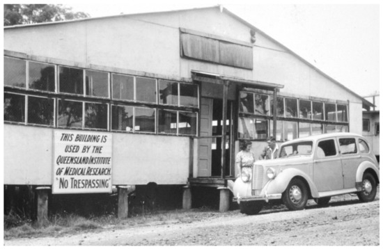
Figure 2. The building in Brisbane that was occupied by the Queensland Institute of Medical Research (QIMR) from 1947 until 1979 (provided with permission from QIMR).
Doherty’s program of virus discovery continued until 1977 with a team that included several other notable scientists, including Harry A. Standfast, Brian H. Kay, Edwin G. Westaway, Barry M. Gorman, and John G. Aaskov. During 1960 and 1961, Doherty isolated 60 strains of 11 viruses from 25,901 mosquitoes of 32 species from Kowanyama (then the Mitchell River Mission aboriginal settlement), Normanton, and Cairns in far north Queensland. These included four novel flaviviruses (Kunjin, Kokobera, Edge Hill, and Stratford), three orthobunyaviruses (Koongol, Wongal, and Maputta) and one orbivirus (Corriparta) [45]. The study also identified two alphaviruses previously unknown in Australia (Sindbis and Getah) and the first isolations of MVEV from mosquitoes [45]. With support from the Rockefeller Foundation, a field station was established at Kowanyama and further expeditions were undertaken to collect arthropods and potential mammalian hosts throughout Queensland. Anopheline mosquitoes and a swamp pheasant (Centropus phasianinus) collected at Kowanyama from 1963 to 1966 yielded three novel viruses (Kowanyama, Trubanaman, and Alfuy viruses) [50]. In 1969–1970, three novel viruses were isolated at Kowanyama (Wongorr and Mitchell River viruses from mosquitoes and the Ngaingan virus from biting midges), three novel viruses were isolated from mosquitoes collected near Charleville in western Queensland (Charleville, Warrego, and Wallal viruses) and two viruses (Belmont and D’Aguilar viruses) were isolated from mosquitoes and biting midges, respectively, collected near Brisbane [51,52]. Further expeditions to Charleville to collect mosquitoes resulted in the isolation of two novel viruses in 1974 (Facey’s Paddock and Murweh viruses) and two novel viruses in 1976 (Parker’s Farm and Little Sussex viruses) [53]. Leanyer virus was also isolated in 1974 from mosquitoes collected near Darwin in the Northern Territory [54]. Viruses were also isolated from wildlife hosts; the Almpiwar virus was isolated from a skink (Cryptoblepharus virgatus) at Kowanyama in 1966 [55] and the Mossman virus was first isolated from a rodent (Rattus leucopus) captured near Mossman in 1970 [56].
In collaboration with Doherty and his QIMR team, expeditions were also conducted to isolate viruses from ticks associated with sea birds. In 1966, Harald N. Johnson from YARU collected soft ticks (Ornithodoros capensis) from sooty tern (Onychoprion fuscatus) colonies on the Great Barrier Reef near Cairns, from which two viruses (Upolu and Johnston Atoll viruses) were isolated [57]. The Saumarez Reef virus was subsequently isolated by Toby D. St. George and colleagues from Australia’s Commonwealth Scientific and Industrial Research Organization (CSIRO) in 1974, from the same species of ticks associated with sooty terns on a coral cay in the southern Coral Sea [58]. In 1972, M. Durno Murray from CSIRO undertook an expedition to the Australian territory of Macquarie Island in the Southern Ocean to collect hard ticks (Ixodes uriae) associated with sea birds, resulting in the isolation of two novel viruses (Nugget and Taggert viruses) [59]. A second CSIRO expedition to Macquarie Island in 1975 yielded two additional novel viruses (Gadget’s Gully and Precarious Point viruses) from hard ticks collected in royal penguin (Eudyptes chrysolophus schlegeli) rookeries [60].
Other research groups in Australia also joined the hunt for arboviruses during the late 1960s and early 1970s, including Ian D. Marshall at the Australian National University in Canberra and Neville F. Stanley at the University of Western Australia. From 1965 to 1975, Marshall and his colleagues conducted surveys for arbovirus activity, particularly RRV and MVEV, in coastal regions of New South Wales and in the Murray River Valley. In addition to these and other known arboviruses, Marshall and colleagues isolated several novel arboviruses from mosquitoes, including Gan Gan, Yacaaba, Tilligerry and Termeil, Paroo River, Picola, and Barmah Forest viruses [61,62] and a novel reovirus, Nelson Bay virus, from a fruit bat (Pteropus poliocephalus) [63]. Like the related alphavirus RRV, the Barmah Forest virus was subsequently shown to be a cause of epidemic polyarthritis in humans [64,65], with infections occurring commonly throughout Australia [66]. The Gan Gan virus also infects humans and is suspected of an association with epidemic polyarthritis [67]. Marshall also conducted a number of collecting trips to Papua New Guinea from 1965 to 1975 funded by the Rockefeller Foundation. During an expedition to the Sepik River District of Papua New Guinea in 1965–1966, he isolated the Joinjakaka virus from a mixed pool of culicine mosquitoes and the Japanaut virus from both culicine mosquitoes and a fruit bat (Syconycteris crassa). In Western Australia, Stanley and colleagues surveyed for arbovirus activity in the Ord River Valley from 1972 to 1976 [68,69]. From 52,000 mosquitoes of 20 species, 195 virus isolates were recovered, including 28 isolates of MVEV and 20 isolates of the Kunjin virus from Cx. annulirostris, suggesting the region may be an endemic focus in Australia [69]. The study also identified eight novel viruses, including Kimberly, Parry’s Creek, Ord River, and Kunnanurra viruses, as well as four unknown isolates (OR379, OR512, OR869, and OR540), which have yet to be characterized [69,70]. Continuing surveillance in Western Australia by others has continued to reveal novel arboviruses, including Oak Vale, Stretch Lagoon, Parry’s Lagoon, and Fitzroy River viruses [71,72,73,74].
In 1968, CSIRO established a new virology laboratory at Long Pocket in Brisbane, headed by Toby D. St. George, to investigate endemic diseases of livestock in northern Australia. In 1967, Doherty and colleagues had isolated bovine ephemeral fever virus (BEFV) from cattle during a major epizootic in Queensland [75] but, despite epidemiological evidence suggesting vector-borne transmission, the virus had never been isolated from insects. This led St. George and colleagues to attempt virus isolations from a location in northern Australia, where serological monitoring of a sentinel herd of cattle indicated that BEFV was likely to be enzootic. For a continuous period from October 1974 until May 1976, insect collections for virus isolation were conducted at Beatrice Hill southeast of Darwin. From the 57,596 mosquitoes and 175,880 biting midges processed, one isolate of BEFV was recovered (from a mosquito pool). However, the collection also yielded 93 other virus isolates from 22 different serological groups, including four novel viruses (CSIRO Village, Marrakai, Beatrice Hill, and Humpty Doo virus) [76]. Most significantly, the collection also yielded a single isolate of a novel serotype of bluetongue virus (BTV serotype 20, BTV-20), a major pathogen of sheep and goats that had previously been regarded as exotic to Australia [77].
The isolation of BTV-20 and the consequences for international trade dramatically changed the landscape with respect to virus discovery and characterization in Australia. An immediate consequence was the approval of government expenditure for the establishment of the $230 million CSIRO Australian Animal Health Laboratory (AAHL) in Geelong, Victoria, providing high-level biosecure containment for laboratory work and live animal studies. The discovery also led to the establishment of a permanent veterinary virology capability at the Berrimah Veterinary Laboratory in Darwin under Geoff P. Gard, and a national program for serological monitoring of sentinel cattle herds and the collection of insect vectors. Efforts to isolate viruses from arthropods and livestock intensified. In the Northern Territory, a second novel serotype of bluetongue virus (BTV-21) was isolated from a healthy sentinel cow at Victoria River Station in 1979 [78], and four other novel arboviruses (Coastal Plains, Berrimah, Adelaide River, and Koolpinyah viruses) were isolated from healthy cattle between 1981 and 1985 [79,80,81,82]. In Queensland, eight novel arboviruses were first isolated between 1976 and 1981 from biting midges (Tibrogargan, Tinaroo, Peaton, Wongabel, and Walkabout Creek viruses), healthy sentinel cattle (the Douglas virus), and soft ticks (Argas robertsi) (Vinegar Hill and Lake Clarendon viruses) [55,83,84,85,86,87]. Surveillance activities by the Berrimah Veterinary Laboratory have continued to the present, with regular reports of the isolation of novel arboviruses. Continuing surveillance by others in northern Australia has also resulted in the isolation from mosquitoes of the Bamaga virus and New Mapoon virus from Cape York [88,89].
The 1990s also saw several significant disease emergence events in Australia, which drew particular attention to bats as reservoir hosts of highly pathogenic viruses. In September 1994, an outbreak of a severe respiratory disease occurred in horses at a stable in Brisbane. Of the 21 affected horses 14 were euthanized or died of the disease. One of two severely affected humans who had contact with the horses also died. Cooperation between the Queensland Government, the newly established CSIRO Australian Animal Health Laboratory, and others resulted in rapid isolation of the Hendra virus, a novel paramyxovirus [90], and the identification of fruit bats as reservoir hosts [91,92]. The Hendra virus has since re-emerged regularly in Australia, with more than 70 confirmed cases in horses and seven infected humans, four of whom have died. In 1996, an injured female fruit bat (Pteropus alecto) was found at Ballina in New South Wales. Tissue homogenates from the euthanized bat were injected into mice, resulting in the isolation of a novel lyssavirus, subsequently named Australian bat lyssavirus (ABLV) [93]. Three fatal human cases of ABLV infection have subsequently been reported [94,95,96]. The virus is now known to occur at low prevalence in five of six families of bats endemic to the Northern Territory, Queensland, and Western Australia [97]. In April 1997, another novel paramyxovirus, the Menangle virus, emerged at a commercial piggery in New South Wales, causing stillbirths with abnormalities of the brain, spinal cord, and skeleton [98]. Two humans exposed to the pigs also developed an influenza-like illness [99]. Fruit bats were again implicated as reservoir hosts [100]. The role of bats in the ecology and emergence of pathogenic viruses has been a major focus of study in Australia since that time, primarily involving research teams led by Hume E. Filed and Linfa Wang.
In all, more than 80 novel RNA viruses representing 9 families and 16 genera have been isolated from humans, livestock, wildlife, and arthropods in Australia and Papua New Guinea, and reported in the literature (Table S1). Many others have been isolated but remain uncharacterized and/or unreported. More complete characterization of these viruses will be facilitated greatly by the use of NGS.
2.4. South and Southeast Asia
South and Southeast Asia have also been a fertile area for virus discovery, particularly novel mosquito-borne and tick-borne flaviviruses and viruses with reservoirs in bat species. Early studies in India, partly funded by the Rockefeller Foundation, by Telford Work and his Indian colleagues from the Virus Research Centre in Pune, including D.P. Murthy, P.N. Bhatt, H. Trapido and K. Pavri, led to the discovery of the Kyasanur Forest disease (KFD) virus [101]. The virus was isolated from sera and tissues collected from a moribund black-faced langur (Presbytis entellus). This followed reports of an epizootic of unknown etiology causing large numbers of deaths in non-human primates and a number of cases of severe febrile illness in villages close to forested areas where dead monkeys had been found. The virus was shown to be closely related to the Russian spring–summer encephalitis (RSSE) serocomplex of flaviviruses, now known as the tick-borne encephalitis (TBE) serocomplex of flaviviruses. The virus was also isolated from some larvae and nymphs of Hemaphysalis spinigera ticks [102]. KFD in humans followed a biphasic course, not unlike TBE, but with some hemorrhagic manifestations not seen in TBE, and without either meningitis or encephalitis. Another tick-borne virus related to the RSSE serocomplex, Langat virus, had been isolated two years earlier from a pool of hard ticks, Ixodes granualtus, collected from forest rats caught near Kuala Lumpur, Malaysia, by C.E. Gordon Smith, then working at the Institute for Medical Research in Kuala Lumpur [103], but it is not known to be a human pathogen.
A number of mosquito-borne flaviviruses were first isolated in Southeast Asia. The most important with respect to human disease are three of the four dengue serotypes. Although dengue serotype 1 had been first isolated independently by Hotta in Japan in 1943 [104], and shortly after by Sabin in Cincinnati in 1945 with material collected in Hawaii [105], the other three serotypes were first isolated from material collected in Southeast Asia. Dengue serotype 2 was also isolated by Sabin in 1945 from material obtained from New Guinea [105] and dengue serotypes 3 and 4 were first isolated in 1956 from human sera and Aedes aegypti and Culex tritaeniorhynchusmosquitoes collected during a major outbreak of epidemic hemorrhagic fever in Manila, Philippines, by W.M. Hammon, A. Rudnick, and colleagues at the University of Pittsburgh [106]. Other novel flaviviruses have been isolated in Malaysia, Thailand, and Papua New Guinea [107]. The Tembusu (TMUV) virus was isolated in Kuala Lumpur in 1957 from various mosquito species [108] and was the first of several closely related viruses, including the ThCAr virus, which was isolated from a pool of Cx. tritaeniorhynchus mosquitoes collected in Chiang Mai, Thailand, in 1992 [109]; the Sitiawan virus, from sick broiler chicks in Malaysia [110]; and the duck Tembusu virus, an infection of ducks and geese causing an egg-drop syndrome in China and Southeast Asia [111,112]. Neutralising antibodies were found in humans in Malaysia [113] but the virus has not been implicated in human disease. Two other mosquito-borne flaviviruses have been described, Jugra virus and Sepik virus. Little is known about the Jugra virus, which was isolated from Aedes spp. and Uranotaenaia spp. mosquitoes and from the blood of a Cynopterus brachyotis fruit bat [108]. The Sepik virus was isolated in 1966 by Ian D. Marshall and colleagues from a pool of Mansonia septempunctata mosquitoes collected in the Sepik District of Papua New Guinea [114]. It was associated with a hospitalized case of febrile illness of unknown origin, with rising neutralising antibody to the virus. The Sepik virus is particularly interesting, as its nucleotide sequence analysis shows it to be the closest known flavivirus to yellow fever virus [115]. A novel lineage of the West Nile virus was isolated in Sarawak, East Malaysia, by D.I.H. Simpson, E.T.W. Bowen, and colleagues, from Cx. pseudovishnui group mosquitoes [116]. Initially called Kunjin virus, it was shown to differ significantly in genomic sequence from the Australian Kunjin viruses, which have been shown to comprise West Nile lineage 1b viruses, and have been described as West Nile lineage 6 virus [107].
Two flaviviruses with no known vector have been isolated in Southeast Asia, both from Cy. brachyotis fruit bats: the Carey Island virus was isolated from a bat in the Jugra Forest, Malaysia, in 1970 by A. Rudnick and colleagues from the Institute of Medical Research, Kuala Lumpur and the International Center for Medical Research, University of California [108], and the Phnom Penh virus was isolated by J.J. Salaun and colleagues in 1969 from the salivary glands and brown of bats [117]. A closely related virus, the Batu Cave virus, is considered to be a variant of Phnom Penh virus.
A considerable number of novel bunyaviruses have been isolated from South and Southeast Asia, particularly by scientists from the National Institute of Virology (formerly the Virus Research Centre) in Pune, including P.N. Bhatt, K. Pavri, K.R. Singh, C.N. Dandawate, F.M. Rodrigues, D.T. Mourya, P.D. Yadev, A.C. Mishra, and many others. Recently reviewed in [118], these viruses include the orthobunyaviruses, Umbre, Kaikalur, Thimiri, and Sathuperi viruses; a nairovirus, Ganjam virus; phleboviruses, Bhanja, and Malsoor viruses; a hantavirus, Thottapalayam virus; and two uncharacterized viruses, Kaisodi and Wanowrie viruses. Additionally, novel bunyaviruses, Batai and Oya viruses, have been isolated in Malaysia, the former from Cx. gelidus mosquitoes [108] and the latter from pigs [119], and the Kaeng Khoi virus was isolated from Tadarida plicata bats in Thailand.
Novel orbiviruses from South and Southeast Asia include the Sathuvachari virus, isolated from starlings (Brahminy myna) collected in Vellore, Tamil Nadu, India, and most closely related to the mosquito-borne orbiviruses [120]; and the Japanaut virus, isolated from a mixed pool of culicine mosquitoes from Papua New Guinea [108].
New rhabdoviruses first isolated in South Asia include the Chandipura virus and Joinjakaka virus—The former, a major human pathogen in India, was isolated from a human infection in 1965 near Nagpur City [121], whereas the latter, isolated in 1966 from a mixed culicine pool in the Sepik District of Papua New Guinea, is not associated with disease in humans or animals. Chandipura infection is characterized by fever, chills, arthralgia, myalgia, vomiting, and weakness.
Two novel alphaviruses were reported in Kuala Lumpur—Bebaru and Getah viruses. Bebaru was first isolated from Cx. (Lophoceraomyia) spp. collected in 1956, but although neutralising antibodies have been found in human sera, it has not been associated with human disease [108]. Getah virus was first isolated from Cx. gelidusmosquitoes collected in 1955 near Kuala Lumpur [108]. It causes a mild disease in horses, characterized by pyrexia, edema of the hind limbs, swelling of the submandibular lymph nodes, and urticarial rash. It also causes a mild disease in pigs, with occasional reproductive problems, including abortion and neonatal infections. Neutralising antibodies have been found in a number of animals and in humans.
The important role of bats as reservoirs of a wide range of viruses was underlined by a number of the viruses described above, and particularly by the discovery of their role as the reservoir of the Nipah virus in Malaysia in 1999 [122] and subsequently their probable role as the origin of severe acute respiratory syndrome coronavirus (SARS-CoV) [123,124]. The Nipah virus, a virus closely related to the Hendra virus in Australia, was first isolated K.B. Chua and S.K. Lam during an outbreak of severe disease of humans and pigs in 1998–1999 in Peninsula Malaysia [125], resulting in 265 human cases with a mortality of 40%, and the culling of over 1 million pigs. Transmission to humans was from infected pigs. The disease in humans was a rapidly progressive encephalitic syndrome, with a significant pulmonary syndrome in some patients [126]. In pigs, the disease was spread via the respiratory tract, and the symptoms were either neural or pulmonary, or both. Subsequent epidemics in Bangladesh and India have substantially expanded our knowledge of the Nipah virus, and have demonstrated that direct transmission of the virus from bats to humans can occur through the consumption of date palm juice, and possibly by other routes, and that mortality rates may often be significantly greater than in Malaysia [127]. Furthermore, evidence of Nipah-like and Hendra-like viruses have been detected either by isolation or serology from other pteropid bats across the geographic range of the genus, and related viruses may be carried by other bat species on other continents. SARS-CoV was first isolated by M. Peiris and colleagues in Hong Kong [128], and contemporaneously in the U.S. [129] and Europe [130]. It was shown to be unrelated to other coronaviruses. Transmission to humans is believed to have been via an intermediate host, such as the Himalayan palm civets (Paguna larvata), through wet markets in southern China.
Continued studies of the Nipah virus in Malaysia, and subsequently in Bangladesh and India, and investigations of SARS-CoV in bats have resulted in an enormous explosion of knowledge of viruses carried by bats, with many examples from most viral families, although in many cases the information is from genomic fragments [131]. Virus isolations have been made from bats, especially frugivorous bats, from India and Malaysia. One of the earliest isolations was a paramyxovirus of the genus Rubulavirus, which was isolated from a Rousettus leschenaultia bat collected near Pune in India [132]. A novel adenovirus from the genus Mastadenovirus was also isolated from the same fruit bat species caught in Maharashtra State [133]. A number of viruses have been isolated from fruit bats in Malaysia by K.B. Chua, S.K. Lam, L.F. Wang, and their colleagues—Tioman virus, a paramyxovirus in the genus Rubulavirusi, isolated from Pteropus hypermelanus and related to the Australian Menangle virus, but not known to cause human disease [134]; Pulau virus, an orthoreovirus related to the Nelson Bay virus of Australia, and not associated with human or animal disease [135]; Melaka virus, an orthoreovirus, causing acute respiratory disease in humans [136]; and Kampar virus, an orthoreovirus related to the Melaka virus, and also causing acute respiratory disease [137]. The importance of orthoreoviruses originating in pteropid bats was assessed in an outpatient clinic, where it was found that pteropine orthoreoviruses are among one of the common causative agents of acute upper respiratory tract infection (URTI), with a cough and sore throat as the most common presenting clinical features [138].
2.5. USSR/Russia
The history of arbovirus research in the USSR began in 1937, when an expedition under the leadership of Lev A. Zilber (at the time, the head of Central Virological Laboratory of Narkomzdrav USSR in Moscow) went to the Russian Far East to study seasonal epidemic encephalitis. This disease with high mortality rates affected forest workers and soldiers stationed in the taiga, mainly those who came from other regions of the USSR. The disease had a pronounced seasonality—the cases started being recorded at the beginning of May, reached the peak in early June, and declined by August. Local doctors designated the disease “spring–summer encephalitis” (SSE) and assumed that it had been caused by some kind of virus. It has also been suggested that there were some similarities between SSE and “summer encephalitis” (Japanese B encephalitis and St. Louis encephalitis) described at the time, it was also assumed to be a toxic form of influenza [139]. However, the etiology and transmission routes of SSE remained unclear. During summer of 1937, Zilber and colleagues isolated at least 29 strains of a new virus from the blood and cerebrospinal fluid of sick people, and from brain tissues of dead patients. The isolated virus had a weak antigenic relationship (in complement fixation tests) with Japanese B encephalitis virus [140]. Based on comparative analysis of epidemiologic data and the seasonal abundance of Ixodes persulcatus ticks in the taiga, Zilber assumed that SSE was transmitted by ticks, in contrast to “summer encephalitis”, which is transmitted by mosquitoes [141]. Several strains of the virus were isolated from the Ix. persulcatus ticks, and their ability to transmit the virus by biting laboratory animals was shown experimentally [142]. One of the first isolated strains (Sofjin) was used for infecting rhesus macaques, which developed the clinical symptoms with signs of central nervous system (CNS) impairment, similar to those in sick people [141,143]. So, the etiological agent of SSE, which was subsequently given the name tick-borne encephalitis, was discovered and is now known by the name tick-borne encephalitis virus (TBEV). In 1938–1939, subsequent expeditions under the leadership of Pavlovsky and Smorodintsev studied in detail various aspects of the ecology, epidemiology, and pathogenesis of TBEV, as well as the protective properties of the first anti-TBE vaccine, obtained from the brain tissue of mice infected by the TBEV strain Sofjin [144,145,146]. Further studies showed that TBEV is also prevalent in other regions of the USSR, including the European part, where the main vector of the virus is Ix. ricinus ticks. At the same time, it was found that TBEV is also an etiological agent of some seasonal encephalitis or febrile illnesses, such as Central European encephalitis or biphasic milk fever [147,148,149]. The strains of TBEV were initially divided into two geographical subtypes (“Far Eastern” and “Central European”). These subtypes differed in severity of the illness and had antigenic differences in virus neutralization tests with serum of convalescents [148]. A third subtype of TBEV (“West Siberian”) was described by Pogodina and her colleagues in 1981 [150]. Genetic data that has been accumulating since the late 1980s confirms the existence of three main TBEV subtypes (or genotypes). The nucleotide difference between genotypes reaches 15–20% when comparing complete genomes [151,152,153].
TBE is the most important arboviral infection in Russia. Despite significant progress in the development of anti-TBEV vaccines, thousands of cases are recorded annually in Russia, mainly in Siberian and Far Eastern regions [154]. In the modern classification, TBEV belongs to the species Tick-borne encephalitis virus of the genus Flavivirus (Flaviviridae) [155]. TBEV is widely distributed within the area of its main arthropod vector—Ix. persulcatus and Ix. ricinus ticks, including Russia, Eastern and Central Europe, Baltic and Scandinavian countries [156,157].
The discovery of TBEV as a causative agent of SSE gave an impetus to studies of similar diseases throughout the USSR. During subsequent years, the major virological centres were established as parts of the Academy of Medical Science of the USSR (AMS USSR), such as the department of neurovirology at the Institute of Neurology (1942), the Institute of Virology (1944), the Institute of Poliomyelitis and Viral Encephalitis (1950), as well as departments of virology at regional medical institutes in Siberia and the Far East. Scientists from these centres were actively involved in the study of various zoonotic viral infections distributed in the USSR. Many participants of the first expeditions subsequently became famous virologists. One of the most notable ones is Michael P. Chumakov, who later headed the Institute of Virology AMS USSR (1950–1954) and the Institute of Poliomyelitis and Viral Encephalitis AMS USSR (1955–1972) in Moscow. Chumakov organized numerous expeditions that aimed to study the etiology of zoonotic human infections. In the 1940s, outbreaks of the disease, designated by local doctors as “atypical tularemia”, “anicteric leptospirosis”, and “Omsk spring–summer fever”, were recorded in several rural regions of the Omsk district in Western Siberia. Clinicians from the Omsk Medical Institute, under the leadership of Ahrem-Akhremovich, described the disease in detail and named it Omsk hemorrhagic fever (OHF), as the patients often developed hemorrhagic diathesis. They also suggested that OHF was transmitted by Dermacentor reticulatesticks, which are highly prevalent in the region [158,159]. In 1947, Chumakov and colleagues investigated the blood of patients with OHF and isolated 40 strains of a new virus, which was similar but different from TBEV in serologic tests. The virus was named Omsk hemorrhagic fever virus (OHFV). Several strains of OHFV were also isolated from De. reticulates ticks collected in the natural foci of OHF [160,161]. In subsequent years, the ecology of OHFV was extensively studied by scientists from the Omsk Medical Institute and the Institute of Poliomyelitis and Viral Encephalitis AMS USSR. The De. reticulatus ticks and their host, a narrow-headed vole (Microtus gregalis), are considered an original natural reservoir of OHFV. The European water vole (Arvicola amphibius), the tundra vole (Microtus oeconomus), and some species of shrews are also involved in the circulation of OHFV [156]. However, the emergence of OHF outbreaks in the 1940s was presumably a consequence of the introduction by humans of muskrats (Ondatra zibethicus) to this region in 1935–1936 [162]. Muskrats are highly susceptible to OHFV and serve as an extremely effective amplifying host. The appearance and growth of the muskrat population in the natural foci of OHF led to an increase in infection rates in other animals and ticks [162,163]. In addition to transmission of OHFV by ticks, humans can get infected while hunting and skinning, by direct contact with blood and excretions of infected animals. Such “muskrat outbreaks” among hunters and their family members have been registered in the region at different times of the year, including winter, which is the season of active hunting for muskrats [164,165]. Based on antigenic relationships, OHFV was assigned to the TBE antigenic complex [166], and later was classified as a separate species, Omsk hemorrhagic fever virus of the genus Flavivirus (family Flaviviridae) [155]. Genome sequence analysis confirmed the close evolutionary relationships of OHFV with TBEV [167,168,169].
In 1944, virologists led by Chumakov studied the etiology of an outbreak of a febrile illness which was accompanied with hemorrhagic manifestations (“acute infectious capillary toxicoses”) in a rural area in the northwest part of the Crimean Peninsula. They designated the disease as Crimean hemorrhagic fever (CHF) and suggested that is transmitted by Hyalomma (plumbeum) marginatum ticks. Despite the absence of virus isolates from specimens from CHF patients or from ticks, the viral etiology of CHF and its zoonotic nature were proven experimentally by infecting volunteers with the blood of CHF patients or a filtered suspension of ticks collected from a hare caught in the focus of the disease [170]. Sporadic cases and outbreaks of CHF were subsequently recorded almost annually in southern regions of the European USSR and Central Asian Soviet republics. The first strains of the CHF virus were isolated by Alexander M. Butenko from Chumakov’s team in 1967, from sera of CHF patients and from Hy. marginatum nymphs isolated in southern Russia [171,172]. Later, the CHF virus was shown to be identical to the Congo virus isolated from a patient with hemorrhagic fever in Zaire (present day Democratic Republic of Congo) and the virus received its present name, the Crimean–Congo hemorrhagic fever virus (CCHFV) [173]. CCHFV is a one of the prototypic nairoviruses and today is assigned to the species Crimean–Congo hemorrhagic fever virus, of the genus Orthonairovirus (Nairoviridae: Bunyavirales).
During spring and summer 1962, Chumakov, together with Libíková from the Institute of Virology in Bratislava (former Cžechoslovakia), investigated an outbreak of fibrile illness in the Kemerovo district in western Siberia. Initially, it was assumed that the patients were affected by TBE, but the sera of the patients did not react with TBEV-specific antigen in serological tests. On the contrary, a new virus, named the Kemerovo virus (KEMV), was isolated from the blood of patients. Several strains of KEMV were also isolated from Ix. persulcatus ticks collected in the region where the outbreak occurred [174,175]. Similar to KEMV, the Tribeč virus and Lipovníc virus were isolated from Ix. ricinus ticks in Czechoslovakia in 1963 [176,177]. Based on morphological studies, KEMV was classified to the genus Orbivirus (family Reoviridae) [178]. The ecology of KEMV in Russia has not been studied sufficiently, but recent research has shown that its prevalence in Ix. persulcatus, Ix. ricinus, Ix. Pavlovsky, and De. reticulatus ticks varies from zero to 10.1% in different regions of the country [179,180].
From the above, it follows that in the period 1930–1960, arboviruses in the USSR were studied mostly as causative agents of human disease. Examinations of arthropods and vertebrates in the natural foci of important human disease often led to exploring some other arboviruses. For example, Butenko isolated the West Nile virus (WNV) and Dhori virus (DHOV) from Hy. marginatum ticks for the first time in the USSR while studying the natural foci of CCHFV in the southern region of Russia in 1964 [181]. In the late 1960s, there was an ecological trend in virology developing in the USSR. The founder of the ecological approach to virology in the USSR was Dmitry K. Lvov, who established the Department of the Ecology of Viruses at the D.I. Ivanovsky Institute of Virology in Moscow (1967), and later headed the Institute (1987–2014). Under his leadership, an ecological and virological survey was organized, aimed to identify the arboviral diversity in hematophagous arthropods and wild animals of the entire USSR. The survey included collecting and examining mosquitoes, ticks, and vertebrate animals (mostly rodents and birds), as well as samples from humans, in different types of biocenoses located in different climatic zones of the USSR.
Lvov and colleagues isolated more than 500 strains of different mosquito-borne viruses, including viruses of the California encephalitis antigenic group (species California encephalitis orthobunyavirus) and Batai and Batai-like viruses (species Bunyamwera orthobunyavirus) in the genus Orthobunyavirus, family Peribunyaviridae[182,183,184,185]. The other mosquito-borne viruses whose circulation was discovered and studied extensively, are the Sindbis virus (SINV) and Getah virus (GETV) (genus Alphavirus, family Togaviridae) [186,187,188].
One of the important subjects of D. Lvov’s research was Ix. uriae ticks, which parasitize on colony-nesting sea birds. In 1969–1974, more than 240 virus strains were isolated from Ix. uriae ticks collected in the nests of sea birds on the coasts and islands in the Sea of Okhotsk, the Bering Sea, and the Barents Sea [189]. The isolated strains were mostly classified as novel bunyaviruses, flaviviruses, and orbiviruses, often based on morphological studies of the virion structure only, because their antigenic relationships with other viruses were not known at the time. Among them, the Sakhalin virus (SAKHV) and Paramushir virus (PRV) were described as a novel bunyaviruses and later classified to the species Sakhalin orthonairovirus (genus Orthonairovirus, family Nairoviridae) [190]. Several other new viruses (Zaliv Terpenia, Comandory, and Rucutama viruses) were discovered and now belong to the species Uukuniemi phlebovirus (genus Phlebovirus, family Phenuiviridae) [191]. The Tyuleniy virus (TYUV) was isolated for the first time, which is one of the prototypic viruses of seabird tick-borne flaviviruses group (genus Flavivirus, family Flaviviridae) [192]. The prevalence of the Okhotsky virus (OKHV) and Aniva virus (ANIV), two newly described viruses belonging to the species Great Island virus (genus Orbivirus, family Reoviridae), have been studied in detail [156,189,193].
Many new viruses were discovered by Lvov and his colleagues while exploring the territories of Central Asia and Transcaucasia. They isolated and studied the Issyk-Kul virus (ISKV), which is associated with bats of the family Vespertionidae and their argasid ticks [194,195]. New viruses, Tamdy (TAMV) and Burana (BURV), were isolated from Hyalomma spp. ticks collected from sheep or cows in pasture lands [196]. Some novel viruses (Artashat, Chim, and Geran viruses) were isolated from argasid ticks collected in rodent burrows [197]. Morphological studies of these viruses by electron microscopy identified them as bunyaviruses. Recently, they were classified as different species of the genus Orthonairovirus (family Nairoviridae) [198].
In total, during the ecological and virological surveys of the 1970s to 2000s, thousands of strains of different arboviruses were isolated, some of which remain to be classified. Based on the studies conducted by Soviet and Russian virologists, we now know that at least 80 zoonotic viruses, assigned to eight viral families, circulate on the territories of the former USSR [156]. Does this number reflect the true diversity of viruses circulating in the vast territory of northern Eurasia? This question can only be answered by additional research aimed at finding new viruses, using new methods and approaches.

