Metal Ion Detection by Glutathione: History
Contributors:

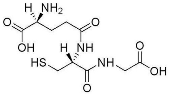
Low cost, sensitive, selective, and rapid methods for heavy metal ion (HMI) detection are of growing demand, and HMI biosensors have great potential in meeting this need due to their timeliness, cost-effectiveness and convenience in operation. The most widely reported peptide probe for HMI detection is glutathione (GSH), especially in case of lead ion (Pb2+) detection. GSH is highly stable, cost-effective, and easy to immobilize on a sensor. 

  • heavy metal ions
  • glutathione
  • biosensor
  • nanoparticle

1. Introduction

In general, heavy metals are the metals with relatively high mass densities (above 5 g/cm3), such as platinum, gold, mercury, and lead. The definition of heavy metals varies from discipline to discipline due to different concerns and perspectives. For physiology and pathology, heavy metals mainly refer to heavy elements with obvious biotoxicity. In terms of toxicity, the most common heavy metals include lead (Pb), mercury (Hg), arsenic (As), cadmium (Cd), and chromium (Cr) [1,2,3,4]. These heavy metal ions (HMIs) accumulate in the human body and cause chronic poisoning, resulting in damage to the immune, gastrointestinal, reproductive, and nervous systems [5,6,7]. Long-term exposure to heavy metals may even cause cancer in humans [8]. Being non-biodegradable, HMIs will continue to exist for decades and even hundreds of years if they are released into the environment [9]. With increasing human activities in mining and manufacturing, etc., HMI contamination and pollution pose great risks to ecological and human health [10,11]. Therefore, it is of great importance to be able to effectively screen for and quantitatively determine HMIs in a range of situations, including food safety, environmental pollution, and clinical diagnosis. Many international organizations and regulatory bodies, such as the World Health Organization (WHO) and the U.S. Environmental Protection Agency (EPA), have listed heavy metals as priority substances for intensive monitoring [1]. In practice, most countries have set standards on the permissible limits for HMI concentrations in water or other mediums to protect the environment and public health.
Traditional HMI measurements are mostly performed using spectroscopic techniques, including atomic absorption spectroscopy (AAS) [4,12], X-ray fluorescence spectrometry (XRF) [4,13], and inductively coupled plasma mass spectrometry (ICP-MS) [4,14], which are gold standards in HMI detection. However, they are expensive, difficult to perform, require complicated pretreatment for the samples, and need to be operated in a central laboratory setting. Thus, traditional spectroscopic techniques cannot meet the needs for pervasive HMI monitoring and screening. In contrast, surface-based biosensors are a good candidate to realize low-cost, sensitive HMI detection with a quick turn-around time. Similar to other biochemical sensors, there are three important components in an HMI sensor: analyte recognition, physical support for probe immobilization, and signal transduction. Various types of sensors have been reported, including nanoparticles [15,16,17], field effect transistors (FETs) [18,19,20], and working electrodes for voltammetric analysis [21]. In terms of signal transduction, there are mainly optical [15] and electrochemical methods [4]. Specific target recognition is always critically important to analyte detection. Antibody [22], nuclei acids [23,24], and peptide/amino acids [25,26] are the most commonly used probes to recognize and bind with HMIs. An antibody as a probe has the best specificity, but faces challenges when detecting HMIs. HMIs are too small to be directly detected with good sensitivity by an antibody, and antibody binding reactions may require a fluid environment different from that HMIs naturally exist in. Two types of functional nuclei acid probes have been developed for metal sensing: aptamers and DNAzymes. It is well-known that screening aptamer sequences to find one that specifically binds with a certain HMI is difficult, in addition to being laborious and time-consuming. Very few metal ions have successfully found their specific aptamers [27,28]. DNAzymes can selectively detect HMIs based on their catalytic activity; however, another nuclei acid sequence is required to be added to work as the substrate–enzyme pair. During metal detection by DNAzyme, additional steps and time are needed to allow for catalytic reactions between the DNAzyme and the substrate. Besides antibodies, aptamers, and DNAzyme, another option is to use a peptide or an amino acid as a probe to bind with HMIs, such as cysteine [29]. The most widely reported peptide probe for HMI detection is glutathione (GSH) [30]. Because there are six possible coordination sites in GSH for binding with metal ions, it has distinct advantages in capturing HMIs when compared with cysteine, especially in case of lead ion (Pb2+) detection. Furthermore, it is highly stable, cost-effective, and easy to immobilize on a sensor. Therefore, GSH, as a promising probe for HMI detection, has received much attention in recent years.

2. Recognition of HMIs by GSH

Glutathione (GSH) is a ubiquitous tripeptide biomolecule (γ-Glu-Cys-Gly) widely found in animals and plants with a concentration from 0.1 mM to 10 mM [31]. GSH is known as an antidote to prevent cells from toxicosis of heavy metal ions by forming complexes as a key step in biological detoxification processes [30].
The chemical structure of GSH is shown in Figure 1. If oxidized, GSH will form disulfide bonds between themselves, and become what is known as glutathione disulphide (GSSG). GSSG lacks the ability to bind with electrodes or nanoparticles. In practice, GSH in its reduced form is used to bind with HMIs. Reduced GSH is a linear tripeptide of L-glutamine, L-cysteine, and glycine, so reduced GSH is also labeled as L-GSH. GSH contains one amino (-NH2), one sulfhydryl (-SH), and two carboxyl (-COOH) groups, all with the ability to bind with HMIs. Therefore, GSH is a versatile ligand with several binding modes.
Figure 1. Glutathione’s Chemical Structure.
GSH has a sulfhydryl group on the cysteinyl portion. The sulfhydryl group is known to coordinate with a wide range of metal ions. For surface-based or nanoparticle-based HMI sensors, GSH is usually immobilized on gold nanoparticles or electrodes. Numerous experiments indicate that there is a strong binding force between gold and the (-SH) group. It is expected that, during probe immobilization, Au–S bonds are formed to link GSH molecules onto the gold surface [32,33,34,35]. As a result, only one amino (-NH2) and two carboxyls (-COOH) are still available to bind with metal ions.
A number of published papers have demonstrated that via the two free (-COOH) groups, strong binding with Pb2+ is preferred over that with other HMIs [35,36,37]. It was found that the solution pH has a strong effect on the affinity of the GSH–Pb2+ complex. At around pH 7, (-COOH) is deprotonated to (-COO-). Known as an oxyphilic ion, Pb2+ can combine with four acetate molecules or eight oxygen atoms [33]. Within a pH range of 4–8, binding between Pb2+ and (-COO-) will predominate over other reactions. Under the same pH condition, (-NH2) is protonated to (-NH3+), which is hypothesized to prevent GSH from combining with other ions due to its charges [33,37]. Therefore, good affinity and selectivity can be achieved for GSH–Pb2+ binding. There exist a number of reports on using GSH to specifically detect Pb2+ from a mixture of multiple HMIs.
Nevertheless, detection of metal ions other than Pb2+ can also be achieved using GSH-based sensors. The GSH support material and the solution pH value play an important role in the HMI–GSH complexation pattern. Research has shown that GSH’s conformational structure is strongly influenced by the solution pH [38]. Various functional groups in GSH can participate in complexation under different working conditions, leading to a certain selectivity. For example, GSH-capped quantum dots (QDs) are often used to detect HMIs other than Pb2+. In [39], GSH immobilized on sulfur- and nitrogen-co-doped carbon QDs presented free (-SH) groups for binding, which showed strong affinity to Hg2+ as opposed to other metal ions (Ag+, Al3+, Ba2+, Cu2+, Pb2+, etc.) and realized specific detection of Hg2+. Simultaneous detection of Cr2+ and Pb2+ was reported using GSH-coated magnetic nanoparticles (NPs) [40], as GSH molecules were immobilized onto Fe3O4 NPs through a hydrogen bond. In this detection scheme, NH4+, Na+, Mg2+, Hg2+ and other MIs caused no obvious responses, except for Cd2+ and Pb2+. This was attributed in part to the negative charges on the functional groups of GSH [40], since the testing was done at a pH of 4.5. Simultaneous detection of multiple HMIs is thus possible by adjusting the solution pH and the GSH immobilization strategy. However, complexation reactions between GSH and HMIs are complicated and difficult to predict, and the detection outcome under particular conditions usually needs to be verified through experimental results.
The foregoing introduces the interactions and formation of complexes between GSH and HMIs, which are the basis for the specific recognition of certain HMIs by GSH-based detection methods. To convert specific binding between GSH and HMIs into a quantifiable signal, a transducer is needed, which can take many different shapes and forms. Transduction schemes may have effects on the sensor performance as well, especially the plasmon-based detection that depends on the interaction between metal ions and nanoparticles. In next section, various GSH-based detection platforms are presented and discussed, with a focus on optical and electrochemical methods.

3. Heavy Metal Ion Detection Platforms Using Glutathione

Based on a signal transduction strategy, most of the GSH-based HMI detection methods can be categorized into two groups: optical methods and electrochemical methods. Then, each method is further divided into subtypes based on the support materials for GSH.

3.1. Optical Techniques

Metallic nanoparticles (NPs) are between 1–100 nm in size with a high surface charge density, a high surface to volume ratio, and, oftentimes, special optical properties due to a localized surface plasmon resonance (LSPR) effect. For these reasons, metallic NPs have been widely used in diverse biological and chemical applications, especially for biochemical detection. Various types of biochemical molecules can be easily incorporated onto the NPs while retaining their biochemical activity. For HMI detection, most metallic NPs are made of gold and some are silver, with gold NPs (GNPs) being the most widely used material in HMI sensors. Other shapes of NPs, such as nanorods and nanostars, have been adopted for sensing [41,42].

As an important discovery originally found in the 1990s, semiconductor quantum dots (QDs) have attracted intense research interest for their excellent luminescence properties. In contrast to traditional fluorescence techniques, QDs have advantages in excitation spectra, photo-luminescence quantum efficiency, and so on [50,51]. Reports of new QD-based sensors are on the rise. Most of the reported QD-based HMI detection schemes utilized II–VI semiconductor QDs. As an example, using Mn-doped ZnS QDs, [52] reported a Pb2+ detection scheme by phosphorescence measurement. Phosphorescence is generated by the energy transfer from the lowest vibrational energy layer of the excited Mn2+ triplet state to the vibrational energy layer of the ground state. Mn–ZnS QDs are a popular II–VI semiconductor QD for biosensor applications. Because Mn–ZnS QDs can be functionalized without using deoxidants and other inducers, they are convenient to work with. Furthermore, Mn–ZnS QDs have a long-lived doped emission, which can set the signal of Pb2+ detection apart from interference by autofluorescence and scattering light.

3.2. HMI Detection Combined with Electrochemical Techniques

Based on our survey of the field, most HMI detection techniques utilized optical techniques to extract the sensing signal. These optical methods have obvious advantages, such as visualization, but may be less advantageous when it comes to field deployability and affordable readout systems. As a result, HMI detection platforms by electrochemical techniques are also worth investigating. Among the electrochemical techniques, field effect transistors (FETs) and working electrodes are the two representative devices.

3.3. Summary of HMI Detection Platforms Using GSH

In summary, GSH has been adopted as the probe molecule in a variety of HMI detection platforms. These detection platforms have demonstrated good detection performance, such as a low LOD and a fast response. Some of them have been applied to practical samples with acceptable outcomes.
The platforms discussed above present commonly reported detection methods, and are not an exhaustive list of all existing GSH-based HMI sensors. Table 1 gives a more comprehensive summary of recently reported HMI detection platforms using GSH. In addition to the examples discussed in the review, Table 1 includes some less-used detection methods with their operational characteristics. As every application has its own unique requirements, different considerations go into choosing a certain detection platform. For example, it is difficult to find a sensor with high sensitivity and a very fast response at the same time. Some trade-off is needed when choosing a detection method. It is our hope that this summary will provide some guidance for choosing a suitable strategy to detect certain HMIs.
Table 1. Recent Achievements in Heavy Metal Ion (HMI) Detection Platforms Using GSH.

Signal Transduction

Sensor Structure

HMI Target

Detection Limit

Linear Range

Response Time

References

Optical methods

Colorimetry

Gold NPs

Pb

0.1 μM

0.1~50 μM

20 ~ 25 min

[43]

Silver NPs

Pb

1 nM

Not mentioned

At least 10 min

[32]

Gold NPs

Cd

4.3 pM

17 pM~16.67 nM

About 17 min

[49]

Localized surface plasmon resonance

Gold NPs

Pb

50 pM

0.1 nM~10 μM

15 min

[33]

Fluorescence

Gold NPs

Pb

0.1 μM

2~350 μM

About 1 min

[44]

Silver NPs

Pb

0.6 pM (200 ppq)

60 pM~2.4 nM (20~800 ppt estimated)

Less than 20 min

[45]

Surface-enhanced Raman scattering

Silver NPs

As3+

10.2 nM (0.76 ppb)

53.7 nM~4.0 μM (4~300 ppb)

At least 2 min

[46]

Whispering gallery mode

Gold NPs

Pb

0.05 nM

2.40~48.26 nM

About 40 min

[35]

Spectrophotometry

Mn-doped ZnS QDs

Pb

2.2 nM (0.45 μg/L)

4.9 nM~0.49 μM (1.0~100 μg/L)

Not mentioned

[52]

Mn-doped ZnS QDs

Pb, Cr, Hg

0.93 μM for mixed HMIs

1 μM~1 mM

Not mentioned

[53]

CdTe QDs

Pb

0.26 nM

0.8~15 nM

About 15 s

[54]

Carbon QDs

Hg

0.05 nM

1 nM~50 μM

At least 20 min

[39]

Silver NPs

Ni2+

75 μM

About 75 μM~1 mM

Not mentioned

[48]

Gold Nanostars

Pb

0.5 μM

About 0.5~4 μM

About 30 min

[42]

Dynamic Light Scattering

Gold Nanorod Chains

Pb

0.025 mM

Not mentioned

Not mentioned

[41]

Electrochemical methods

Square Wave Anodic Stripping Voltammetry, SWASV

Magnetic NPs

Cd, Pb

1.6 nM (0.182 μg/L);

0.8 nM (0.172 μg/L)

4.4~879 nM (0.5~100 μg/L),

2.3~460 nM (0.5~100 μg/L)

210 s

210 s

[40]

Glassy-Carbon Electrode

Cd

0.05 nM

2~20 nM

120 s

[56]

Carbon Paste Electrode

Cd

8.5 nM (2 ppb)

4.2~420 nM (1~100 ppb)

Longer than 7 min

[58]

Differtial pulsed anodic striping voltammetry, DPASV

Screen-Printed Carbon Nanofiber Electrode

Pb, Cd

~0.1 nM (~3 μg/L)

About 0.3~4.5 nM (about 10~150 μg/L)

120 s

[57]

FET (Drain current)

Field effect Transistor

Pb

10 nM

10 nM~10 μM

1–2 s

[34]

FET (Pulse-driven capacitance)

Gate Capacitance

Pb

<4.8 nM

(<1 ppb)

24~96 nM (5~20 ppb)

1–2 s

[19]

This entry is adapted from the peer-reviewed paper 10.3390/app9030489