Submitted Successfully!
To reward your contribution, here is a gift for you: A free trial for our video production service.
Thank you for your contribution! You can also upload a video entry or images related to this topic.
Version Summary Created by Modification Content Size Created at Operation
1 + 1622 word(s) 1622 2021-02-02 10:16:03 |
2 format correct Meta information modification 1622 2021-02-18 09:14:38 |

Video Upload Options

Do you have a full video?

Confirm

Are you sure to Delete?
Cite
If you have any further questions, please contact Encyclopedia Editorial Office.
Saitoh, T. DNA Damage and Multiple Myeloma. Encyclopedia. Available online: https://encyclopedia.pub/entry/7294 (accessed on 25 April 2024).
Saitoh T. DNA Damage and Multiple Myeloma. Encyclopedia. Available at: https://encyclopedia.pub/entry/7294. Accessed April 25, 2024.
Saitoh, Takayuki. "DNA Damage and Multiple Myeloma" Encyclopedia, https://encyclopedia.pub/entry/7294 (accessed April 25, 2024).
Saitoh, T. (2021, February 16). DNA Damage and Multiple Myeloma. In Encyclopedia. https://encyclopedia.pub/entry/7294
Saitoh, Takayuki. "DNA Damage and Multiple Myeloma." Encyclopedia. Web. 16 February, 2021.
DNA Damage and Multiple Myeloma
Edit

Multiple myeloma (MM) is an incurable plasma cell malignancy characterized by genomic instability. MM cells present various forms of genetic instability, including chromosomal instability, microsatellite instability, and base-pair alterations, as well as changes in chromosome number. The tumor microenvironment and an abnormal DNA repair function affect genetic instability in this disease. In addition, states of the tumor microenvironment itself, such as inflammation and hypoxia, influence the DNA damage response, which includes DNA repair mechanisms, cell cycle checkpoints, and apoptotic pathways. Unrepaired DNA damage in tumor cells has been shown to exacerbate genomic instability and aberrant features that enable MM progression and drug resistance.

multiple myeloma DNA repair genomic instability DNA damage response base excision repair homologous recombination

1. Introduction

Multiple myeloma (MM), which accounts for 1% of all cancers and approximately 10% of all hematologic malignancies, is characterized by the clonal proliferation of plasma cells [1][2]. The development and introduction of therapeutics, such as the immunomodulatory drugs thalidomide and lenalidomide and the proteasome inhibitor (PI) bortezomib, have led to improved prognosis of MM [1]. Before the introduction of alkylating agents in MM treatment, the median survival time of symptomatic patients was <1 year, and the introduction of melphalan in the 1960s resulted in improved survival [3][4]. High-dose melphalan (HDM) followed by autologous stem cell transplantation (ASCT) has become a standard of care for younger patients after bortezomib-based induction regimens [5][6]. In addition, the introduction of novel agents, particularly bortezomib combined with lenalidomide plus dexamethasone, has improved the outcome of patients who are ineligible for ASCT [7]. Recently, carfilzomib [8], pomalidomide [9], panobinostat [10], ixazomib [11], elotuzumab [12], daratumumab [13], isatuximab [14], and selinexor [15] have been approved by the Food and Drug Administration (FDA) for the treatment of relapsed MM and promise to improve outcomes. Currently, numerous combination therapies are available and include immunomodulatory drugs, PIs, histone deacetylase inhibitors, and monoclonal antibodies. However, MM remains an incurable disease, and new therapeutic strategies are still needed [1].

MM cells present genomic instability [16], whose molecular basis is not fully understood. Recently, it has been reported that the DNA damage response (DDR) may influence genomic changes in MM [17][18][19][20]. An abnormal DNA repair function may provide an alternative explanation for aneuploidy and chromosomal rearrangements. Furthermore, the tumor microenvironment may be mutagenic and constitute a significant source of genetic instability, affecting genomic stability and tumor resistance to treatment (Figure 1) [21][22][23]. This review provides an overview of the DDR, with a special focus on its function on MM. We will also discuss the role of DNA repair in regulating the metabolism and progression of MM cells.

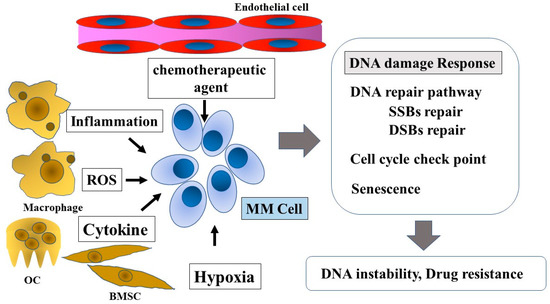
Figure 1. DNA damage response in multiple myeloma and the tumor microenvironment. Tumor microenvironment factors, such as chemotherapeutic agents, inflammation, ROS, cytokines, genotoxic stress, and hypoxia, influence the DNA damage response in MM cells. The DNA damage response affects disease progression and drug resistance. ROS, reactive oxygen species; OC, osteoclast; BMSC, bone marrow stromal cell; MM Cell, multiple myeloma cell; SSBs, single-strand breaks; DSBs, double-strand breaks.

2. Genomic Instability in MM

Most cancers are characterized by genomic instability [24][25][26], as cells often display chromosomal translocation, aneuploidy, and unchecked cell proliferation. Genomic instability is also a hallmark of MM cells, manifesting largely as whole chromosome- or translocation-based aneuploidy [22][27]. MM cells present various forms of genomic instability, including chromosomal instability (CIN), microsatellite instability, and increased frequency of mutations [28]. Especially, copy number and structural changes due to CIN are common features of MM. Numerical CIN is associated with copy number alterations (CNAs) of entire chromosomes or amplifications and deletions of chromosome arms [29]. The MM genome is also characterized by aberrations caused by structural CIN, such as chromosomal rearrangements, inversions, or complex reassembly of different parts of the chromosomes [29]. Centrosomes play important roles in processes that ensure proper segregation of chromosomes in human cells [30]. Chromosome abnormalities may be associated with centrosome amplification or alterations in the spindle assembly checkpoint. A high centrosome index—prompted by an abnormally high expression of genes encoding the main centrosome proteins—showed an independent prognostic factor in MM [31]. Moreover, dysregulations of the cell-cycle also affect CIN, the most prominent example of which is dysregulation of cyclin D expression—present in both hyperdiploid and non-hyperdiploid MM [28][32]. Furthermore, CIN is affected by the tumor microenvironment, such as hypoxia [33]. Depending on the genes affected, CIN can contribute to a further increase in genome instability [28].

MM patients are broadly grouped into hyperdiploid or non-hyperdiploid groups, depending on the number of chromosomes present in the MM cells. Hyperdiploid tumors are characterized by trisomies of one or more of the odd-numbered chromosomes 3, 7, 9, 11, 15, or 17, while the majority of non-hyperdiploid tumors display a translocation involving the IGH locus on chromosome 14 and one of the five recurrent translocation partners on chromosomes 4, 6, 11, 16, and 20. Five chromosomal partners account for the majority of primary IGH translocations: 11q13 (CCND1), 6p21 (CCND3), 4p16 (FGFR3 and NSD2), and 16q23 (MAF) [34][35]. These events are present in most patients with monoclonal gammopathy of undetermined significance (MGUS), and secondary genetic alterations occur with an increased incidence in disease progression from MGUS to MM [36]. These secondary events include translocations, deletions, and chromosome gains, involving genes such as MYCKRASNRAS, and TP53, which are involved in the DDR. Secondary events are detected in the late stage of the disease and affect its progression [36][37]. Among these alterations, t (4:14) and del(17p) were associated with poor outcomes [38]. The complexity of the genomic alteration characteristics is correlated with different grades of CIN. These observations strongly implicate CIN as an important biological and prognostic marker in MM [28][39].

Current treatment modalities of MM include alkylating agents, PIs, and anthracyclines, all of which induce excessive DNA damage [23]. DNA-damaging agents, including chemotherapy, can induce mutations and destabilize the genome, resulting in secondary malignancies or allowing the selection of resistant cancer cell clones [23][40]. Melphalan is a nitrogen mustard known to induce mono-alkylation of adenine and guanine, and interstrand DNA crosslinks (ICLs) involving guanine. The number of melphalan-induced ICLs is correlated with its concentration [41]. Cyclophosphamide is also an alkylating agent that induces ICLs. In a cohort of patients with MM treated with HDM and ASCT, polymorphisms in DNA repair genes, including poly(ADP-ribose) polymerase1 (PARP1), RAD51PCNAOGG1XPCBRCA1ERCC1BARD1, and TP53BP1, were associated with the outcome and overall survival [42]. These genes are significantly enriched in genes involved in homologous recombination repair (HRR) and nucleotide excision repair (NER), necessary for ICL repair. Bortezomib is the first PI approved by the FDA for the treatment of newly diagnosed and relapsed/refractory MM. This drug can bind to and form a complex with the active site of the threonine hydroxyl group in the β5 subunit of the proteasome and block its chymotrypsin-like activity [43]. Recent studies on proteasome inhibition in MM cells have revealed that the accumulation of unfolded proteins in the endoplasmic reticulum, the so-called endoplasmic reticulum stress, triggers that of several pro-apoptotic factors and cell stressors, such as reactive oxygen species (ROS). PI affects DNA repair via depletion of the free ubiquitin pool that is critical for further protein ubiquitination for building DNA repair foci through protein recruitment and degradation. Bortezomib treatment may prevent DNA resection by inhibiting the proteasomal degradation of proteins involved in chromatin relaxation, thus preventing the recruitment of replication protein A (RPA) onto single-stranded DNA (ssDNA) [44][45][46]. In vitro studies showed that bortezomib can possess synergistic anti-myeloma effects with melphalan by sensitizing MM cells to chemotherapeutic agents [47][48]. In clinical studies, bortezomib in combination with melphalan is efficacious for MM, both in the untreated and relapsed settings [49][50]. In the ASCT setting, the Intergroupe Francophone Du Myeloma (IFM) Phase II study showed significantly better outcomes in the ASCT recipients who received bortezomib–HDM conditioning compared with those treated with HDM only [51]. However, a recent analysis of the IFM 2014-02 trial Phase III study, in which 300 randomized patients received upfront ASCT to bortezomib and HDM, showed no difference in CR rate, PFS, and OS [52]. Doxorubicin is one of the most effective chemotherapy drugs for the treatment of many cancers, including lung cancers, leukemia, lymphoma, and MM [53]. This chemotherapeutic exerts its effects on cancer cells by intercalating into DNA, which results in DNA synthesis inhibition. Moreover, doxorubicin is a DNA topoisomerase II inhibitor, leading to DNA strand breaks by forming a cleavable complex with DNA and DNA topoisomerase II [54] and ROS formation in cells. Alkylating agents and DNA topoisomerase II inhibitors pose the risk of secondary cancers, such as therapy-related acute myeloid leukemia. Compared with de novo acute myeloid leukemia, the therapy-related disease is associated with poor prognosis and abnormal karyotypes, including the −5, −7, abnl(17p), complex karyotypes, and monosomal karyotypes [55].

DNA damage requires effective DNA repair capacity, which may be limited in tumor cells. Compared to normal cells, cancer cells present a higher accumulation of DNA damage and replication stress (RS) due to faulty cell cycle checkpoint activation [23][56]. Sources of RS include fragile sites, replication-transcription complex collision, secondary DNA structures, depletion of replication factors and nucleotides, and oncogenic stress [57]. Furthermore, DNA repair activity is required to counteract oxidative DNA damage in tumor cells induced by the tumor microenvironment. Cancer cells utilize mutagenic repair pathways for their advantage and to escape death [23]. Such repair pathways in cancer cells may represent treatment targets for their sensitization to drugs. Indeed, DNA repair inhibition has been used as a strategy for cancer treatment. Therapeutic strategies that target DNA damage include the use of PARP inhibitors for BRCA1/BRCA2-mutated cancers [58]. Thus, DNA repair pathways, including HRR, may be the target of drugs used to treat MM and play a role in resistance [59][60]. Next, we provide an overview of the DDR and major DNA repair pathways.

3. DNA Repair Pathways

Mammalian cells have six major DNA repair pathways involved in the DDR: the base excision repair (BER), NER, and mismatch repair (MMR) pathways repair nucleotide lesions on ssDNA; the HR and NHEJ pathways are involved in DSB repair; and the Fanconi anemia (FA) pathway repairs ICL lesions in co-operation with the NER and HR pathways [23][40][56].

References

  1. Rajkumar, S.V. Multiple myeloma: 2020 update on diagnosis, risk-stratification and management. Am. J. Hematol. 2020, 95, 548–567.
  2. Rajkumar, S.V.; Dimopoulos, M.A.; Palumbo, A.; Blade, J.; Merlini, G.; Mateos, M.V.; Kumar, S.; Hillengass, J.; Kastritis, E.; Richardson, P.; et al. International Myeloma Working Group updated criteria for the diagnosis of multiple myeloma. Lancet Oncol. 2014, 15, e538–e548.
  3. Bergsagel, D.E.; Sprague, C.C.; Austin, C.; Griffith, K.M. Evaluation of new chemotherapeutic agents in the treatment of multiple myeloma. IV. L-Phenylalanine mustard (NSC-8806). Cancer Chemother. Rep. 1962, 21, 87–99.
  4. Osgood, E.E. The survival time of patients with plasmocytic myeloma. Cancer Chemother. Rep. 1960, 9, 1–10.
  5. Attal, M.; Harousseau, J.L.; Stoppa, A.M.; Sotto, J.J.; Fuzibet, J.G.; Rossi, J.F.; Casassus, P.; Maisonneuve, H.; Facon, T.; Ifrah, N.; et al. A prospective, randomized trial of autologous bone marrow transplantation and chemotherapy in multiple myeloma. Intergroupe Français du Myélome. N. Engl. J. Med. 1996, 335, 91–97.
  6. Child, J.A.; Morgan, G.J.; Davies, F.E.; Owen, R.G.; Bell, S.E.; Hawkins, K.; Brown, J.; Drayson, M.T.; Selby, P.J. Medical Research Council Adult Leukaemia Working Party. High-dose chemotherapy with hematopoietic stem-cell rescue for multiple myeloma. N. Engl. J. Med. 2003, 348, 1875–1883.
  7. Durie, B.G.M.; Hoering, A.; Abidi, M.H.; Rajkumar, S.V.; Epstein, J.; Kahanic, S.P.; Thakuri, M.; Reu, F.; Reynolds, C.M.; Sexton, R.; et al. Bortezomib with lenalidomide and dexamethasone versus lenalidomide and dexamethasone alone in patients with newly diagnosed myeloma without intent for immediate autologous stem-cell transplant (SWOG S0777): A randomised, open-label, phase 3 trial. Lancet 2017, 389, 519–527.
  8. Stewart, A.K.; Rajkumar, S.V.; Dimopoulos, M.A.; Masszi, T.; Špička, I.; Oriol, A.; Hájek, R.; Rosiñol, L.; Siegel, D.S.; Mihaylov, G.G.; et al. ASPIRE Investigators. Carfilzomib, lenalidomide, and dexamethasone for relapsed multiple myeloma. N. Engl. J. Med. 2015, 372, 142–152.
  9. Lacy, M.Q.; Hayman, S.R.; Gertz, M.A.; Dispenzieri, A.; Buadi, F.; Kumar, S.; Greipp, P.R.; Lust, J.A.; Russell, S.J.; Dingli, D.; et al. Pomalidomide (CC4047) plus low-dose dexamethasone as therapy for relapsed multiple myeloma. J. Clin. Oncol. 2009, 27, 5008–5014.
  10. San-Miguel, J.F.; Hungria, V.T.; Yoon, S.S.; Beksac, M.; Dimopoulos, M.A.; Elghandour, A.; Jedrzejczak, W.W.; Günther, A.; Nakorn, T.N.; Siritanaratkul, N.; et al. Panobinostat plus bortezomib and dexamethasone versus placebo plus bortezomib and dexamethasone in patients with relapsed or relapsed and refractory multiple myeloma: A multicentre, randomised, double-blind phase 3 trial. Lancet Oncol. 2014, 15, 1195–1206.
  11. Moreau, P.; Masszi, T.; Grzasko, N.; Bahlis, N.J.; Hansson, M.; Pour, L.; Sandhu, I.; Ganly, P.; Baker, B.W.; Jackson, S.R.; et al. TOURMALINE-MM1 Study Group. Oral Ixazomib, Lenalidomide, and Dexamethasone for Multiple Myeloma. N. Engl. J. Med. 2016, 374, 1621–1634.
  12. Lonial, S.; Dimopoulos, M.; Palumbo, A.; White, D.; Grosicki, S.; Spicka, I.; Walter-Croneck, A.; Moreau, P.; Mateos, M.V.; Magen, H.; et al. ELOQUENT-2 Investigators. Elotuzumab Therapy for Relapsed or Refractory Multiple Myeloma. N. Engl. J. Med. 2015, 373, 621–631.
  13. Lonial, S.; Weiss, B.M.; Usmani, S.Z.; Singhal, S.; Chari, A.; Bahlis, N.J.; Belch, A.; Krishnan, A.; Vescio, R.A.; Mateos, M.V.; et al. Daratumumab monotherapy in patients with treatment-refractory multiple myeloma (SIRIUS): An open-label, randomised, phase 2 trial. Lancet 2016, 387, 1551–1560.
  14. Attal, M.; Richardson, P.G.; Rajkumar, S.V.; San-Miguel, J.; Beksac, M.; Spicka, I.; Leleu, X.; Schjesvold, F.; Moreau, P.; Dimopoulos, M.A.; et al. ICARIA-MM study group. Isatuximab plus pomalidomide and low-dose dexamethasone versus pomalidomide and low-dose dexamethasone in patients with relapsed and refractory multiple myeloma (ICARIA-MM): A randomised, multicentre, open-label, phase 3 study. Lancet 2019, 394, 2096–2107.
  15. Chari, A.; Vogl, D.T.; Gavriatopoulou, M.; Nooka, A.K.; Yee, A.J.; Huff, C.A.; Moreau, P.; Dingli, D.; Cole, C.; Lonial, S.; et al. Oral Selinex-or-Dexamethasone for Triple-Class Refractory Multiple Myeloma. N. Engl. J. Med. 2019, 381, 727–738.
  16. Negrini, S.; Gorgoulis, V.G.; Halazonetis, T.D. Genomic instability—An evolving hallmark of cancer. Nat. Rev. Mol. Cell Biol. 2010, 11, 220–228.
  17. Shammas, M.A.; Shmookler Reis, R.J.; Koley, H.; Batchu, R.B.; Li, C.; Munshi, N.C. Dysfunctional homologous recombination medi-ates genomic instability and progression in myeloma. Blood 2009, 113, 2290–2297.
  18. Walters, D.K.; Wu, X.; Tschumper, R.C.; Arendt, B.K.; Huddleston, P.M.; Henderson, K.J.; Dispenzieri, A.; Jelinek, D.F. Evidence for ongoing DNA damage in multiple myeloma cells as revealed by constitutive phosphorylation of H2AX. Leukemia 2011, 25, 1344–1353.
  19. Herrero, A.B.; San Miguel, J.; Gutierrez, N.C. Deregulation of DNA double-strand break repair in multiple myeloma: Implications for genome stability. PLoS ONE 2015, 10, e0121581.
  20. Cottini, F.; Hideshima, T.; Suzuki, R.; Tai, Y.T.; Bianchini, G.; Richardson, P.G.; Anderson, K.C.; Tonon, G. Synthetic Lethal Approaches Exploiting DNA Damage in Aggressive Myeloma. Cancer Discov. 2015, 5, 972–987.
  21. Yuan, J.; Glazer, P.M. Mutagenesis induced by the tumor microenvironment. Mutat. Res. 1998, 400, 439–446.
  22. Cagnetta, A.; Lovera, D.; Grasso, R.; Colombo, N.; Canepa, L.; Ballerini, F.; Calvio, M.; Miglino, M.; Gobbi, M.; Lemoli, R.; et al. Mechanisms and Clinical Applications of Genome Instability in Multiple Myeloma. BioMed Res. Int. 2015, 2015, 943096.
  23. Tshering, D. Lama-Sherpa 1, Lalita A Shevde An Emerging Regulatory Role for the Tumor Microenvironment in the DNA Damage Response to Double-Strand Breaks. Mol. Cancer Res. 2020, 18, 185–193.
  24. Chatterjee, N.; Walker, G.C. Mechanisms of DNA damage, repair, and mutagenesis. Environ. Mol. Mutagenesis 2017, 58, 235–263.
  25. Yao, Y.; Dai, W. Genomic Instability and Cancer. J. Carcinog. Mutagenesis 2014, 5, 1000165.
  26. Lengauer, C.; Kinzler, K.; Vogelstein, B. Genetic instabilities in human cancers. Nature 1998, 396, 643–649.
  27. Chng, W.J.; Fonseca, R. Centrosomes and myeloma; aneuploidy and proliferation. Environ. Mol. Mutagenesis 2009, 50, 697–707.
  28. Neuse, C.J.; Lomas, O.C.; Schliemann, C.; Shen, Y.J.; Manier, S.; Bustoros, M.; Ghobrial, I.M. Genome instability in multiple myeloma. Leukemia 2020, 34, 2887–2897.
  29. Manier, S.; Salem, K.Z.; Park, J.; Landau, D.A.; Getz, G.; Ghobrial, I.M. Genomic complexity of multiple myeloma and its clinical implications. Nat. Rev. Clin. Oncol. 2017, 14, 100–113.
  30. Pihan, G.A.; Wallace, J.; Zhou, Y.; Doxsey, S.J. Centrosome abnormalities and chromosome instability occur together in pre-invasive carcinomas. Cancer Res. 2003, 63, 1398–1404.
  31. Chng, W.J.; Ahmann, G.J.; Henderson, K.; Santana-Davila, R.; Greipp, P.R.; Gertz, M.A.; Lacy, M.Q.; Dispenzieri, A.; Kumar, S.; Rajkumar, S.V.; et al. Clinical implication of centrosome amplification in plasma cell neoplasm. Blood 2006, 107, 3669–3675.
  32. Bergsagel, P.L.; Kuehl, W.M.; Zhan, F.; Sawyer, J.; Barlogie, B.; Shaughnessy, J., Jr. Cyclin D dysregulation: An early and unifying pathogenic event in multiple myeloma. Blood 2005, 106, 296–303.
  33. Bindra, R.S.; Glazer, P.M. Genetic instability and the tumor microenvironment: Towards the concept of microenvironment-induced mutagenesis. Mutat. Res. 2005, 569, 75–85.
  34. Kumar, S.; Fonseca, R.; Ketterling, R.P.; Dispenzieri, A.; Lacy, M.Q.; Gertz, M.A.; Hayman, S.R.; Buadi, F.K.; Dingli, D.; Knudson, R.A.; et al. Trisomies in multiple myeloma: Impact on survival in patients with high-risk cytogenetics. Blood 2012, 119, 2100–2105.
  35. Fonseca, R.; Bergsagel, P.L.; Drach, J.; Shaughnessy, J.; Gutierrez, N.; Stewart, A.K.; Morgan, G.; Van Ness, B.; Chesi, M.; Minvielle, S.; et al. International Myeloma Working Group. International Myeloma Working Group molecular classification of multiple myeloma: Spotlight review. Leukemia 2009, 23, 2210–2221.
  36. Morgan, G.J.; Walker, B.A.; Davies, F.E. The genetic architecture of multiple myeloma. Nat. Rev. Cancer 2012, 12, 335–348.
  37. Gabrea, A.; Leif Bergsagel, P.; Michael Kuehl, W. Distinguishing primary and secondary translocations in multiple myeloma. DNA Repair 2006, 5, 1225–1233.
  38. Fonseca, R.; Blood, E.; Rue, M.; Harrington, D.; Oken, M.M.; Kyle, R.A.; Dewald, G.W.; Van Ness, B.; Van Wier, S.A.; Henderson, K.J.; et al. Clinical and biologic implications of recurrent genomic aberrations in myeloma. Blood 2003, 101, 4569–4575.
  39. Chung, T.H.; Mulligan, G.; Fonseca, R.; Chng, W.J. A novel measure of chromosome instability can account for prognostic difference in multiple myeloma. PLoS ONE 2013, 8, e66361.
  40. Tubbs, A.; Nussenzweig, A. Endogenous DNA Damage as a Source of Genomic Instability in Cancer. Cell 2017, 168, 644–656.
  41. Spanswick, V.J.; Craddock, C.; Sekhar, M.; Mahendra, P.; Shankaranarayana, P.; Hughes, R.G.; Hochhauser, D.; Hartley, J.A. Repair of DNA interstrand crosslinks as a mechanism of clinical resistance to melphalan in multiple myeloma. Blood 2002, 100, 224–229.
  42. Dumontet, C.; Landi, S.; Reiman, T.; Perry, T.; Plesa, A.; Bellini, I.; Barale, R.; Pilarski, L.M.; Troncy, J.; Tavtigian, S.; et al. Genetic polymorphisms associated with outcome in multiple myeloma patients receiving high-dose melphalan. Bone Marrow Transplant. 2010, 45, 1316–1324.
  43. Chen, D.; Frezza, M.; Schmitt, S.; Kanwar, J.; Dou, Q.P. Bortezomib as the first proteasome inhibitor anticancer drug: Current status and future perspectives. Curr. Cancer Drug Targets 2011, 11, 239–253.
  44. Gourzones-Dmitriev, C.; Kassambara, A.; Sahota, S.; Rème, T.; Moreaux, J.; Bourquard, P.; Hose, D.; Pasero, P.; Constantinou, A.; Klein, B. DNA repair pathways in human multiple myeloma: Role in oncogenesis and potential targets for treatment. Cell Cycle 2013, 12, 2760–2773.
  45. Jacquemont, C.; Taniguchi, T. Proteasome function is required for DNA damage response and fanconi anemia pathway acti-vation. Cancer Res. 2007, 67, 7395–7405.
  46. Murakawa, Y.; Sonoda, E.; Barber, L.J.; Zeng, W.; Yokomori, K.; Kimura, H.; Niimi, A.; Lehmann, A.; Zhao, G.Y.; Hochegger, H.; et al. Inhibitors of the proteasome suppress homologous DNA recombination in mammalian cells. Cancer Res. 2007, 67, 8536–8543.
  47. Yarde, D.N.; Oliveira, V.; Mathews, L.; Wang, X.; Villagra, A.; Boulware, D.; Shain, K.H.; Hazlehurst, L.A.; Alsina, M.; Chen, D.T.; et al. Targeting the Fanconi anemia/BRCA pathway circumvents drug resistance in multiple myeloma. Cancer Res. 2009, 69, 9367–9375.
  48. Popat, R.; Maharaj, L.; Oakervee, H.; Cavenagh, J.; Joel, S. Schedule dependent cytotoxicity of bortezomib and melphalan in multiple myeloma. Br. J. Haematol. 2013, 160, 111–114.
  49. San Miguel, J.F.; Schlag, R.; Khuageva, N.K.; Dimopoulos, M.A.; Shpilberg, O.; Kropff, M.; Spicka, I.; Petrucci, M.T.; Palumbo, A.; Samoilova, O.S.; et al. VISTA Trial Investigators. Bortezomib plus melphalan and prednisone for initial treatment of multiple myeloma. N. Engl. J. Med. 2008, 359, 906–917.
  50. Popat, R.; Oakervee, H.; Williams, C.; Cook, M.; Craddock, C.; Basu, S.; Singer, C.; Harding, S.; Foot, N.; Hallam, S.; et al. Bortezomib, low-dose intravenous melphalan, and dexamethasone for patients with relapsed multiple myeloma. Br. J. Haematol. 2009, 144, 887–894.
  51. Roussel, M.; Moreau, P.; Huynh, A.; Mary, J.-Y.; Danho, C.; Caillot, D.; Hulin, C.; Fruchart, C.; Marit, G.; Pégourié, B.; et al. Intergroupe Francophone du Myélome (IFM) Bortezomib and high-dose melphalan as conditioning regimen before autologous stem cell transplantation in patients with de novo multiple myeloma: A phase 2 study of the Intergroupe Francophone du Myelome (IFM). Blood 2010, 115, 32–37.
  52. Roussel, M.; Hebraud, B.; Lauwers-Cances, V.; Macro, M.; Leleu, X.; Hulin, C.; Karlin, L.; Royer, B.; Perrot, A.; Moreau, P.; et al. Bortezomib and High-Dose Melphalan vs. High-Dose Melphalan As Conditioning Regimen before Autologous Stem Cell Transplantation in De Novo Multiple Myeloma Patients: A Phase 3 Study of the Intergroupe Francophone Du Myelome (IFM 2014-02). Blood 2017, 130 (Suppl. 1), 398.
  53. Wakharde, A.A.; Awad, A.H.; Bhagat, A.; Karuppayil, S.M. Synergistic Activation of Doxorubicin against Cancer: A Review. Am. J. Clin. Microbiol. Antimicrob. 2018, 1, 1–6.
  54. Taymaz-Nikerel, H.; Karabekmez, M.E.; Eraslan, S.; Kırdar, B. Doxorubicin induces an extensive transcriptional and metabolic rewiring in yeast cells. Sci. Rep. 2018, 8, 13672.
  55. Kayser, S.; Döhner, K.; Krauter, J.; Köhne, C.H.; Horst, H.A.; Held, G.; von Lilienfeld-Toal, M.; Wilhelm, S.; Kündgen, A.; Götze, K.; et al. German-Austrian AMLSG. The impact of therapy-related acute myeloid leukemia (AML) on outcome in 2853 adult patients with newly diagnosed AML. Blood 2011, 117, 2137–2145.
  56. Ciccia, A.; Elledge, S.J. The DNA damage response: Making it safe to play with knives. Mol. Cell 2010, 40, 179–204.
  57. Gaillard, H.; García-Muse, T.; Aguilera, A. Replication stress and cancer. Nat. Rev. Cancer 2015, 15, 276–289.
  58. Singh, N.; Pay, S.L.; Bhandare, S.B.; Arimpur, U.; Motea, E.A. Therapeutic Strategies and Biomarkers to Modulate PARP Activity for Targeted Cancer Therapy. Cancers 2020, 12, 972.
  59. Gkotzamanidou, M.; Terpos, E.; Bamia, C.; Munshi, N.C.; Dimopoulos, M.A.; Souliotis, V.L. DNA repair of myeloma plasma cells correlates with clinical outcome: The effect of the nonhomologous end-joining inhibitor SCR7. Blood 2016, 128, 1214–1225.
  60. Herrero, A.B.; Gutiérrez, N.C. Targeting ongoing DNA damage in multiple myeloma: Effects of DNA damage response inhibitors on plasma cell survival. Front. Oncol. 2017, 7, 98.
More
Information
Contributor MDPI registered users' name will be linked to their SciProfiles pages. To register with us, please refer to https://encyclopedia.pub/register :
View Times: 509
Revisions: 2 times (View History)
Update Date: 18 Feb 2021
1000/1000