Submitted Successfully!
To reward your contribution, here is a gift for you: A free trial for our video production service.
Thank you for your contribution! You can also upload a video entry or images related to this topic.
Version Summary Created by Modification Content Size Created at Operation
1 + 2017 word(s) 2017 2021-11-08 04:30:45 |
2 format correction Meta information modification 2017 2021-11-17 04:51:25 |

Video Upload Options

Do you have a full video?

Confirm

Are you sure to Delete?
Cite
If you have any further questions, please contact Encyclopedia Editorial Office.
Lin, Z. Autophagy Regulation by miRNAs and Ubiquitination System. Encyclopedia. Available online: https://encyclopedia.pub/entry/16076 (accessed on 19 April 2024).
Lin Z. Autophagy Regulation by miRNAs and Ubiquitination System. Encyclopedia. Available at: https://encyclopedia.pub/entry/16076. Accessed April 19, 2024.
Lin, Zhenghong. "Autophagy Regulation by miRNAs and Ubiquitination System" Encyclopedia, https://encyclopedia.pub/entry/16076 (accessed April 19, 2024).
Lin, Z. (2021, November 17). Autophagy Regulation by miRNAs and Ubiquitination System. In Encyclopedia. https://encyclopedia.pub/entry/16076
Lin, Zhenghong. "Autophagy Regulation by miRNAs and Ubiquitination System." Encyclopedia. Web. 17 November, 2021.
Autophagy Regulation by miRNAs and Ubiquitination System
Edit

MicroRNAs (miRNAs) are non-coding single-stranded RNA molecules encoded by endogenous genes with ~22 nucleotides which are involved in the regulation of post-transcriptional gene expression. Ubiquitination and deubiquitination are common post-translational modifications in eukaryotic cells and important pathways in regulating protein degradation and signal transduction, in which E3 ubiquitin ligases and deubiquitinases (DUBs) play a decisive role. MiRNA and ubiquitination are involved in the regulation of most biological processes, including autophagy.

microRNAs E3 ubiquitin ligases deubiquitinases autophagy

1. Introduction

A living cell is a complex dynamic system which can respond and adapt to environmental changes and stress all of the time. Among living cells, proteins play a key role in all physiological and pathological cellular functions. Therefore, it is of great significance to understand the synthesis, degradation, modification, and related regulation of proteins.
Ubiquitin proteasome system (UPS) is one of the main pathways regulating protein degradation in eukaryotic cells and is also a key regulatory mechanism in a variety of biological processes [1]. Ubiquitination is a reversible post-translational modification that occurs under the continuous action of E1 ubiquitin activating enzyme, E2 ubiquitin coupling enzyme, and E3 ubiquitin ligase [2][3]. Proteins labeled with ubiquitin are broken down by proteasome into smaller peptides, amino acids, and ubiquitin that can be reused [4]. In addition, ubiquitination can also serve as a marker to activate certain signals, such as autophagy and immune response [5].
MicroRNA (miRNA) is an evolutionarily conserved small non-coding RNA that is involved in the regulation of gene expression during the translation phase and is considered to be abnormally expressed in a variety of human diseases [6][7][8][9]. MiRNA can inhibit the expression of target genes at the translation level or directly lead to the degradation of mRNA through complementary binding with target mRNA [10][11][12].

2. Overview of miRNA

In the eukaryotic genome, only a small number of genes encode proteins, and about 97% of the transcribed products are non-coding RNAs [13]. MicroRNAs (miRNAs) are ~22 nt small noncoding RNAs that are known to play an important role in the post-transcriptional regulation of messenger RNA (mRNA) [14][15]. It is estimated that more than 60% of human genes are regulated by miRNAs [16]. At the same time, studies have shown that the sequence and structure of miRNAs are highly evolutionarily conserved among different species, suggesting that miRNAs have a critical regulatory function [11][17][18].
MiRNAs are usually transcribed in the nucleus by RNA polymerase II (polII), and the initial product is a large primary miRNAs (pri-miRNA) with a 5′ 7-methyl guanosine cap and a 3′ poly adenosine tail [14]. Pri-miRNA was originally processed by Drosha in the nucleus to form a precursor miRNA (pre-miRNA) of ~70 nt that forms a hairpin, which was exported to the cytoplasm via nuclear transport receptor exportin-5 and the cofactor Ran-GTP [19]. It is then cleaved by the RNase III enzyme Dicer into a double stranded RNA of ~22 nt [20]. Under the action of the Argonaute (AGO) proteins, one strand of this duplex is selected as a mature miRNA and is then incorporated into the miRNA-induced silencing complex (miRISC) [21][22].
MiRISC directs the miRNA to binding sites in the target mRNAs, which usually leads to gene repression [22]. If the miRNA is completely complementary to the target site, the binding of these miRNAs often leads to degradation of the target mRNA. MiRNAs that are not completely complementary to the target mRNA usually inhibit the expression of the target gene at the protein translation level without affecting the mRNA stability (Figure 1) [10][23].
Figure 1. The biogenesis of microRNA (miRNA). MiRNA is transcribed by RNA polymerase II (pol II) in the nucleus as a pre-miRNA, processed by Drosha to a pre-miRNA. Pre-miRNA is then exported from the nucleus to the cytoplasm by exportin 5 (XPO5). In the cytoplasm, Dicer cleaves pre-miRNA to produce the miRNA duplex, and one strand of the resulting duplex is loaded onto the Argonaute (AGO) protein to form a miRNA-induced silencing complex (miRISC), which targets mRNAs for regulation. MiRNAs that form perfect duplexes with their targets direct degradation and those that support partial duplexes inhibit protein expression.
A miRNA can have multiple target genes, and several miRNAs can jointly regulate the same gene [15][23]. Therefore, the wide variety of biological functions of miRNAs is not surprising. Although the important roles of miRNAs have been demonstrated in several studies, the research on miRNAs is still in its infancy and only a small part of their biological functions has been elucidated [9][24][25][26][27].

3. Overview of Ubiquitin-Proteasome System (UPS)

Ubiquitin (Ub) is a 76-amino-acid protein highly conserved among all eukaryotes. It covalently binds to the lysine residues of the substrate protein and acts as a signal molecule to mediate its degradation or regulate its biological functions [2]. Ubiquitin contains seven lysine residues (K6, K11, K27, K29, K33, K48, and K63) and one N-terminal methionine residue, each of which can be attached to another ubiquitin moiety [3]. As a consequence, the modification of ubiquitin can form ubiquitin chains of different lengths and linkage types. The lysine binding sites of ubiquitin determine different cellular functions and protein fates, including degradation, signal transduction, and altered subcellular localization [5]. Among them, K48- and K11-linked chains mediate the recognition and degradation of ubiquitinated substrate proteins by 26S proteasome, whereas other linked sites, such as K63, do not lead to degradation but regulate other cellular processes, such as DNA repair, mitochondrial genetics or NF-κB signaling pathways [28]. The physiological functions of other atypical ubiquitination modifications (K6, K27, K29, and K33) are unknown but have been of interest to researchers.
Ubiquitin binding is a multistep reaction that requires the sequential action of three enzymes (E1 Ub-activating enzyme, the E2 Ub-conjugating enzyme, and the E3 ubiquitin ligase) [1]. In the presence of ATP, E1 activates Ub and transfers it to E2, whose active site cysteine forms a thioester bond with the C-terminal carboxyl group of Ub. The E3 enzyme mediates the last step of Ub transfer through simultaneous interaction with a Ub-loaded E2 enzyme and a specific substrate, and finally forms the ubiquitinated substrate (Figure 2) [4][29]. Given the substrate recognition and substrate specificity of E3 ligase, its role in ubiquitin modification is particularly critical. There are more than 600 known E3 ligases in human, and they can be grouped into three categories according to their conserved domains: HECT E3 (homologous to the E6-associated protein carboxyl terminus) [30], RING finger E3s [31], and RBR (RING between RING) type E3s [32].
Figure 2. The overview of the ubiquitin-proteasome system. Ubiquitin is activated and bound to E1 in an ATP-dependent manner. Then, the activated ubiquitin is transferred to the E2, while the substrate protein to be degraded is specifically targeted by E3 ubiquitin ligase, and ubiquitin is ligated to the substrate. Ubiquitinated substrate proteins are recognized by the 26S proteasome and degraded into small peptides and amino acids. In contrast, DUB reverses ubiquitination by removing the polyubiquitin chains of proteins and maintains intracellular ubiquitin levels. In addition, some ubiquitination modifications that do not lead to degradation induce related biological effects through signal recognition, such as kinase activation, localization changes.
Ubiquitination is a reversible process that can be reversed by a specific group of enzymes called deubiquitinases (DUBs) [33]. There are about 100 DUBs encoded by the human genome, which are mainly divided into six classes: ubiquitin-specific Proteases (USPs); ubiquitin carboxy-terminal hydrolases (UCHs); ovarian-tumor proteases (OTUs); Machado-joseph disease Protein Domain Proteases (MJDs), JAMM/MPN domain-associated metallopeptidases and monocyte chemotactic protein-induced protein (MCPIP). The most abundant sub-family of DUBs is the USPs with over 50 members [34][35].

4. Overview of Autophagy System

Autophagy is a stress-responsive catabolic process that degrades intracellular components through lysosomal enzymes [36]. In normal physiological state, only a small amount of autophagy occurs in cells to maintain homeostasis. When cells are stimulated by intracellular and extracellular factors such as starvation, hypoxia, pathogen invasion, etc. [37], a large number of autophagy can be induced through the transduction of cell signaling pathways [38]. Thus, autophagy is a pivotal actor in development, immune response, as well as metabolic regulation and has been shown to be associated with cellular modifications related to senescence, with most studies now suggesting that a reduced autophagic potential is one of the factors of cell senescence [36][39][40]. Autophagy not only removes protein aggregates, but also damages organelles and plays a role in quality control of the cytoplasm such as mitophagy [41]. In the case of damaged mitochondria, mitophagy removes malfunctioning mitochondria to maintain the population at an optimal state. In recent years, mitophagy has received increasing attention since mitochondrial dysfunction is at the foundation of numerous diseases and a growing number of studies also suggested mitophagy as a therapeutic target [42][43].
Autophagy is composed of several closely related steps including autophagy initiation, autophagosome maturation, and autophagolysosome fusion, which involves many important autophagy-related proteins and complexes [44]. These core autophagy proteins include the following parts: (1) the ATG1/ULK1 complex, including ATG1, ATG11, ATG13, ATG17, ATG29, and ATG31, is the only core protein complex with serine/threonine kinase activity in autophagy signaling pathway. The ULK1 complex acts as a bridge connecting upstream energy receptors mTOR and AMPK with downstream autophagosomes in vivo [45][46], and plays an important role in autophagy initiation [47][48]; (2) the PI3K complex, including Vps34, Vps15, ATG6/Beclin1, and ATG14, catalyze the conversion of the lipid molecule PI to PI3P, thereby recruiting the protein that binds to PI3P [49]. Vps34 is the class III PI3K in mammals. Vps34 is activated by binding to Vps15 and further binds to Beclin1 to form the Vps34-Vps15-Beclin1 complex. During autophagy, Vps34-Vps15-Beclin1 binds to a variety of autophagy-related proteins. For example, ATG14 is combined with to Vps34-Vps15-Beclin1 to form ATG14-Vps34-Vps15-Beclin1, which is involved in the formation of autophagic vesicles [50]; the (3) ATG9 and WIPI/ATG2-ATG18 complex. ATG9 is a transmembrane protein with six transmembrane domains, which may play a role in regulating autophagy by affecting vesicular transport [51]. ATG9 circulates in autophagic vesicles and cytoplasm, depending on ATG17 or ATG11 complex to locate PAS, and ATG2-ATG18 complex to leave PAS [52][53]. In mammals, specific silence of mATG9 gene can inhibit the formation of autophagic vesicles and protein degradation, and inhibit the occurrence of autophagy; and (4) ubiquitin-like systems ATG12-ATG5 and ATG8/LC3. There are two ubiquitin-like binding pathways involved in autophagosome formation. Both ATG8 and ATG12 are ubiquitin-like proteins, ATG12 can covalently bind with ATG5, and ATG8 can covalently bind with the lipid molecule phosphatidylethanolamine PE [54][55]. Similar to the ubiquitin system, ATG12 is transmitted by ATG7 to ATG10, which eventually binds to the lysine side chain of ATG5 and forms a complex with ATG16, promoting the exposure of membrane-binding sites on ATG5. Similarly, ATG7 transfers ATG8 to ATG3 [56]. With the help of ATG12-ATG5-ATG16, LC3 conjugates to lipid molecule phosphatidylethanolamine (PE), promoting isolation membrane expansion and autophagic vesicle completion [57]. These key proteins in the complex autophagy regulatory network are regulated by a variety of molecular signals, including ubiquitin ligases, deubiquitinases and miRNAs
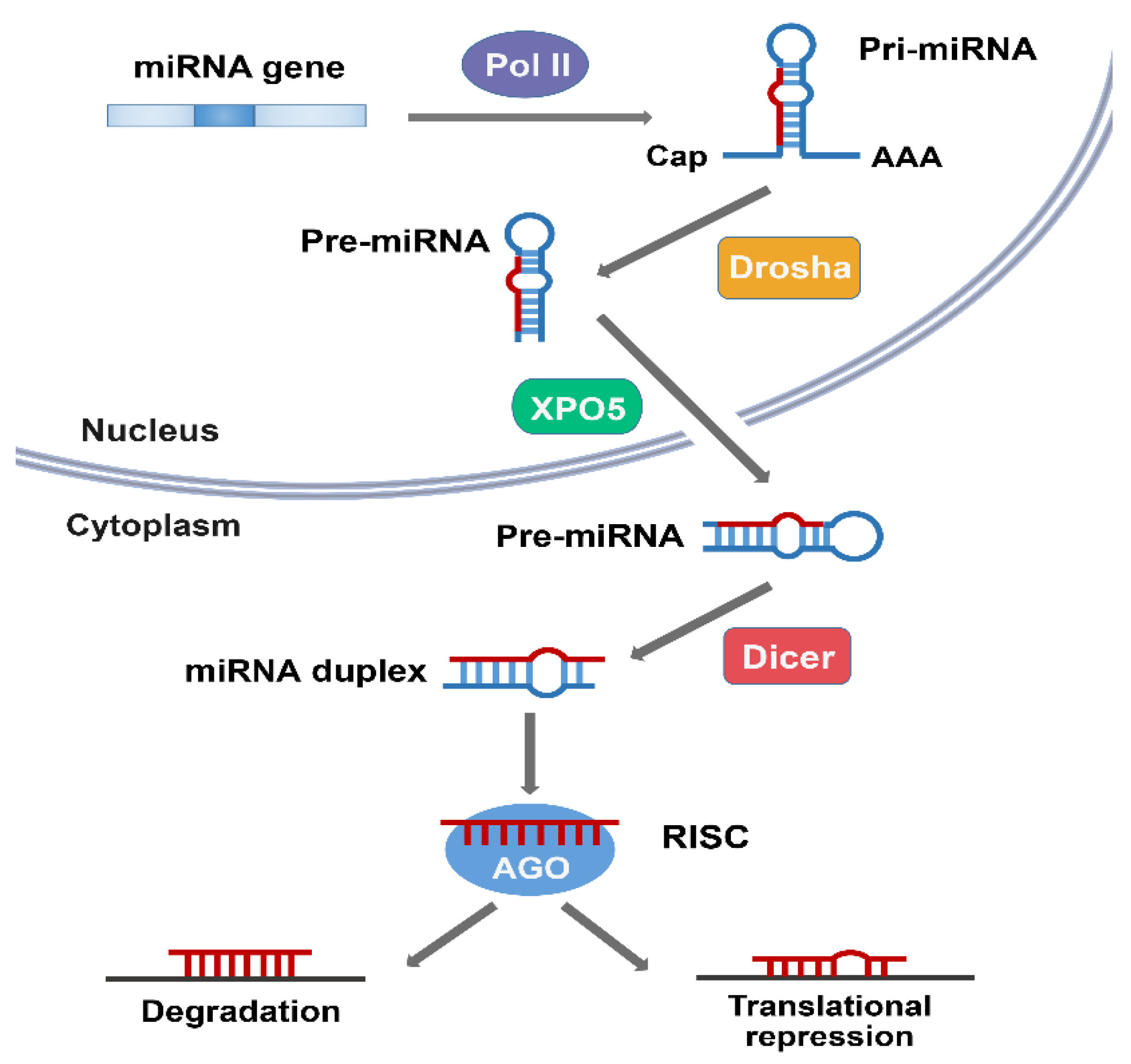
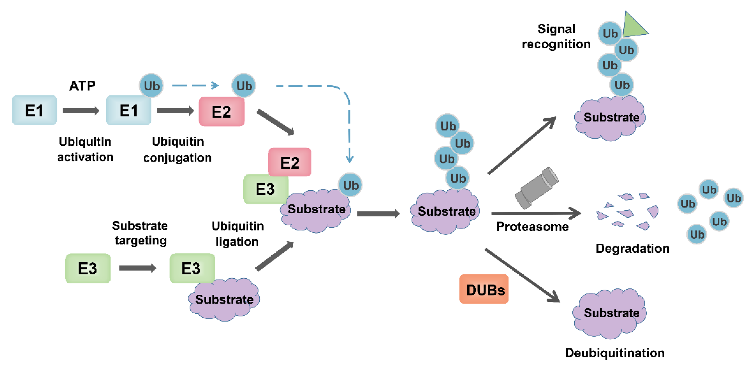
 [58].

5. MiRNAs Are Involved in Autophagy via Regulation of E3 Ubiquitin Ligases

E3 ubiquitin ligase specifically recognizes substrates and induces substrate protein degradation, and most of the key proteins involved in autophagy are regulated by ubiquitin ligase. Therefore, it is of great significance to understand the ubiquitination mechanism in autophagy. At the same time, miRNA often acts as an upstream regulator of E3 ubiquitin ligase, co-regulating autophagy with ubiquitin ligase (Table 1).
Table 1. MiRNAs and E3 ligases involved in autophagy.

MiRNA/E3

Target

Function

References

Mir-30a

MARCH5

MARCH5 mRNA acts as ceRNA of ATG5

[59]

Mir-200a

MARCH7

MARCH7 mRNA acts as ceRNA of ATG7

[60]

Mir-233

TRIM37

Promotes autophagy by inhibiting MTORC1

[61]

Mir-34a-5p

SYVN1

Induces autophagy

[62]

Mir-146a

TRAF6

Inhibits autophagy via ULK1 protein

[63]

Mir-27

NEDD4

Attenuates autophagy through Notch1

[64]

TRIM65

Mir-138-5P

Upregulates ATG7 by inhibiting miRISC

[65]

6. MiRNAs Are Involved in Autophagy via DUBs Regulation

As part of the ubiquitin-proteasome system, the role of deubiquitinases is equally important. Deubiquitinases stabilize substrate proteins and participate in the regulation of autophagy related signaling pathways by removing ubiquitin chains of substrates. There was also an interesting crosstalk between MiRNA and deubiquitinases (Table 2).
Table 2. MiRNAs and DUBs involved in autophagy.

MiRNA/E3

Target

Function

References

Mir-29c

USP22

Inhibits autophagy

[66]

Mir-6825-5p

USP22


Mir-6845-5p

Inhibits SIRT1-mediated autophagy

[67]

Mir-6886-3p


Mir-26b

USP9X

Suppresses Autophagy by inhibiting p53

[68]

Mir-26a

USP15

Activates autophagy

[69]

References

  1. Nandi, D.; Tahiliani, P.; Kumar, A.; Chandu, D. The ubiquitin-proteasome system. J. Biosci. 2006, 31, 137–155.
  2. Swatek, K.N.; Komander, D. Ubiquitin modifications. Cell Res. 2016, 26, 399–422.
  3. Finley, D. Recognition and processing of ubiquitin-protein conjugates by the proteasome. Annu. Rev. Biochem. 2009, 78, 477–513.
  4. Zheng, N.; Shabek, N. Ubiquitin Ligases: Structure, Function, and Regulation. Annu. Rev. Biochem. 2017, 86, 129–157.
  5. Berndsen, C.E.; Wolberger, C. New insights into ubiquitin E3 ligase mechanism. Nat. Struct. Mol. Biol. 2014, 21, 301–307.
  6. Ambros, V. microRNAs: Tiny regulators with great potential. Cell 2001, 107, 823–826.
  7. Bushati, N.; Cohen, S.M. microRNA functions. Annu. Rev. Cell Dev. Biol. 2007, 23, 175–205.
  8. Simonson, B.; Das, S. MicroRNA Therapeutics: The Next Magic Bullet? Mini Rev. Med. Chem. 2015, 15, 467–474.
  9. Akkoc, Y.; Gozuacik, D. MicroRNAs as major regulators of the autophagy pathway. Biochim. Biophys. Acta Mol. Cell Res. 2020, 1867, 118662.
  10. Filipowicz, W.; Bhattacharyya, S.N.; Sonenberg, N. Mechanisms of post-transcriptional regulation by microRNAs: Are the answers in sight? Nat. Rev. Genet. 2008, 9, 102–114.
  11. Krol, J.; Loedige, I.; Filipowicz, W. The widespread regulation of microRNA biogenesis, function and decay. Nat. Rev. Genet. 2010, 11, 597–610.
  12. Pasquinelli, A.E.; Hunter, S.; Bracht, J. MicroRNAs: A developing story. Curr. Opin. Genet. Dev. 2005, 15, 200–205.
  13. Shabalina, S.A.; Spiridonov, N.A. The mammalian transcriptome and the function of non-coding DNA sequences. Genome Biol. 2004, 5, 105.
  14. Chekulaeva, M.; Filipowicz, W. Mechanisms of miRNA-mediated post-transcriptional regulation in animal cells. Curr. Opin. Cell Biol. 2009, 21, 452–460.
  15. Fabian, M.R.; Sonenberg, N.; Filipowicz, W. Regulation of mRNA translation and stability by microRNAs. Annu. Rev. Biochem. 2010, 79, 351–379.
  16. Bentwich, I. Prediction and validation of microRNAs and their targets. FEBS Lett. 2005, 579, 5904–5910.
  17. Friedman, J.M.; Jones, P.A. MicroRNAs: Critical mediators of differentiation, development and disease. Swiss Med. Wkly. 2009, 139, 466–472.
  18. Mohr, A.M.; Mott, J.L. Overview of microRNA biology. Semin. Liver Dis. 2015, 35, 3–11.
  19. Han, J.; Pedersen, J.S.; Kwon, S.C.; Belair, C.D.; Kim, Y.K.; Yeom, K.H.; Yang, W.Y.; Haussler, D.; Blelloch, R.; Kim, V.N. Posttranscriptional crossregulation between Drosha and DGCR8. Cell 2009, 136, 75–84.
  20. Yan, H.; Liang, F.S. miRNA inhibition by proximity-enabled Dicer inactivation. Methods 2019, 167, 117–123.
  21. Schwarz, D.S.; Hutvagner, G.; Du, T.; Xu, Z.; Aronin, N.; Zamore, P.D. Asymmetry in the assembly of the RNAi enzyme complex. Cell 2003, 115, 199–208.
  22. Sheu-Gruttadauria, J.; MacRae, I.J. Phase Transitions in the Assembly and Function of Human miRISC. Cell 2018, 173, 946–957.e16.
  23. Bartel, D.P. MicroRNAs: Target recognition and regulatory functions. Cell 2009, 136, 215–233.
  24. Ali Syeda, Z.; Langden, S.S.S.; Munkhzul, C.; Lee, M.; Song, S.J. Regulatory Mechanism of MicroRNA Expression in Cancer. Int. J. Mol. Sci. 2020, 21, 143.
  25. Barwari, T.; Joshi, A.; Mayr, M. MicroRNAs in Cardiovascular Disease. J. Am. Coll. Cardiol. 2016, 68, 2577–2584.
  26. Davalos, V.; Esteller, M. MicroRNAs and cancer epigenetics: A macrorevolution. Curr. Opin. Oncol. 2010, 22, 35–45.
  27. Trang, P.; Weidhaas, J.B.; Slack, F.J. MicroRNAs as potential cancer therapeutics. Oncogene 2008, 27 (Suppl. 2), 52–57.
  28. Erpapazoglou, Z.; Walker, O.; Haguenauer-Tsapis, R. Versatile roles of k63-linked ubiquitin chains in trafficking. Cells 2014, 3, 1027–1088.
  29. Jarome, T.J.; Devulapalli, R.K. The Ubiquitin-Proteasome System and Memory: Moving Beyond Protein Degradation. Neuroscientist 2018, 24, 639–651.
  30. Weber, J.; Polo, S.; Maspero, E. HECT E3 Ligases: A Tale With Multiple Facets. Front. Physiol. 2019, 10, 370.
  31. Deshaies, R.J.; Joazeiro, C.A. RING domain E3 ubiquitin ligases. Annu. Rev. Biochem. 2009, 78, 399–434.
  32. Spratt, D.E.; Walden, H.; Shaw, G.S. RBR E3 ubiquitin ligases: New structures, new insights, new questions. Biochem. J. 2014, 458, 421–437.
  33. Eletr, Z.M.; Wilkinson, K.D. Regulation of proteolysis by human deubiquitinating enzymes. Biochim. Biophys. Acta 2014, 1843, 114–128.
  34. Magraoui, F.E.; Reidick, C.; Meyer, H.E.; Platta, H.W. Autophagy-Related Deubiquitinating Enzymes Involved in Health and Disease. Cells 2015, 4, 596–621.
  35. Nijman, S.M.; Luna-Vargas, M.P.; Velds, A.; Brummelkamp, T.R.; Dirac, A.M.; Sixma, T.K.; Bernards, R. A genomic and functional inventory of deubiquitinating enzymes. Cell 2005, 123, 773–786.
  36. Mizushima, N.; Komatsu, M. Autophagy: Renovation of cells and tissues. Cell 2011, 147, 728–741.
  37. Mazure, N.M.; Pouyssegur, J. Hypoxia-induced autophagy: Cell death or cell survival? Curr. Opin. Cell Biol. 2010, 22, 177–180.
  38. Yu, L.; Chen, Y.; Tooze, S.A. Autophagy pathway: Cellular and molecular mechanisms. Autophagy 2018, 14, 207–215.
  39. Rajendran, P.; Alzahrani, A.M.; Hanieh, H.N.; Kumar, S.A.; Ben Ammar, R.; Rengarajan, T.; Alhoot, M.A. Autophagy and senescence: A new insight in selected human diseases. J. Cell. Physiol. 2019, 234, 21485–21492.
  40. Rubinsztein, D.C.; Marino, G.; Kroemer, G. Autophagy and aging. Cell 2011, 146, 682–695.
  41. Klionsky, D.J.; Codogno, P. The mechanism and physiological function of macroautophagy. J. Innate Immun. 2013, 5, 427–433.
  42. Killackey, S.A.; Philpott, D.J.; Girardin, S.E. Mitophagy pathways in health and disease. J. Cell Biol. 2020, 219, e202004029.
  43. Strappazzon, F. A global view of the miRNA-mitophagy connexion. Prog. Mol. Biol. Transl. Sci. 2020, 172, 37–54.
  44. Xie, Z.; Klionsky, D.J. Autophagosome formation: Core machinery and adaptations. Nat. Cell Biol. 2007, 9, 1102–1109.
  45. Alers, S.; Loffler, A.S.; Wesselborg, S.; Stork, B. Role of AMPK-mTOR-Ulk1/2 in the regulation of autophagy: Cross talk, shortcuts, and feedbacks. Mol. Cell. Biol. 2012, 32, 2–11.
  46. Nazio, F.; Strappazzon, F.; Antonioli, M.; Bielli, P.; Cianfanelli, V.; Bordi, M.; Gretzmeier, C.; Dengjel, J.; Piacentini, M.; Fimia, G.M.; et al. mTOR inhibits autophagy by controlling ULK1 ubiquitylation, self-association and function through AMBRA1 and TRAF6. Nat. Cell Biol. 2013, 15, 406–416.
  47. Hosokawa, N.; Hara, T.; Kaizuka, T.; Kishi, C.; Takamura, A.; Miura, Y.; Iemura, S.; Natsume, T.; Takehana, K.; Yamada, N.; et al. Nutrient-dependent mTORC1 association with the ULK1-Atg13-FIP200 complex required for autophagy. Mol. Biol. Cell 2009, 20, 1981–1991.
  48. Militello, R.D.; Colombo, M.I. A membrane is born: Origin of the autophagosomal compartment. Curr. Mol. Med. 2011, 11, 197–203.
  49. Abrahamsen, H.; Stenmark, H.; Platta, H.W. Ubiquitination and phosphorylation of Beclin 1 and its binding partners: Tuning class III phosphatidylinositol 3-kinase activity and tumor suppression. FEBS Lett. 2012, 586, 1584–1591.
  50. Zhong, Y.; Wang, Q.J.; Li, X.; Yan, Y.; Backer, J.M.; Chait, B.T.; Heintz, N.; Yue, Z. Distinct regulation of autophagic activity by Atg14L and Rubicon associated with Beclin 1-phosphatidylinositol-3-kinase complex. Nat. Cell Biol. 2009, 11, 468–476.
  51. Mizushima, N.; Yoshimori, T.; Ohsumi, Y. The role of Atg proteins in autophagosome formation. Annu. Rev. Cell Dev. Biol. 2011, 27, 107–132.
  52. Kotani, T.; Kirisako, H.; Koizumi, M.; Ohsumi, Y.; Nakatogawa, H. The Atg2-Atg18 complex tethers pre-autophagosomal membranes to the endoplasmic reticulum for autophagosome formation. Proc. Natl. Acad. Sci. USA 2018, 115, 10363–10368.
  53. Proikas-Cezanne, T.; Takacs, Z.; Donnes, P.; Kohlbacher, O. WIPI proteins: Essential PtdIns3P effectors at the nascent autophagosome. J. Cell Sci. 2015, 128, 207–217.
  54. Ichimura, Y.; Kirisako, T.; Takao, T.; Satomi, Y.; Shimonishi, Y.; Ishihara, N.; Mizushima, N.; Tanida, I.; Kominami, E.; Ohsumi, M.; et al. A ubiquitin-like system mediates protein lipidation. Nature 2000, 408, 488–492.
  55. Rogov, V.; Dotsch, V.; Johansen, T.; Kirkin, V. Interactions between autophagy receptors and ubiquitin-like proteins form the molecular basis for selective autophagy. Mol. Cell 2014, 53, 167–178.
  56. Noda, N.N.; Satoo, K.; Fujioka, Y.; Kumeta, H.; Ogura, K.; Nakatogawa, H.; Ohsumi, Y.; Inagaki, F. Structural basis of Atg8 activation by a homodimeric E1, Atg7. Mol. Cell 2011, 44, 462–475.
  57. Kabeya, Y.; Mizushima, N.; Yamamoto, A.; Oshitani-Okamoto, S.; Ohsumi, Y.; Yoshimori, T. LC3, GABARAP and GATE16 localize to autophagosomal membrane depending on form-II formation. J. Cell Sci. 2004, 117, 2805–2812.
  58. Botti-Millet, J.; Nascimbeni, A.C.; Dupont, N.; Morel, E.; Codogno, P. Fine-tuning autophagy: From transcriptional to posttranslational regulation. Am. J. Physiol. Cell Physiol. 2016, 311, C351–C362.
  59. Hu, J.; Meng, Y.; Zhang, Z.; Yan, Q.; Jiang, X.; Lv, Z.; Hu, L. MARCH5 RNA promotes autophagy, migration, and invasion of ovarian cancer cells. Autophagy 2017, 13, 333–344.
  60. Hu, J.; Zhang, L.; Mei, Z.; Jiang, Y.; Yi, Y.; Liu, L.; Meng, Y.; Zhou, L.; Zeng, J.; Wu, H.; et al. Interaction of E3 Ubiquitin Ligase MARCH7 with Long Noncoding RNA MALAT1 and Autophagy-Related Protein ATG7 Promotes Autophagy and Invasion in Ovarian Cancer. Cell. Physiol. Biochem. 2018, 47, 654–666.
  61. Brigant, B.; Demont, Y.; Ouled-Haddou, H.; Metzinger-Le Meuth, V.; Testelin, S.; Garcon, L.; Metzinger, L.; Rochette, J. TRIM37 is highly expressed during mitosis in CHON-002 chondrocytes cell line and is regulated by miR-223. Bone 2020, 137, 115393.
  62. Tian, F.; Wang, J.; Zhang, Z.; Yang, J. LncRNA SNHG7/miR-34a-5p/SYVN1 axis plays a vital role in proliferation, apoptosis and autophagy in osteoarthritis. Biol. Res. 2020, 53, 9.
  63. Tang, Y.; Tao, Y.; Wang, L.; Yang, L.; Jing, Y.; Jiang, X.; Lei, L.; Yang, Z.; Wang, X.; Peng, M.; et al. NPM1 mutant maintains ULK1 protein stability via TRAF6-dependent ubiquitination to promote autophagic cell survival in leukemia. FASEB J. 2021, 35, e21192.
  64. Che, F.; Chen, J.; Wan, C.; Huang, X. MicroRNA-27 Inhibits Autophagy and Promotes Proliferation of Multiple Myeloma Cells by Targeting the NEDD4/Notch1 Axis. Front. Oncol. 2020, 10, 571914.
  65. Pan, X.; Chen, Y.; Shen, Y.; Tantai, J. Knockdown of TRIM65 inhibits autophagy and cisplatin resistance in A549/DDP cells by regulating miR-138-5p/ATG7. Cell Death Dis. 2019, 10, 429.
  66. Huang, L.; Hu, C.; Cao, H.; Wu, X.; Wang, R.; Lu, H.; Li, H.; Chen, H. MicroRNA-29c Increases the Chemosensitivity of Pancreatic Cancer Cells by Inhibiting USP22 Mediated Autophagy. Cell. Physiol. Biochem. 2018, 47, 747–758.
  67. Xiong, H.; Ni, Z.; He, J.; Jiang, S.; Li, X.; He, J.; Gong, W.; Zheng, L.; Chen, S.; Li, B.; et al. LncRNA HULC triggers autophagy via stabilizing Sirt1 and attenuates the chemosensitivity of HCC cells. Oncogene 2017, 36, 3528–3540.
  68. Chen, E.; Li, E.; Liu, H.; Zhou, Y.; Wen, L.; Wang, J.; Wang, Y.; Ye, L.; Liang, T. miR-26b enhances the sensitivity of hepatocellular carcinoma to Doxorubicin via USP9X-dependent degradation of p53 and regulation of autophagy. Int. J. Biol. Sci. 2021, 17, 781–795.
  69. Liang, H.; Su, X.; Wu, Q.; Shan, H.; Lv, L.; Yu, T.; Zhao, X.; Sun, J.; Yang, R.; Zhang, L.; et al. LncRNA 2810403D21Rik/Mirf promotes ischemic myocardial injury by regulating autophagy through targeting Mir26a. Autophagy 2020, 16, 1077–1091.
More
Information
Contributor MDPI registered users' name will be linked to their SciProfiles pages. To register with us, please refer to https://encyclopedia.pub/register :
View Times: 405
Revisions: 2 times (View History)
Update Date: 17 Nov 2021
1000/1000