Submitted Successfully!
To reward your contribution, here is a gift for you: A free trial for our video production service.
Thank you for your contribution! You can also upload a video entry or images related to this topic.
Version Summary Created by Modification Content Size Created at Operation
1 + 3323 word(s) 3323 2021-10-29 11:44:02 |
2 format corrected. Meta information modification 3323 2021-11-03 05:05:50 |

Video Upload Options

Do you have a full video?

Confirm

Are you sure to Delete?
Cite
If you have any further questions, please contact Encyclopedia Editorial Office.
Grimm, M. Medium-Chain Length Fatty Acids Enhance Aβ Degradation. Encyclopedia. Available online: https://encyclopedia.pub/entry/15567 (accessed on 25 April 2024).
Grimm M. Medium-Chain Length Fatty Acids Enhance Aβ Degradation. Encyclopedia. Available at: https://encyclopedia.pub/entry/15567. Accessed April 25, 2024.
Grimm, Marcus. "Medium-Chain Length Fatty Acids Enhance Aβ Degradation" Encyclopedia, https://encyclopedia.pub/entry/15567 (accessed April 25, 2024).
Grimm, M. (2021, October 30). Medium-Chain Length Fatty Acids Enhance Aβ Degradation. In Encyclopedia. https://encyclopedia.pub/entry/15567
Grimm, Marcus. "Medium-Chain Length Fatty Acids Enhance Aβ Degradation." Encyclopedia. Web. 30 October, 2021.
Medium-Chain Length Fatty Acids Enhance Aβ Degradation
Edit

The accumulation of amyloid β-protein (Aβ) is one of the major pathological hallmarks of Alzheimer’s disease. Insulin-degrading enzyme (IDE), a zinc-metalloprotease, is a key enzyme involved in Aβ degradation, which, in addition to Aβ production, is critical for Aβ homeostasis. Here, we demonstrate that saturated medium-chain fatty acids (MCFAs) increase total Aβ degradation whereas longer saturated fatty acids result in an inhibition of its degradation, an effect which could not be detected in IDE knock-down cells. Further analysis of the underlying molecular mechanism revealed that MCFAs result in an increased exosomal IDE secretion, leading to an elevated extracellular and a decreased intracellular IDE level whereas gene expression of IDE was unaffected in dependence of the chain length. Additionally, MCFAs directly elevated the enzyme activity of recombinant IDE, while longer-chain length fatty acids resulted in an inhibited IDE activity. The effect of MCFAs on IDE activity could be confirmed in mice fed with a MCFA-enriched diet, revealing an increased IDE activity in serum. Our data underline that not only polyunsaturated fatty acids such as docosahexaenoic acid (DHA), but also short-chain fatty acids, highly enriched, for example in coconut oil, might be beneficial in preventing or treating Alzheimer’s disease.

Aβ degradation insulin-degrading enzyme Alzheimer´s disease fatty acids medium-chain fatty acids lauric acid myristic acid coconut oil

1. Introduction

In general, lipids seem to play an important role in the pathogenesis of AD. There are extensive alterations in the lipid and fatty acid (FA) composition of human brain tissue affected by the disease. This might influence Aβ production, since a strong effect of the lipid environment on proteolytic APP-processing is well established [1][2]. In contrast, the impact of lipid homeostasis on Aβ-degrading mechanisms is largely unknown. Therefore, in the present study, we analyzed the effect of FA acyl chain length on Aβ degradation. This is of particular interest as medium-chain FAs (MCFAs), similarly to PUFAs, have been reported to be advantageous in mild cognitive impairment (MCI) and early stages of AD. MCFAs are saturated FAs (SFAs) consisting of 6 to 12 carbon atoms, which can be nutritionally administered as medium-chain triglycerides (MCTs) or coconut oil. The effects of MCFAs in helping ameliorate the cognitive decline caused by AD are generally attributed to the elevation of circulating ketone bodies, compensating for the impaired cerebral glucose metabolism [3][4][5][6][7][8][9][10][11]. However, we recently found that the MCFA decanoic acid (10:0) promotes neuronal health independent of ketone levels by reducing oxidative stress levels [12]. The results of the present study revealed that MCFAs might additionally act by stimulating IDE-dependent Aβ degradation, while saturated very long-chain FAs (VLCFAs) have the opposite effect.

2. Analysis on Results

2.1. FA Acyl Chain Length Affects IDE-Dependent Aβ Degradation

To analyze the effect of FA carbon chain length on total Aβ degradation, mouse Neuro2a cells (Neuro2a control) were treated with SFAs of increasing length with PC as a constant headgroup (PC10:0, PC12:0, PC14:0, PC16:0, PC18:0, PC20:0, PC22:0 and PC24:0). After preincubation with the different PC species (10 µM) for 18 h, cells were incubated with the phospholipids along with synthetic human Aβ40 peptides (0.5 µg/mL) for a further 6 h. Afterwards, the remaining, non-degraded human Aβ40 in the cell culture supernatant was quantified by western blot analysis. As shown in Supplementary Figure S2A, there was an 80.5 ± 1.0% reduction in synthetic human Aβ40 concentrations corresponding to 19.5 ± 1.0% remaining human Aβ40 after the 6 h incubation period. Considering the linear correlation (R2 = 0.9952, p < 0.001) between the quantified band intensities and synthetic human Aβ40 in the range of 0.5–0 µg/mL (Supplementary Figure S2B), one can assume that the obtained signals are within the linear range of detection.
The level of remaining human Aβ40 in the medium of cells treated with the single PC species were normalized to that in the supernatant of cells treated with the control lipid PC16:0 (set at 100%). PC16:0 contains palmitic acid (16:0), one of the most abundant SFAs within the human brain [13]. Importantly, the control lipid revealed no significant effect on total Aβ degradation compared to the solvent EtOH (p = 0.552) (Supplementary Figure S3A). As illustrated in Figure 1A and Supplementary Figure S3B, a strong effect of FA acyl chain length on total Aβ degradation was observed. In comparison to PC16:0, PC10:0 and PC14:0 significantly reduced the level of remaining non-degraded human Aβ40 in the cell culture supernatant (p = 0.030 and p = 0.005, respectively), indicating that PC10:0 and PC14:0 increase Aβ degradation. Remaining human Aβ40 tended to be decreased in the presence of PC12:0 as well (p = 0.092). In contrast, the level of remaining human Aβ40 was slightly increased by PC20:0 and significantly elevated by PC22:0 and PC24:0 (p = 3.3 × 10⁻⁸ and p = 1.9 × 10⁻⁴, respectively), again, compared to PC16:0 (Figure 1A). Based on these data, a significant correlation between the FA acyl chain length and the level of remaining human Aβ40 peptides was found (R2 = 0.76, p = 0.005) (Figure 1B), and the percentage reduction of the supplemented Aβ40 peptides as a function of treatment was additionally calculated (Supplementary Figure S3C).
Figure 1. Effect of FA acyl chain length on IDE-dependent Aβ degradation. (A) Analysis of the effect of increasing FA carbon chain length (PC10:0, PC12:0, PC14:0, PC18:0, PC20:0, PC22:0 and PC24:0) on total Aβ degradation in mouse Neuro2a control cells compared to PC16:0 (set at 100%) (n ≥ 3). (B) Pearson correlation between the FA acyl chain length and the level of remaining human Aβ40 peptides (R2 = 0.76 and R = 0.87). (C) Pooled analysis of the effect of the examined phospholipids on Aβ degradation (PC10:0–14:0 and PC20:0–24:0 compared to PC16:0–18:0 (set at 100%), respectively) by Western Blot (n > 11). (D) Investigation of the effects of FA acyl chain length on total Aβ degradation in stably transfected Neuro2a IDE-knockdown cells (Neuro2a IDE KD) (n ≥ 9). Statistical significance was set as * p ≤ 0.05; ** p ≤ 0.01 and *** p ≤ 0.001. n.s.: not significant.
Since PC10:0/PC12:0/PC14:0, PC16:0/PC18:0 and PC20:0/PC22:0/24:0 had similar effects on total Aβ degradation (Figure 1A, Supplementary Figure S3B), phospholipids were pooled into three groups throughout the study: PC16:0–18:0 containing palmitic acid (16:0) and stearic acid (18:0) as the major SFAs in human brain tissue [13] (control, set as 100%), PC10:0–14:0 containing MCFAs/shorter-chained FAs and PC20:0–24:0 containing VLCFAs. Evaluation of the grouped data revealed a significant reduction of remaining human Aβ40 to 66.8 ± 3.7% in cells treated with PC10:0–14:0 compared to PC16:0–18:0 (p = 6.9 × 10⁻⁶). In contrast, PC20:0–24:0 significantly increased the level of remaining human Aβ40 peptides to 147.7 ± 8.1% in comparison to PC16:0–18:0 (p = 1.46 × 10⁻⁴); hence, the comparison of PC10:0–14:0 to PC20:0–24:0 also revealed a significant difference (p = 1.96 × 10⁻⁷) (Figure 1C). These opposite effects on Aβ40 degradation were further verified using enzyme-linked immunosorbent assay (ELISA), resulting in a significant difference in remaining Aβ40 peptides in the medium of cells treated with PC10:0–14:0 (95.3 ± 2.6%) compared to PC20:0–24:0 (105.3 ± 3.4%) (p = 0.035; Figure S3D). These data indicate that saturated MCFAs stimulate total Aβ degradation, while saturated VLCFAs seem to have the opposite effect. Importantly, LDH activity in the cell culture supernatant, the cellular uptake of propidium iodide and total cell numbers were found to be unaffected by the different treatments (LDH activity: p = 0.633; propidium iodide uptake (−Triton): p = 0.795; propidium iodide uptake (+Triton): p = 0.358) (Supplementary Figure S3E,F). Accordingly, the observed effects are not based on alterations in membrane integrity and thus cell viability caused by the different lipids.
In order to investigate whether the effects of FA acyl chain length on total Aβ degradation are dependent on IDE, the experiment was replicated by using stably transfected Neuro2a IDE-knockdown cells (Neuro2a IDE KD). IDE protein level and total Aβ degradation is strongly reduced in this cell line compared to the mock-transfected control cells (Neuro2a control) used above (Supplementary Figure S4A,B) [14][15]. As shown in Figure 1D, neither PC10:0–14:0 nor PC20:0-24:0 significantly affected total Aβ degradation in the Neuro2a IDE KD cells compared to PC16:0–18:0 (p = 0.129 and p = 0.969, respectively). In this cell line, none of the used single PC species significantly altered the total Aβ degradation in comparison to PC16:0 (Supplementary Figure S4C,D). Accordingly, and in contrast to the corresponding control cells, Neuro2a IDE KD cells displayed no significant correlation between the FA acyl chain length and the level of remaining human Aβ40 peptides (R2 = 0.17, p = 0.31) (Supplementary Figure S4E). These results indicate that saturated MCFAs/shorter-chained FAs and saturated VLCFAs affect Aβ degradation conducted by IDE.

2.2. FA Acyl Chain Length Affects the Secretion of IDE and Its Catalytic Activity

MCFAs are endogenous activators of the peroxisome proliferator activated receptor γ (PPARγ). PPARγ binds to a functional peroxisome proliferator-response element (PPRE) in the IDE promoter region and has been reported to promote IDE gene transcription in primary neurons [16][17]. For these reasons, we analyzed whether IDE gene expression is affected by FA acyl chain length under the conditions chosen in the present study. RT-PCR analysis revealed that the treatment of Neuro2a control cells with both PC10:0–14:0 and PC20:0–24:0 had no impact on IDE gene expression compared to control (p = 0.763 and p = 0.717, respectively) (Figure 2A). This finding demonstrates that the FA acyl chain length-dependent effects on IDE-dependent Aβ degradation (Figure 1) are not based on alterations in IDE gene expression.
Figure 2. Effect of FA acyl chain length on IDE secretion and catalytic activity. (A) Real time PCR analysis of the effect of FA carbon chain length (PC10:0–14:0 and PC20:0–24:0 compared to PC16:0–18:0, respectively) on Ide gene expression in Neuro2a control cells (n ≥ 11). (B) Analysis of the impact of PC10:0–14:0 and PC20:0–24:0 on the cellular sorting of IDE in Neuro2a control cells. Extracellular and intracellular IDE protein levels were examined by Western Blot (n ≥ 9). (C,D) Examination of the direct effect of the FA acyl chain length on the catalytic activity of IDE by using the fluorogenic substrate Mca-RPPGFSAFK(Dnp)-OH (n ≥ 12) (C) or human Aβ40 peptides (n = 5) (D). (E) Effect of MCFAs on IDE activity investigated in the serum of APPswe/PS1ΔE9 mice fed with a diet enriched in coconut oil containing high amounts of MCFAs or an isocaloric control diet (n = 7). Statistical significance was set as * p ≤ 0.05, ** p ≤ 0.01 and *** p ≤ 0.001. n.s.: not significant.
As already mentioned, IDE seems to be released into the extracellular space in association with exosomes, where it plays a major role in the catabolism of secreted Aβ peptides [18][19]. To further clarify the mechanism by which FA acyl chain length affects IDE-dependent Aβ degradation, we analyzed the impact of PC10:0–14:0 and PC20:0–24:0 on the cellular sorting of IDE. Western Blot analysis revealed that the cellular content of β-actin as control showed no significant difference for all treatments (PC10:0–14:0 vs. PC16:0–18:0: p = 0.716; PC20:0–24:0 vs. PC16:0–18:0: p = 0.952) (Supplementary Figure S5A). In contrast, a significantly elevated extracellular IDE protein level (138.9 ± 7.8%, p = 2.3 × 10⁻⁴), along with a significantly reduced intracellular IDE protein content (76.1 ± 4.4%, p = 0.040), were observed in cells treated with PC10:0–14:0 compared to PC16:0–18:0 (Figure 2B). Thus, the stimulated Aβ degradation in cells treated with PC10:0–14:0 might be due to an increased IDE release into the extracellular compartment. To exclude that this effect is caused by general alterations in protein secretion, SEAP activity was measured in the medium of treated cells transiently expressing SEAP. SEAP activity in the cell culture supernatant and hence protein secretion through the constitutive, unregulated secretory pathway was slightly (below 5%) altered for PC20:0–24:0 vs. PC16:0–18:0 (PC10:0–14:0 vs. PC16:0–18:0: p = 0.242; PC20:0–24:0 vs. PC16:0–18:0: 104.6 ± 1.49%, p = 0.045) (Supplementary Figure S5B). Accordingly, PC10:0–14:0 seems to specifically stimulate the release of IDE into the extracellular space resulting in an increased clearance of the supplemented human Aβ40 peptides. In contrast, PC20:0–24:0 had no effect on IDE sorting since neither the extracellular nor the intracellular IDE protein level was altered in cells incubated with PC20:0–24:0 compared to control (p = 1.000 and p = 0.571, respectively) (Figure 2B). This indicates that the reduced Aβ degradation in cells treated with PC20:0–24:0 should be attributed to another mechanism of action.
For this reason, the direct effect of the FA acyl chain length on the catalytic activity of IDE was assessed. Recombinant human IDE was in vitro-incubated with the different PC species prior to the addition of the fluorogenic substrate Mca-RPPGFSAFK(Dnp)-OH and the subsequent measurement of the resulting fluorescence. IDE activity was significantly increased to 134.9 ± 7.6% in the presence of PC10:0–14:0 in comparison to PC16:0–18:0 (p = 0.0133) and significantly reduced in the presence of PC20:0–24:0 (56.0 ± 6.9%, p = 0.0015). Thus, the comparison of PC10:0–14:0 to PC20:0–24:0 showed a significant difference as well (p < 0.001) (Figure 2C). The experiment was replicated utilizing synthetic human Aβ40 instead of the fluorogenic substrate, leading to similar results. ELISA measurement showed PC10:0–14:0 to slightly reduce the remaining human Aβ40 peptides compared to PC16:0–18:0 (89.6 ± 4.1%). However, this effect did not reach significance (p = 0.537). In contrast, PC20:0–24:0 significantly increased the non-degraded human Aβ40 compared to both PC16:0–18:0 (133.2 ± 8.2%, p = 0.011) and PC10:0–14:0 (p = 0.002) (Figure 2D). Thus, the catalytic activity of IDE seems to be directly stimulated by saturated MCFAs/shorter-chained FAs, while it is inhibited by saturated VLCFAs.
To examine whether MCFAs also affect IDE in live animals, APPswe/PS1ΔE9 mice were fed with a diet enriched with coconut oil, containing high amounts of MCFAs, or an isocaloric control diet for 10 weeks. Afterwards, IDE activity was measured in serum samples, representing the extracellular environment. As already mentioned, IDE has been detected in human serum and cerebrospinal fluid [20][21]. It is one of the major proteases involved in the cleavage of the fluorogenic substrate Mca-RPPGFSAFK (Dnp)-OH in human serum reflecting the degradation of Aβ [21]. Since this substrate is not specific to any particular protease, IDE activity was calculated by subtracting the unspecific background of the substrate turnover determined in the presence of the IDE-inhibitor NEM. As illustrated in Figure 2E, the catalytic activity of IDE was significantly increased to 142.4 ± 5.0% in the serum of mice fed with the coconut oil-enriched diet compared to control chow (p = 4.32 × 10⁻³). These data show that the supplementation of saturated MCFAs also increases the activity of IDE in the extracellular compartment in an ex vivo model.

3. Current Insights

The relationship between Aβ generation and degradation determines the cerebral Aβ accumulation, which is one of the major histopathological hallmarks of AD [22][23]. While it is well established that Aβ production is strongly affected by lipids and FAs [1][2], less is known about the impact of the lipid environment on Aβ degradation. Therefore, in the present study, we analyzed the effect of FA acyl chain length on the enzymatic degradation of the peptide.
Neuro2a cells were exposed to saturated FAs with an acyl chain length ranging from 10 to 24 C-atoms before total Aβ degradation was measured. PC represents the most abundant phospholipid in mammalian cellular membranes [24], palmitic acid (16:0) and stearic acid (18:0) are the major SFAs in human brain tissue [13]. Correspondingly, FAs were applied as PCs containing identical FAs in the sn1- and sn2-position, and PC16:0 or PC16:0–18:0 (for the grouped evaluation) were chosen as controls. This experimental setup enabled us to analyze the impact of the FA acyl chain length regardless of possible effects of the choline headgroup or the glycerophosphoric acid. Liposomes containing the used phospholipids are efficiently taken up by cells. This results in the incorporation of the supplemented PCs into cellular membranes, probably affecting, e.g., membrane fluidity and structure, or their phospholipase A-dependent hydrolysis into lysolipids and free FAs [25].
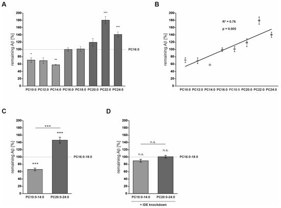
In Neuro2a cells, PCs containing MCFAs and shorter-chained FAs (PC10:0–14:0) significantly stimulated the degradation of exogenous human Aβ40 peptides conducted by IDE, one of the major Aβ-degrading proteases [20][26][27]. The elevated Aβ degradation in the presence of PC10:0–14:0 was accompanied by changes in IDE sorting and a direct stimulating effect on IDE activity (Figure 3). PC10:0–14:0 was found to specifically increase the release of IDE into the extracellular space, a process which occurs at least partially in association with exosomes [19][28]. IDE exosomally released by Neuro2a has been reported to be proteolytically active as the inhibition of exosome release leads to increased endogenous Aβ levels in the cell culture supernatant [19]. Considering that both exosome release in general and exosome-associated IDE secretion are known to be strongly affected by various lipids [28][14][29][30][31][32], one can assume that saturated MCFAs and shorter-chained FAs stimulate the exosomal IDE secretion into the extracellular compartment. This might lead to the elevated degradation of synthetic human Aβ40 peptides added to the medium of cells treated with PC10:0–14:0, which could be further strengthened by the increased IDE activity in presence of these PC species. In this context, a recent publication of Song et al. should be mentioned which reports that IDE is not secreted from cultured cells, but rather released nonspecifically as a consequence of reduced membrane integrity. In this study, only ~1% of total cellular IDE was released from HEK-293 and BV-2 cells. In the conditioned medium of intact Neuro2a cells, the enzyme was almost undetectable, even after concentrating the samples 10-fold [33]. In contrast, we and others [19] found measurable amounts of IDE in the cell culture supernatant of this cell line. Possible reasons for these divergent results include differences in the used growth medium, the cell to medium ratios, the methods for the concentration and detection of IDE as well as clonal heterogeneity. Further, Song et al. found LDH and two other measured cytosolic markers (glyceraldehyde dehydrogenase (GAPDH) and pitrilysin) to be released at the same relative levels as IDE from HEK-293 and BV-2 cells treated with lovastatin, implying that IDE is released nonspecifically by lysed cells [33]. However, unlike lovastatin, PC10:0–14:0 seems to specifically stimulate the secretion of IDE since it did not affect the release of SEAP, a marker for the constitutive secretory pathway [34]. Additionally, two established indicators for cytolysis, the activity of LDH in the conditioned cell culture medium and the cellular uptake of PI [35][36], were unaltered by the different phospholipids. Altogether, these data make a disruption of plasma membrane integrity very unlikely as the cause for the increased IDE release from cells treated with PC10:0–14:0. This is in line with other studies reporting a release of IDE into the cell culture supernatant in the absence of detectable LDH [20][19]. The reason for these differences is unclear but could be based on differences in the cell lines used and the cytotoxic potential of the applied stimulants.
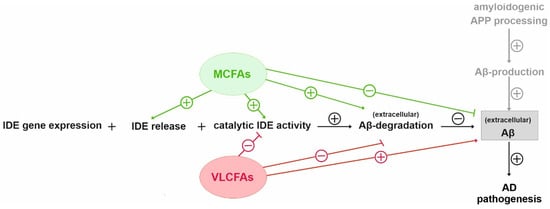
Figure 3. Schematic overview of the proposed effects of MCFAs and VLCFAs on IDE dependent Aβ degradation. The increased Aβ degradation in the presence of PC10:0–14:0 seemed to be based on changes in IDE sorting and a direct stimulating effect on IDE activity. In contrast, PC20:0–24:0 decreased the IDE-dependent Aβ degradation, probably by directly inhibiting the catalytic activity of the enzyme.
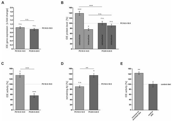
On the other hand, the saturated VLFAs contained in PC20:0–24:0 significantly reduced the IDE-dependent Aβ degradation, probably by directly inhibiting the catalytic activity of the enzyme (Figure 3). In line with these data, an in vitro-inhibition of IDE-dependent insulin degradation by long-chain free FAs (C16–C20) with IC50-values ranging from 10 to 50 µM has been reported in the study by Hamel et al., mentioned above [37]. A direct interaction between FAs and IDE is further supported by the suspected “cytosolic fatty-acid binding proteins signature” within the enzyme and our previous observation that different FAs distinctly bind to recombinant IDE [37][14]. Altogether, our cell culture data indicate that saturated MCFAs and shorter-chained FAs are more beneficial than saturated longer-chained FAs and VLCFAs with respect to IDE-dependent Aβ degradation.
An increased IDE activity was also observed in the serum of mice upon dietary supplementation of MCFAs in the form of coconut oil. We would like to point out that the effects obtained with transgenic mice fed with coconut oil are comparable to the effects observed in cell culture. By utilizing an IDE knock-down, we could confirm that the major effect of coconut oil is linked to the Aβ degrading enzyme IDE. Due to limitation of samples size in mice, this confirmation which would be ideally done by a specific inhibitor of IDE (e.g., 6bK) was not possible. Therefore, we cannot rule out that, in vivo, other proteases besides IDE are affected by coconut oil, which is a potential caveat in our study. In addition, we suggest further studies investigating whether mutations in the “cytosolic fatty acid binding protein signature” would reinforce the notion that MCFAs regulate IDE. In order to address this question, further research is warranted.

References

  1. Grimm, M.O.; Mett, J.; Grimm, H.S.; Hartmann, T. APP Function and Lipids: A Bidirectional Link. Front. Mol. Neurosci. 2017, 10, 63.
  2. Mett, J.; Hartmann, T.; Grimm, M.O.W. The Effects of Glycerophospholipids and Fatty Acids on APP Processing: Implications for Alzheimer’s Disease. In Handbook of Lipids in Human Function; Elsevier: Urbana, IL, USA, 2016; pp. 377–421.
  3. Fernando, W.M.; Martins, I.J.; Goozee, K.G.; Brennan, C.S.; Jayasena, V.; Martins, R.N. The role of dietary coconut for the prevention and treatment of Alzheimer’s disease: Potential mechanisms of action. Br. J. Nutr. 2015, 114, 1–14.
  4. Chatterjee, P.; Fernando, M.; Fernando, B.; Dias, C.B.; Shah, T.; Silva, R.; Williams, S.; Pedrini, S.; Hillebrandt, H.; Goozee, K.; et al. Potential of coconut oil and medium chain triglycerides in the prevention and treatment of Alzheimer’s disease. Mech. Ageing Dev. 2020, 186, 111209.
  5. Henderson, S.T.; Vogel, J.L.; Barr, L.J.; Garvin, F.; Jones, J.J.; Costantini, L.C. Study of the ketogenic agent AC-1202 in mild to moderate Alzheimer’s disease: A randomized, double-blind, placebo-controlled, multicenter trial. Nutr. Metab. 2009, 6, 31.
  6. Rebello, C.J.; Keller, J.N.; Liu, A.G.; Johnson, W.D.; Greenway, F.L. Pilot feasibility and safety study examining the effect of medium chain triglyceride supplementation in subjects with mild cognitive impairment: A randomized controlled trial. BBA Clin. 2015, 3, 123–125.
  7. Reger, M.A.; Henderson, S.T.; Hale, C.; Cholerton, B.; Baker, L.D.; Watson, G.S.; Hyde, K.; Chapman, D.; Craft, S. Effects of beta-hydroxybutyrate on cognition in memory-impaired adults. Neurobiol. Aging 2004, 25, 311–314.
  8. Taylor, M.K.; Sullivan, D.K.; Mahnken, J.D.; Burns, J.M.; Swerdlow, R.H. Feasibility and efficacy data from a ketogenic diet intervention in Alzheimer’s disease. Alzheimers Dement. Transl. Res. Clin. Interv. 2018, 4, 28–36.
  9. Ota, M.; Matsuo, J.; Ishida, I.; Takano, H.; Yokoi, Y.; Hori, H.; Yoshida, S.; Ashida, K.; Nakamura, K.; Takahashi, T.; et al. Effects of a medium-chain triglyceride-based ketogenic formula on cognitive function in patients with mild-to-moderate Alzheimer’s disease. Neurosci. Lett. 2019, 690, 232–236.
  10. Hu Yang, I.; De la Rubia Orti, J.E.; Selvi Sabater, P.; Sancho Castillo, S.; Rochina, M.J.; Manresa Ramon, N.; Montoya-Castilla, I. Coconut Oil: Non-Alternative Drug Treatment against Alzheimer S Disease. Nutr. Hosp. 2015, 32, 2822–2827.
  11. de la Rubia Orti, J.E.; Garcia-Pardo, M.P.; Drehmer, E.; Sancho Cantus, D.; Julian Rochina, M.; Aguilar, M.A.; Hu Yang, I. Improvement of Main Cognitive Functions in Patients with Alzheimer’s Disease after Treatment with Coconut Oil Enriched Mediterranean Diet: A Pilot Study. J. Alzheimers Dis. 2018, 65, 577–587.
  12. Mett, J.; Muller, U. The medium-chain fatty acid decanoic acid reduces oxidative stress levels in neuroblastoma cells. Sci. Rep. 2021, 11, 6135.
  13. Sastry, P.S. Lipids of nervous tissue: Composition and metabolism. Prog. Lipid Res. 1985, 24, 69–176.
  14. Grimm, M.O.; Mett, J.; Stahlmann, C.P.; Haupenthal, V.J.; Blumel, T.; Stotzel, H.; Grimm, H.S.; Hartmann, T. Eicosapentaenoic acid and docosahexaenoic acid increase the degradation of amyloid-beta by affecting insulin-degrading enzyme. Biochem. Cell Biol. 2016, 94, 534–542.
  15. Grimm, M.O.; Stahlmann, C.P.; Mett, J.; Haupenthal, V.J.; Zimmer, V.C.; Lehmann, J.; Hundsdorfer, B.; Endres, K.; Grimm, H.S.; Hartmann, T. Vitamin E: Curse or Benefit in Alzheimer’s Disease? A Systematic Investigation of the Impact of alpha-, gamma- and delta-Tocopherol on Ass Generation and Degradation in Neuroblastoma Cells. J. Nutr. Health Aging 2015, 19, 646–656.
  16. Liberato, M.V.; Nascimento, A.S.; Ayers, S.D.; Lin, J.Z.; Cvoro, A.; Silveira, R.L.; Martinez, L.; Souza, P.C.; Saidemberg, D.; Deng, T.; et al. Medium chain fatty acids are selective peroxisome proliferator activated receptor (PPAR) gamma activators and pan-PPAR partial agonists. PLoS ONE 2012, 7, e36297.
  17. Du, J.; Zhang, L.; Liu, S.; Zhang, C.; Huang, X.; Li, J.; Zhao, N.; Wang, Z. PPARgamma transcriptionally regulates the expression of insulin-degrading enzyme in primary neurons. Biochem. Biophys. Res. Commun. 2009, 383, 485–490.
  18. Vekrellis, K.; Ye, Z.; Qiu, W.Q.; Walsh, D.; Hartley, D.; Chesneau, V.; Rosner, M.R.; Selkoe, D.J. Neurons regulate extracellular levels of amyloid beta-protein via proteolysis by insulin-degrading enzyme. J. Neurosci. 2000, 20, 1657–1665.
  19. Bulloj, A.; Leal, M.C.; Xu, H.; Castano, E.M.; Morelli, L. Insulin-degrading enzyme sorting in exosomes: A secretory pathway for a key brain amyloid-beta degrading protease. J. Alzheimers. Dis. 2010, 19, 79–95.
  20. Qiu, W.Q.; Walsh, D.M.; Ye, Z.; Vekrellis, K.; Zhang, J.; Podlisny, M.B.; Rosner, M.R.; Safavi, A.; Hersh, L.B.; Selkoe, D.J. Insulin-degrading enzyme regulates extracellular levels of amyloid beta-protein by degradation. J. Biol. Chem. 1998, 273, 32730–32738.
  21. Liu, Z.; Zhu, H.; Fang, G.G.; Walsh, K.; Mwamburi, M.; Wolozin, B.; Abdul-Hay, S.O.; Ikezu, T.; Leissring, M.A.; Qiu, W.Q. Characterization of insulin degrading enzyme and other amyloid-beta degrading proteases in human serum: A role in Alzheimer’s disease? J. Alzheimers Dis. 2012, 29, 329–340.
  22. Nalivaeva, N.N.; Turner, A.J. Targeting amyloid clearance in Alzheimer’s disease as a therapeutic strategy. Br. J. Pharmacol. 2019, 176, 3447–3463.
  23. Saido, T.; Leissring, M.A. Proteolytic degradation of amyloid beta-protein. Cold Spring Harb. Perspect. Med. 2012, 2, a006379.
  24. Kadowaki, H.; Grant, M.A. Relationship of membrane phospholipid composition, lactosylceramide molecular species, and the specificity of CMP-N-acetylneuraminate:lactosylceramide alpha 2,3-sialyltransferase to the molecular species composition of GM3 ganglioside. J. Lipid Res. 1995, 36, 1274–1282.
  25. Werlein, A.; Peters, A.; Ngoune, R.; Winkler, K.; Putz, G. Interference of phosphatidylcholines with in-vitro cell proliferation—No flock without black sheep. Biochim. Biophys. Acta 2015, 1848, 1599–1608.
  26. Farris, W.; Mansourian, S.; Chang, Y.; Lindsley, L.; Eckman, E.A.; Frosch, M.P.; Eckman, C.B.; Tanzi, R.E.; Selkoe, D.J.; Guenette, S. Insulin-degrading enzyme regulates the levels of insulin, amyloid beta-protein, and the beta-amyloid precursor protein intracellular domain in vivo. Proc. Natl. Acad. Sci. USA 2003, 100, 4162–4167.
  27. Miller, B.C.; Eckman, E.A.; Sambamurti, K.; Dobbs, N.; Chow, K.M.; Eckman, C.B.; Hersh, L.B.; Thiele, D.L. Amyloid-beta peptide levels in brain are inversely correlated with insulysin activity levels in vivo. Proc. Natl. Acad. Sci. USA 2003, 100, 6221–6226.
  28. Tamboli, I.Y.; Barth, E.; Christian, L.; Siepmann, M.; Kumar, S.; Singh, S.; Tolksdorf, K.; Heneka, M.T.; Lutjohann, D.; Wunderlich, P.; et al. Statins promote the degradation of extracellular amyloid -peptide by microglia via stimulation of exosome-associated insulin-degrading enzyme (IDE) secretion. J. Biol. Chem. 2010, 285, 37405–37414.
  29. Llorente, A.; van Deurs, B.; Sandvig, K. Cholesterol regulates prostasome release from secretory lysosomes in PC-3 human prostate cancer cells. Eur. J. Cell Biol. 2007, 86, 405–415.
  30. Phuyal, S.; Hessvik, N.P.; Skotland, T.; Sandvig, K.; Llorente, A. Regulation of exosome release by glycosphingolipids and flotillins. FEBS J. 2014, 281, 2214–2227.
  31. Record, M.; Carayon, K.; Poirot, M.; Silvente-Poirot, S. Exosomes as new vesicular lipid transporters involved in cell-cell communication and various pathophysiologies. Biochim. Biophys. Acta 2014, 1841, 108–120.
  32. Trajkovic, K.; Hsu, C.; Chiantia, S.; Rajendran, L.; Wenzel, D.; Wieland, F.; Schwille, P.; Brugger, B.; Simons, M. Ceramide triggers budding of exosome vesicles into multivesicular endosomes. Science 2008, 319, 1244–1247.
  33. Song, E.S.; Rodgers, D.W.; Hersh, L.B. Insulin-degrading enzyme is not secreted from cultured cells. Sci. Rep. 2018, 8, 2335.
  34. Gorr, S.U.; Moore, Y.R. Sorting of a constitutive secretory protein to the regulated secretory pathway of exocrine cells. Biochem. Biophys. Res. Commun. 1999, 257, 545–548.
  35. Kumar, P.; Nagarajan, A.; Uchil, P.D. Analysis of Cell Viability by the Lactate Dehydrogenase Assay. Cold Spring Harb. Protoc. 2018, 2018, pdb.prot095497.
  36. Crowley, L.C.; Scott, A.P.; Marfell, B.J.; Boughaba, J.A.; Chojnowski, G.; Waterhouse, N.J. Measuring Cell Death by Propidium Iodide Uptake and Flow Cytometry. Cold Spring Harb. Protoc. 2016, 2016, pdb-prot087163.
  37. Hamel, F.G.; Upward, J.L.; Bennett, R.G. In vitro inhibition of insulin-degrading enzyme by long-chain fatty acids and their coenzyme A thioesters. Endocrinology 2003, 144, 2404–2408.
More
Information
Contributor MDPI registered users' name will be linked to their SciProfiles pages. To register with us, please refer to https://encyclopedia.pub/register :
View Times: 295
Entry Collection: Neurodegeneration
Revisions: 2 times (View History)
Update Date: 03 Nov 2021
1000/1000