Submitted Successfully!
To reward your contribution, here is a gift for you: A free trial for our video production service.
Thank you for your contribution! You can also upload a video entry or images related to this topic.
Version Summary Created by Modification Content Size Created at Operation
1 n/a + 1574 word(s) 1574 2020-06-01 10:32:02 |
2 format correct Meta information modification 1574 2020-06-03 03:39:18 | |
3 format correct -14 word(s) 1560 2020-10-28 03:57:55 |

Video Upload Options

Do you have a full video?

Confirm

Are you sure to Delete?
Cite
If you have any further questions, please contact Encyclopedia Editorial Office.
Han, P.; Ivanovski, S. Salivary Diagnosis and Monitoring: COVID-19. Encyclopedia. Available online: https://encyclopedia.pub/entry/969 (accessed on 23 April 2024).
Han P, Ivanovski S. Salivary Diagnosis and Monitoring: COVID-19. Encyclopedia. Available at: https://encyclopedia.pub/entry/969. Accessed April 23, 2024.
Han, Pingping, Sašo Ivanovski. "Salivary Diagnosis and Monitoring: COVID-19" Encyclopedia, https://encyclopedia.pub/entry/969 (accessed April 23, 2024).
Han, P., & Ivanovski, S. (2020, June 01). Salivary Diagnosis and Monitoring: COVID-19. In Encyclopedia. https://encyclopedia.pub/entry/969
Han, Pingping and Sašo Ivanovski. "Salivary Diagnosis and Monitoring: COVID-19." Encyclopedia. Web. 01 June, 2020.
Salivary Diagnosis and Monitoring: COVID-19
Edit

The coronavirus disease 2019 (COVID-19) outbreak, caused by the novel severe acute respiratory syndrome coronavirus 2 (SARS-CoV-2), has become a global ongoing pandemic. Timely, accurate and non-invasive SARS-CoV-2 detection in both symptomatic and asymptomatic patients, as well as determination of their immune status, will facilitate effective large-scale pandemic control measures to prevent the spread of COVID-19. Saliva is a biofluid whose anatomical source and location is of particularly strategic relevance to COVID-19 transmission and monitoring. This review focuses on the role of saliva as both a foe (a common mode of viral transmission via salivary droplets and potentially aerosols) and a friend (as a non-invasive diagnostic tool for viral detection and immune status surveillance) in combating COVID-19.

COVID-19 salivary diagnostics salivary bioaerosols transmission

1. The Versatility of Saliva for COVID-19 Diagnosis

The timing (highest viral titres) and specimen collection sources can significantly influence the diagnostic sensitivity of SARS-CoV-2 detection tests. One study reported that oropharyngeal swabs (n = 398) were more often used than nasopharyngeal swabs (n = 8) in China during the COVID19 outbreak; however, SARS-CoV-2 RNA was detected in only 32% of oropharyngeal swabs [1]. On 19 March 2020, the World Health Organisation (WHO) recommended that both upper (nasopharyngeal and oropharyngeal swabs) and lower (sputum, bronchoalveolar, or lavage endotracheal aspirate) respiratory specimens should be collected; however, upper respiratory samples may fail to detect early viral infection and the collection of lower respiratory specimens increases biosafety risk to healthcare workers via aerosol/droplets formation. As the SARS-CoV-2 virus shedding progresses, additional samples sources, such as stool, saliva, and blood, can be used as alternatives, or combined with respiratory specimens. However, only 15% of patients hospitalised with pneumonia had detectable SARS-CoV-2 RNA in serum [2], and 55% of patients showed positive SARS-CoV-2 RNA in fecal samples [3]. Conversely, in saliva samples, it was reported from different clinical studies that 87%, 91.6%, and 100% of COVID-19 patients were identified as being viral positive, respectively [4][5][6], suggesting that saliva is a powerful specimen source for the diagnosis of the SARS-CoV-2 virus.

Saliva also represents an attractive biofluid source option for the detection of SARS-CoV-2, due to being non-invasive, easy-to-access, and low-cost, as well as having the ability to “mirror” systemic and local disease status [7]. It is well-known that saliva harbors a wide range of circulatory components (Figure 1), such as pro-inflammatory cytokines [8][9], chemokines [10], matrix metalloproteinases [11][12], mitochondrial DNA [13], genomic DNA [14], bacteria [15], SARS-CoV and SARS-CoV-2 virus [16][4][5], SARS-CoV antibodies [16], miRNAs [17], and extracellular vesicles (EVs) [18]. Furthermore, saliva samples can be stored at –80 °C for several years with little degradation [19]. It is preferable to aliquot and freeze the samples to avoid freeze–thaw cycles. For salivary RNA research, it was discovered that saliva samples can be stored in Trizol for more than two years at –80 °C without adding RNase inhibitors [20][21], suggesting such specimens can be used for future diagnostics. Thus, saliva may be a valuable specimen to collect in COVID-19 patients at different time points during disease onset progression and follow-up. Indeed, saliva may be useful for both diagnosing the presence and sequelae of COVID-19 infection, as well as identifying and tracking the development of immunity to the virus.

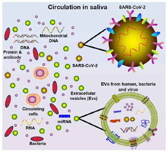
Figure 1. Schematic diagram of saliva components, including cells, mitochondrial DNA, DNA, protein/antibody, bacteria, miRNA, extracellular vesicles (EVs, from multiple oral cavity resident species), and SARS-CoV-2 virus.

2. Salivary Diagnostics for COVID-19

Saliva has been widely investigated as a potential diagnostic tool for chronic systemic and local (oral) diseases [7], with less attention given to its utility in acute infectious diseases, such as COVID-19. The salivary gland can be infected by SARS-CoV-2 virus resulting in the subsequent release of viral particles or antibodies into saliva, as evidenced in Rhesus macaque primates where salivary gland epithelial cells were the first target cells for SARS-CoV infection [16]. This is likely to be facilitated by the high expression of hACE2 (SARS-CoV-2 receptor) on the epithelial cells of the oral mucosa, as demonstrated using single-cell RNA sequencing [22].

Saliva and throat wash (by gargling 10 mL saline) samples from 17 SARS-CoV patients were found to be SARS-CoV RNA positive, with the highest detection rate a median of four days after disease onset and during lung lesion development [23]. Saliva samples from 75 patients successfully validated saliva as a viable biosample source for COVID-19 detection when compared to nasopharyngeal or oropharyngeal swabs [24].

At present, only three clinical studies (Table 1) and one animal model have investigated the use of salivary diagnostics for COVID-19. SARS-CoV-2 was detected in self-collected saliva (by asking the patients to expectorate saliva) in 11 out of 12 confirmed cases [5]. Another recent study found that 100% of COVID-19 patients (n = 25) were detected as viral positive in drooling saliva samples [6]. Further, in a cohort of COVID-19 positive patients, it has been demonstrated that 87% of posterior oropharyngeal (deep throat) saliva samples were detected viral positive (n = 23), and serial respiratory viral load of SARS-CoV-2 was detected from week 1 and up to 25 days after symptom onset, while serum (n = 16) samples showed positive RT-qPCR detection only 14 days after symptom onset [4]. Additionally, Kim et al. demonstrated that SARS-CoV-2-infected ferret animals shed virus in nasal washes, saliva, urine, and feces up to eight days post-infection and ferret-to-ferret transmission occurred only two days post-contact [25]. Notwithstanding the limitations of small sample size and lack of detailed saliva collection methodology, these studies nevertheless imply that saliva is a promising non-invasive alternative specimen for SARS-CoV-2 diagnosis. Further investigations are required to explore the potential role of saliva for COVID-19 detection in both symptomatic and asymptomatic patients.

Table 1. Current clinical research finding using salivary diagnosis for COVID-19.

Sample Size
Age (Years)
Sample Source Diagnosis Technique Diagnosis Efficiency in Saliva Reference
To et al. 10 Female, 13 Male
Median: 62 (37–75)
Posterior oropharyngeal saliva RT-qPCR 87% of patients were viral positive [4]
To et al. 5 Female, 7 Male
Median: 62.5 (35–75)
Saliva from throat RT-qPCR 91.7% of patients were positive [5]
Azzi et al. 8 Female, 17 Male
Mean ± standard deviation: 61.5 ± 11.2
Drooling saliva RT-qPCR 100% of patients were viral positive [6]

In summary, the current gold standard diagnostic test is RT-qPCR to detect SARS-CoV-2 RNA which takes approximately 48 h to obtain the test results. More new tests with higher sensitivity and specificity need to be appropriately validated before being implemented into the current routine diagnosis.

3. Salivary Immunity Monitoring for COVID-19

From early reports on the clinical characteristics of COVID-19, it is now apparent that not all people exposed to SARS-CoV-2 are infected and not all infected patients develop severe symptoms [26]. Indeed, three broad presentations of SARS-CoV-2 infection can be characterised: (i) an asymptomatic incubation stage with or without detectable virus; (ii) non-severe symptomatic presentation with confirmed presence of virus; and (iii) a severe respiratory symptomatic stage with high viral load [27]. Determining the immune status of an individual is likely to become increasingly critical as the COVID-19 pandemic progresses, because from a prevention perspective, individuals at stage I (the stealth carriers or the super spreaders), are particularly important because they may spread the virus unknowingly.

Two stages of the immune response during COVID-19 disease progression have been proposed [28]: (1) Immune-defense-based protective phase: elimination of SARS-CoV-2 virus by an individual’s adaptive immune response; and (2) inflammation-driven phase: when the protective immune response is impaired and prolonged propagated virus load leads to an adverse inflammatory response in organs with high hACEs expression. Indeed, a likely pathogenic mechanism of SARS-CoV-2 is overactivation of T cells with an increase in CD4+ T Helper cells and enhanced cytotoxicity of CD4+ and CD8+ T cells [29], which leads to an imbalanced pro-inflammatory and anti-inflammatory cytokine response and severe immune injury in susceptible patients [30]. Although this concept needs to be confirmed by more clinical research, it may provide useful research directions to tackle COVID-19.

During the previous SARS outbreak, a common transmission pattern hypothesis was that SARS-CoV virus silently infected asymptomatic patients, which may have led to population immunity against infection (herd immunity) that may explain the eradication of the virus, although this is yet to be confirmed [31]. Although a study suggests that coronavirus antibodies are highly prevalent in the general population after exposure to four non-SARS coronavirus strains [32], there is no definitive evidence on whether permanent immunity would be generated against other CoV species, such as SARS-COV-2. Notably, after SARS-CoV infection in a murine model, the production of SARS-CoV-specific serum IgG and secretory immunoglobulin A (sIgA) were detected in saliva following intranasal immunisation [33].

In relation to COVID-19, intensive care unit (ICU) patients had higher plasma levels of pro-inflammatory cytokines, including IL-2, IL-7, IL-10, GSCF, IP10, MCP-1, MIP-1A, and TNF-α, compared with non-ICU patients [2], suggesting the emergence of a robust immune-inflammatory response in severe symptomatic COVID-19 patients. Importantly, several studies have demonstrated that COVID-19 patients developed IgG and IgM antibodies against SARS-CoV-2 in blood samples. Both IgG and IgM antibodies against the SARS-CoV-2 nucleoprotein and spike receptor-binding domain were increased in serum at day 10 after symptom onset for up to three weeks [4]. A point-of-care lateral flow immunoassay (LFIA) test product (VivaDiag COVID-19 IgM/IgG Rapid Test) was designed to detect IgM and IgG in blood samples of COVID-19 patients in 15 min [34]. However, the sensitivity of the VivaDiag COVID-19 IgM/IgG Rapid Test was only 18.4% in blood samples of acute COVID-19 patients from the emergency department [35], suggesting that serological tests require more research before being deemed suitable for routine diagnosis. Additionally, the seroconversion rate for total antibodies, IgM, and IgG were shown to be 93.1%, 82.7%, and 64.7%, respectively, in hospitalised COVID-19 patients, peaking 7–14 days after symptom onset [36]. Given the non-invasive and cost-effective nature of saliva collection, it would be important to investigate whether this immunity detection is feasible in saliva samples as a tool for facilitating the testing of COVID-19 immunity at the population-level.

References

  1. Wang, W.; Xu, Y.; Gao, R.; Lu, R.; Han, K.; Wu, G.; Tan, W. Detection of SARS-CoV-2 in Different Types of Clinical Specimens. JAMA 2020.
  2. Huang, C.; Wang, Y.; Li, X.; Ren, L.; Zhao, J.; Hu, Y.; Zhang, L.; Fan, G.; Xu, J.; Gu, X.; et al.et al. Clinical features of patients infected with 2019 novel coronavirus in Wuhan, China. Lancet 2020, 395, 497–506.
  3. Yongjian Wu; Cheng Guo; Lantian Tang; Zhongsi Hong; Jianhui Zhou; Xin Dong; Huan Yin; Qiang Xiao; Yanping Tang; Xiujuan Qu; et al.Liangjian KuangXiaomin FangNischay MishraJiahai LuHong ShanGuanmin JiangXi Huang Prolonged presence of SARS-CoV-2 viral RNA in faecal samples. The Lancet Gastroenterology & Hepatology 2020, 5, 434-435, 10.1016/s2468-1253(20)30083-2.
  4. Kelvin Kai-Wang To; Owen Tak-Yin Tsang; Wai-Shing Leung; Anthony Raymond Tam; Tak-Chiu Wu; David Christopher Lung; Cyril Chik-Yan Yip; Jian-Piao Cai; Jacky Man-Chun Chan; Thomas Shiu-Hong Chik; Daphne Pui-Ling Lau; Chris Yau-Chung Choi; Lin-Lei Chen; Wan-Mui Chan; Kwok-Hung Chan; Jonathan Daniel Ip; Anthony Chin-Ki Ng; Rosana Wing-Shan Poon; Cui-Ting Luo; Vincent Chi-Chung Cheng; Jasper Fuk-Woo Chan; Ivan Fan-Ngai Hung; Zhiwei Chen; Honglin Chen; Kwok-Yung Yuen; Temporal profiles of viral load in posterior oropharyngeal saliva samples and serum antibody responses during infection by SARS-CoV-2: an observational cohort study. The Lancet Infectious Diseases 2020, 20, 565-574, 10.1016/s1473-3099(20)30196-1.
  5. Kelvin Kai-Wang To; Owen Tak-Yin Tsang; Cyril Chik-Yan Yip; Kwok-Hung Chan; Tak-Chiu Wu; Jacky Man-Chun Chan; Wai-Shing Leung; Thomas Shiu-Hong Chik; Chris Yau-Chung Choi; Darshana H Kandamby; David Christopher Lung; Anthony Raymond Tam; Rosana Wing-Shan Poon; Agnes Yim-Fong Fung; Ivan Fan-Ngai Hung; Vincent Chi-Chung Cheng; Jasper Fuk-Woo Chan; Kwok-Yung Yuen; Consistent Detection of 2019 Novel Coronavirus in Saliva. Clinical Infectious Diseases 2020, null, , 10.1093/cid/ciaa149.
  6. Azzi, L.; Carcano, G.; Gianfagna, F.; Grossi, P.; Gasperina, D.D.; Genoni, A.; Fasano, M.; Sessa, F.; Tettamanti, L.; Carinci, F.; et al. SALIVA IS A RELIABLE TOOL TO DETECT SARS-CoV-2. J. Infect. 2020.
  7. Tina Pfaffe; Justin J. Cooper-White; Peter Beyerlein; Karam Kostner; Chamindie Punyadeera; Diagnostic Potential of Saliva: Current State and Future Applications. Clinical Chemistry 2011, 57, 675-687, 10.1373/clinchem.2010.153767.
  8. Nurcan Buduneli; Denis F. Kinane; Host-derived diagnostic markers related to soft tissue destruction and bone degradation in periodontitis. Journal of Clinical Periodontology 2011, 38, 85-105, 10.1111/j.1600-051x.2010.01670.x.
  9. Kinane, D.F.; P, P.M.; Loos, B.G. Host-response: Understanding the cellular and molecular mechanisms of hostmicrobial interactions: Consensus of the 7th European Workshop on Periodontology. J. Clin. Periodontol. 2011, 38, 44–48. [Google Scholar] [CrossRef] [PubMed]
  10. T A Silva; G P Garlet; S.Y. Fukada; J S Silva; F Q Cunha; Chemokines in oral inflammatory diseases: apical periodontitis and periodontal disease.. Journal of Dental Research 2007, 86, 306–319.
  11. J.L. Ebersole; Julie L. Schuster; Jason Stevens; Dolph Dawson; Richard J. Kryscio; Yushin Lin; Mark V. Thomas; Craig S. Miller; Patterns of Salivary Analytes Provide Diagnostic Capacity for Distinguishing Chronic Adult Periodontitis from Health. Journal of Clinical Immunology 2012, 33, 271-279, 10.1007/s10875-012-9771-3.
  12. W.V. Giannobile; Khalaf F. Al-Shammari; David P. Sarment; Matrix molecules and growth factors as indicators of periodontal disease activity. Periodontology 2000 2003, 31, 125-134, 10.1034/j.1600-0757.2003.03108.x.
  13. Wei-Wen Jiang; Brett Masayesva; Marianna Zahurak; Andre L. Carvalho; Eli Rosenbaum; Elizabeth Mambo; Shaoyu Zhou; Khalid Minhas; Nicole Benoit; William H. Westra; et al.Anthony AlbergDavid SidranskyWayne KochJoseph Califano Increased Mitochondrial DNA Content in Saliva Associated with Head and Neck Cancer. Clinical Cancer Research 2005, 11, 2486-2491, 10.1158/1078-0432.ccr-04-2147.
  14. Thais Francini Garbieri; Daniel Thomas Brozoski; Thiago J. Dionísio; Carlos F. Santos; Lucimara Neves; Human DNA extraction from whole saliva that was fresh or stored for 3, 6 or 12 months using five different protocols. Journal of Applied Oral Science 2017, 25, 147-158, 10.1590/1678-77572016-0046.
  15. Toru Takeshita; Shinya Kageyama; Michiko Furuta; Hidenori Tsuboi; Kenji Takeuchi; Yukie Shibata; Yoshihiro Shimazaki; Sumio Akifusa; Toshiharu Ninomiya; Yutaka Kiyohara; et al.Yoshihisa Yamashita Bacterial diversity in saliva and oral health-related conditions: the Hisayama Study. Scientific Reports 2016, 6, 22164, 10.1038/srep22164.
  16. Li Liu; Qiang Wei; Xavier Alvarez; Haibo Wang; Yanhua Du; Hua Zhu; Hong Jiang; Jingying Zhou; Pokman Lam; Linqi Zhang; et al.Andrew LacknerChuan QinZhiwei Chen Epithelial Cells Lining Salivary Gland Ducts Are Early Target Cells of Severe Acute Respiratory Syndrome Coronavirus Infection in the Upper Respiratory Tracts of Rhesus Macaques. Journal of Virology 2011, 85, 4025-4030, 10.1128/jvi.02292-10.
  17. Alireza Tahamtan; Christopher S. Inchley; Mona Marzban; Masoumeh Tavakoli-Yaraki; Majid Teymoori-Rad; Britt Nakstad; Vahid Salimi; The role of microRNAs in respiratory viral infection: friend or foe?. Reviews in Medical Virology 2016, 26, 389-407, 10.1002/rmv.1894.
  18. Marta Cristaldi; Rodolfo Mauceri; Olga Di Fede; Giovanna Giuliana; Giuseppina Campisi; Vera Panzarella; Salivary Biomarkers for Oral Squamous Cell Carcinoma Diagnosis and Follow-Up: Current Status and Perspectives. Frontiers in Physiology 2019, 10, 1476, 10.3389/fphys.2019.01476.
  19. Silvia Chiappin; Giorgia Antonelli; Rosalba Gatti; Elio Franco De Palo; Saliva specimen: A new laboratory tool for diagnostic and basic investigation. Clinica Chimica Acta 2007, 383, 30-40, 10.1016/j.cca.2007.04.011.
  20. Pratibala Pandit; Justin J. Cooper-White; Chamindie Punyadeera; High-Yield RNA-Extraction Method for Saliva. Clinical Chemistry 2013, 59, 1118-1122, 10.1373/clinchem.2012.197863.
  21. Pingping Han; Saso Ivanovski; Effect of Saliva Collection Methods on the Detection of Periodontium-Related Genetic and Epigenetic Biomarkers-A Pilot Study.. International Journal of Molecular Sciences 2019, 20, 4729, 10.3390/ijms20194729.
  22. Hao Xu; Liang Zhong; Jiaxin Deng; Jiakuan Peng; Hongxia Dan; Xin Zeng; Taiwen Li; Qianming Chen; High expression of ACE2 receptor of 2019-nCoV on the epithelial cells of oral mucosa. International Journal of Oral Science 2020, 12, 1-5, 10.1038/s41368-020-0074-x.
  23. Wei-Kung Wang; Shey-Ying Chen; I-Jung Liu; Yee-Chun Chen; Hui-Ling Chen; Chao-Fu Yang; Pei-Jer Chen; Shiou-Hwei Yeh; Chuan-Liang Kao; Li-Min Huang; et al.Po-Ren HsuehJann-Tay WangWang-Hwei ShengChi‐Tai FangChien‐Ching HungSzu-Min HsiehChan-Ping SuWen-Chu ChiangJyh-Yuan YangJih-Hui LinHsien-Ping HuYu-Ping ChiangJin-Town WangPan-Chyr YangSui-Yuan Changmembers of the SARS Research Group of the National Taiwan UniversityNational Taiwan University Hospital Detection of SARS-associated Coronavirus in Throat Wash and Saliva in Early Diagnosis. Emerging Infectious Diseases 2004, 10, 1213-1219, 10.3201/eid1007.031113.
  24. Researchers Validate Viability of Saliva in COVID-19 Detection. Available online: https://www.mpo-mag.com/contents/view_breaking-news/2020-04-03/researchers-discover-that-saliva-can-detect-covid-19/ (accessed on 4 March 2020).
  25. Young-Il Kim; Seong-Gyu Kim; Se-Mi Kim; Eun-Ha Kim; Su-Jin Park; Kwang-Min Yu; Jae-Hyung Chang; Seunghun Lee; Mark Anthony B. Casel; Jihye Um; et al.Min-Suk SongHye Won JeongVan Dam LaiYeonjae KimBum Sik ChinJun-Sun ParkKi-Hyun ChungSuan-Sin FooHaryoung PooIn-Pil MoOk-Jun LeeRichard J. WebbyJae U. JungYoung Ki ChoiEun Ji Kim Infection and Rapid Transmission of SARS-CoV-2 in Ferrets. Cell Host & Microbe 2020, 27, 704-709.e2, 10.1016/j.chom.2020.03.023.
  26. Nanshan Chen; Min Zhou; Xuan Dong; Jieming Qu; Fengyun Gong; Yang Han; Yang Qiu; Jingli Wang; Ying Liu; Yuan Wei; et al.Jia'an XiaTing YuXinxin ZhangLi Zhang Epidemiological and clinical characteristics of 99 cases of 2019 novel coronavirus pneumonia in Wuhan, China: a descriptive study. The Lancet 2020, 395, 507-513, 10.1016/s0140-6736(20)30211-7.
  27. Dawei Wang; Bo Hu; Chang Hu; Fangfang Zhu; Xing Liu; Jing Zhang; Binbin Wang; Hui Xiang; Zhenshun Cheng; Yong Xiong; et al.Yan ZhaoYirong LiXinghuan WangZhiyong Peng Clinical Characteristics of 138 Hospitalized Patients With 2019 Novel Coronavirus–Infected Pneumonia in Wuhan, China. JAMA 2020, 323, 1061, 10.1001/jama.2020.1585.
  28. Yufang Shi; Ying Wang; Changshun Shao; Jianan Huang; Jianhe Gan; Xiaoping Huang; Enrico Bucci; Mauro Piacentini; Giuseppe Ippolito; Gerry Melino; et al. COVID-19 infection: the perspectives on immune responses. Cell Death & Differentiation 2020, 27, 1451-1454, 10.1038/s41418-020-0530-3.
  29. Zhe Xu; Lei Shi; Yijin Wang; Jiyuan Zhang; Lei Huang; Chao Zhang; Shuhong Liu; Peng Zhao; Hongxia Liu; Li Zhu; et al.Yanhong TaiChangqing BaiTingting GaoJinwen SongPeng XiaJinghui DongJingmin ZhaoFu-Sheng Wang Pathological findings of COVID-19 associated with acute respiratory distress syndrome. The Lancet Respiratory Medicine 2020, 8, 420-422, 10.1016/s2213-2600(20)30076-x.
  30. Ong, E.Z.; Chan, Y.F.Z.; Leong, W.Y.; Lee, N.M.Y.; Kalimuddin, S.; Mohideen, S.M.H.; Chan, K.S.; Tan, A.T.; Bertoletti, A.; Ooi, E.E.; et al. Antonio Bertoletti, and Jenny Guek Hong Low. A dynamic immune response shapes COVID-19 progression. Cell Host Microbe 2020.
  31. Didier Raoult; Alimuddin Zumla; Franco Locatelli; Giuseppe Ippolito; Guido Kroemer; Coronavirus infections: Epidemiological, clinical and immunological features and hypotheses.. Cell Stress 2020, 4, 66-75, 10.15698/cst2020.04.216.
  32. Emily G. Severance; Ioannis Bossis; Faith B. Dickerson; Cassie R. Stallings; Andrea E. Origoni; Anne Sullens; Robert H. Yolken; Raphael P. Viscidi; Development of a Nucleocapsid-Based Human Coronavirus Immunoassay and Estimates of Individuals Exposed to Coronavirus in a U.S. Metropolitan Population. Clinical and Vaccine Immunology 2008, 15, 1805-1810, 10.1128/cvi.00124-08.
  33. Baojing Lu; Yi Huang; Li Huang; Bao Li; Zhenhua Zheng; Ze Chen; Jianjun Chen; Qinxue Hu; Hanzhong Wang; Effect of mucosal and systemic immunization with virus-like particles of severe acute respiratory syndrome coronavirus in mice. Immunology 2010, 130, 254-261, 10.1111/j.1365-2567.2010.03231.x.
  34. Li, Z.; Yi, Y.; Luo, X.; Xiong, N.; Liu, Y.; Li, S.; Sun, R.; Wang, Y.; Hu, B.; Chen, W.; et al. Development and Clinical Application of a Rapid IgM-IgG Combined Antibody Test for SARS-CoV-2 Infection Diagnosis. J. Med. Virol. 2020.
  35. Cassaniti, I.I.; Novazzi, F.; Giardina, F.; Salivaro, F.; Sachs, M.; Perlini, S.; Bruno, R.; Mojoli, F.; Baldanti, F. Performance of VivaDiag COVID-19 IgM/IgG Rapid Test is inadequate for diagnosis of COVID-19 in acute patients referring to emergency room department. J. Med. Virol. 2020.
  36. Zhao, J.; Yuan, Q.; Wang, H.; Liu, W.; Liao, X.; Su, Y.-Y.; Wang, X.; Yuan, J.; Li, T.; Li, J.-X.; et al. Antibody responses to SARS-CoV-2 in patients of novel coronavirus disease 2019. Clin. Infect. Dis. 2020.
More
Information
Subjects: Pathology
Contributors MDPI registered users' name will be linked to their SciProfiles pages. To register with us, please refer to https://encyclopedia.pub/register : ,
View Times: 948
Entry Collection: COVID-19
Revisions: 3 times (View History)
Update Date: 28 Oct 2020
1000/1000