Submitted Successfully!
To reward your contribution, here is a gift for you: A free trial for our video production service.
Thank you for your contribution! You can also upload a video entry or images related to this topic.
Version Summary Created by Modification Content Size Created at Operation
1 + 1487 word(s) 1487 2021-08-10 05:57:43 |
2 format correct Meta information modification 1487 2021-08-24 02:53:05 |

Video Upload Options

Do you have a full video?

Confirm

Are you sure to Delete?
Cite
If you have any further questions, please contact Encyclopedia Editorial Office.
Mateos, M. Diabetic Macular Oedema. Encyclopedia. Available online: https://encyclopedia.pub/entry/13486 (accessed on 18 April 2024).
Mateos M. Diabetic Macular Oedema. Encyclopedia. Available at: https://encyclopedia.pub/entry/13486. Accessed April 18, 2024.
Mateos, Milagros. "Diabetic Macular Oedema" Encyclopedia, https://encyclopedia.pub/entry/13486 (accessed April 18, 2024).
Mateos, M. (2021, August 24). Diabetic Macular Oedema. In Encyclopedia. https://encyclopedia.pub/entry/13486
Mateos, Milagros. "Diabetic Macular Oedema." Encyclopedia. Web. 24 August, 2021.
Diabetic Macular Oedema
Edit

Diabetic macular oedema (DMO) is one of the leading causes of vision loss associated with diabetic retinopathy (DR). New insights in managing this condition have changed the paradigm in its treatment, with intravitreal injections of antivascular endothelial growth factor (anti-VEGF) having become the standard therapy for DMO worldwide. However, there is no single standard therapy for all patients DMO refractory to anti-VEGF treatment; thus, further investigation is still needed.

diabetic macular oedema diabetic retinopathy macular oedema rho-associated kinases

1. Introduction

Diabetic retinopathy (DR) is one of the feared chronic complications of Diabetes Mellitus (DM), a pandemic disease that affects approximately 463 million adults nowadays. Its prevalence has been rising in recent years, exemplifying its steady increase; by 2045, 700 million adults are expected to live with diabetes. A total of 40% of diabetic patients over 40 years old suffer from DR. [1].

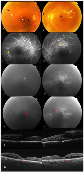
Diabetic macular oedema (DMO) is associated with an abnormal increase in fluid volume in the macula, whether infiltrating retinal layers or collecting in the subretinal space. It is a significant cause of vision loss in those patients since it primarily affects central vision. DMO may occur in any disease stage and is more frequent in insulin-dependent type 2 DM [1].

The pathogenesis of DMO is multifactorial and complex, with a combination of pathological conditions related to retinal hypoxia, vascular permeability, angiogenesis, and inflammation processes leading to the development of the characteristic retinal alterations in patients with DMO [2][3][4][5].

Since the 1970s, focal/grid laser treatment has been considered the only available treatment for the management of this and it was accepted that in general, it only stabilised functional loss but did not improve it [6]. However, the breakthrough of intravitreal antivascular endothelial growth factor (VEGF) therapy has now replaced laser treatment as several trials have shown improved outcomes [7][8], which is nowadays the treatment of choice. However, anti-VEGF therapy has several limitations since it requires multiple visits and injections, increasing the burden placed on the healthcare system. Furthermore, DMO persists in more than 40% of patients even after numerous intravitreal injections according to the protocol I trial [4]. The efficacy of anti-VEGF treatment may be less pronounced in the clinical practice setting where undertreatment is very frequent [9][10][11] ( Figure 1 ). Moreover, there are certain doubts about the long-term effect of the inhibition of a factor (VEGF) that is essential, among other things, to keep the choriocapillaris in good condition.

Figure 1. Retinal alterations despite sustained intravitreal therapy for DMO. Color fundus photography (A,B), fluorescein angiography (FA) (C,H), and spectral-domain optical coherence tomography (SD-OCT) (I,L) of a patient with nonproliferative diabetic retinopathy and refractory diabetic macular oedema (rDMO). (A,B) Hard exudates (top left and right, white arrows), microaneurysms and retinal hemorrhages (black arrows). (C,D) Early phase of FA showing multiple hyperfluorescent spots predominantly from areas with microaneurysmatic changes (green arrows) and hyperreflectivity of focal laser scars (yellow stars). (EH) Late-phase of FA of the same patient showing diffuse leakage from microaneurysm (green stars), circinate areas (blue stars) towards and within the foveal avascular zone corresponding to rDMO (red stars). (I,J) SD-OCT displaying multiple intraretinal cysts and subfoveal neuroretinal detachment (white asterisk). (K,L) B-scans of the same patient, 14 years after multiple treatments, showing refractory cystoid macular oedema, multiple hyperreflective dots and disruption of external retinal layers (red arrowheads).

2. Pathophysiology of Diabetic Macular Oedema

The inner BRB is composed of endothelial cells of retinal vessels sealed by tight junctions, where pericytes also seem to play an important role, and the astrocytes create a complex structure formed by the interaction of different scaffolding and transmembrane and signalling proteins [2][12][13].

As mentioned, the inner BRB is highly regulated by a complex neuro-glio-vascular system made by endothelial cells and their basal lamina, surrounded by pericytes, astrocytes, Müller cells and microglia, which interact with them, and other retinal components selectively controlling the molecular transport across this barrier [2][14][15].

BRB breakdown can result from a disruption of the tight junctions, by upregulation of substance transport across RPE cells or retinal vascular endothelial cells, by degenerative changes to the barrier-forming cells or to the regulatory cells (pericytes and glia cells) [16]. BRB breakdown can be related to localized structural defects, such as microaneurysm, or to a diffuse retinal vascular leakage in which diffusible factors can be involved.

Capillary abnormalities and obstruction in DR induce retinal hypoxia, which is also one of the hinges of DME pathogenesis [2][17]. In hypoxic conditions, hypoxia-inducible factor 1α (HIF-1α) promotes the expression of angiogenic factors, including VEGF. Therefore, HIF production promotes retinal vessels formation in an attempt to improve retinal oxygenation but contributing to pro and antiangiogenic factors disbalance and its deleterious consequences for retinal cells and BRBs [2][14][18][19]. VEGF and HIF-1α have shown an intravitreal concentration up to 10 times higher in diabetic patients than in nondiabetic ones [17][14].

3. Therapeutic Options for DMO

Choosing between different anti-VEGF therapies to initiate DMO treatment requires the consideration of many factors, including cost and efficacy [20]. Switching strategy implementation must be taken into account in cases unresponsive to the initial regime with a monthly intravitreal antiangiogenic drug at three or six months follow-up when either a persistent CMT on OCT, a visual acuity under 20/40, or an improvement of VA of less than a line from baseline, is present [21].

A non-negligible percentage of DMO patients do not respond to antiangiogenic treatment, although there is a lack of consensus regarding when to consider a DMO to be persistent, resistant or refractory [22][23][24]. For example, DRCRN and RISE/RIDE papers defined persistent DMO as those eyes under monthly intravitreal treatment for at least 6 months with a central subfield thickness (CST) measured by OCT of above 250 µm [8][22]. In contrast, other research groups considered refractory DMO to occur when there was no response to the last three monthly anti-VEGF injections, considering a lack of response to be a worsening of BCVA according to two early treatment diabetic retinopathy studies (ETDRS) or when there was a reduction of less than 10% or 50 µm in CST [25][23][26][27].

Whether one criterion or the other is considered, resistant or persistent DMO is still considered a relevant proportion of DMO patients. In protocol T from DRCRN, 40% of patients suffer from persistent DMO within 24 weeks of treatment [22][28], and 30–40% of patients did not improve visual function in three years of therapy in the RISE/RIDE study [29]. In this framework, from the 1970s, intravitreal corticosteroid treatment has been studied in DMO with positive outcomes due to the potential role of inflammation on its pathogenesis and because some of them seem to also have anti-VEGF properties [30][31][32]. Triamcinolone acetonide (TA) was the first corticosteroid used for this purpose [33].

Bearing in mind the inflammatory component in DMO, corticosteroids, either used alone or in combination with specific anti-VEGF therapy, are a current therapy that can help us individualise treatments [33][20][26]. Intravitreal dexamethasone implant (0.7 mg) (Ozurdex ® , Allergan, Inc., Irvine, CA, USA) treatment has been demonstrated to produce an improvement in structural parameters, mainly CMT and CST, alone or in combination with anti-VEGF therapy [25][23][26][32][34]. Its posology is more convenient since it consists of a biodegradable solid polymer drug-delivery system whose efficacy has been demonstrated up to six months after its administration [31][35]. Fluocinolone acetonide (FA) (0.19 mg) is the content of another nonbiodegradable implant (Iluvien ® , Alimera Sciences, Alpharetta, GA, USA) which can release the drug for up to three years, and FAME studies demonstrated its efficacy particularly in long-term chronic DMO [36][33].

4. Rho Kinase and ROCK Pathways

As mentioned, inflammatory factors play a key role in DMO pathogenesis, and their own signalling pathways and expression are influenced by Rho kinases. In vitro assays have also been conducted to prove this relationship using human retinal Müller glial cells and microvascular endothelial cells, ex vivo retinal explants, bovine retinal endothelial cells and rat retinal cells [37][38][39][40][41]. Results of recent research indicate that ROCK-1 activation induces focal vascular constrictions, endoluminal blebbing, retinal hypoxia, and remodelling of RPE cells contributing to outer barrier breakdown [37][42]. The blebbing-induced closure reversed by ROCK inhibitors, could open a window for intervention in case of macular ischemia.

Its contribution also has been demonstrated in simvastatin-induced nitric-oxide-mediated dilatation of retinal arterioles [43]. The eNOS enzyme and its phosphorylation appear to be downregulated in retinal endothelial cells, and fasudil treatment also has been proved to have a positive effect in reversing the decreased expression of this enzyme, but the production of nitric oxide (NO) is required to be maintained for fasudil to work [44].

ROCK-1 translocation and blebbing and an increased level of activated Rho in endothelial cells were also found in diabetic rat retinas producing endothelial impairment and the DMO [42][44]. Rat and human models have shown that under hyperglycaemic conditions and ROCK-1 activation, which occurred in the RPE, occludin, ZO-1 and claudin-5 protein levels are reduced, destabilising tight junctions in endothelial cells and causing inner-BRB disruption [9][45].

The eNOS enzyme and its phosphorylation appear to be downregulated in diabetic retinal endothelial cells, and fasudil treatment has also proven to have a positive effect in reversing the decreased expression of this enzyme, but the production of nitric oxide (NO) is required to be maintained for fasudil to work [46][47][48][49][50][51][52].

References

  1. Ajlan, R.S.; Silva, P.S.; Sun, J.K. Vascular Endothelial Growth Factor and Diabetic Retinal Disease. Semin. Ophthalmol. 2016, 31, 40–48.
  2. Daruich, A.; Matet, A.; Moulin, A.; Kowalczuk, L.; Nicolas, M.; Sellam, A.; Rothschild, P.R.; Omri, S.; Gélizé, E.; Jonet, L.; et al. Mechanisms of Macular Edema: Beyond the Surface; Elsevier: Amsterdam, The Netherlands, 2018; Volume 63.
  3. Rübsam, A.; Parikh, S.; Fort, P.E. Role of Inflammation in Diabetic Retinopathy. Int. J. Mol. Sci. 2018, 19, 942.
  4. Nguyen, Q.D.; Brown, D.M.; Marcus, D.M.; Boyer, D.S.; Patel, S.; Feiner, L.; Gibson, A.; Sy, J.; Rundle, A.C.; Hopkins, J.J.; et al. Ranibizumab for Diabetic Macular Edema: Results from 2 Phase Iii Randomized Trials: RISE and RIDE. Ophthalmology 2012, 119, 789–801.
  5. Bloomgarden, Z.T. Diabetic Retinopathy and Diabetic Neuropathy. Diabetes Care 2007, 30, 760–765.
  6. Early Treatment Diabetic Retinopathy Study Research Group. Early Photocoagulation for Diabetic Retinopathy: ETDRS Report Number 9. Ophthalmology 1991, 98, 766–785.
  7. Dugel, P.U.; Layton, A.; Varma, R. Diabetic Macular Edema Diagnosis and Treatment in the Real World: An Analysis of Medicare Claims Data (2008 to 2010). Ophthalmic Surg. Lasers Imaging Retin. 2016, 47, 258–267.
  8. Heier, J.S.; Korobelnik, J.F.; Brown, D.M.; Schmidt-Erfurth, U.; Do, D.V.; Midena, E.; Boyer, D.S.; Terasaki, H.; Kaiser, P.K.; Marcus, D.M.; et al. Intravitreal Aflibercept for Diabetic Macular Edema: 148-Week Results from the VISTA and VIVID Studies. Ophthalmology 2016, 123, 2376–2385.
  9. Arima, M.; Nakao, S.; Yamaguchi, M.; Feng, H.; Fujii, Y.; Shibata, K.; Wada, I.; Kaizu, Y.; Ahmadieh, H.; Ishibashi, T.; et al. Claudin-5 Redistribution Induced by Inflammation Leads to Anti-VEGF–Resistant Diabetic Macular Edema. Diabetes 2020, 69, 981–999.
  10. Yoshida, S.; Murakami, T.; Nozaki, M.; Suzuma, K.; Baba, T.; Hirano, T.; Sawada, O.; Sugimoto, M.; Takamura, Y.; Tsuiki, E. Review of Clinical Studies and Recommendation for a Therapeutic Flow Chart for Diabetic Macular Edema. Graefe’s Arch. Clin. Exp. Ophthalmol. 2021, 259, 815–836.
  11. Haritoglou, C.; Maier, M.; Neubauer, A.S.; Augustin, A.J. Current Concepts of Pharmacotherapy of Diabetic Macular Edema. Expert Opin. Pharmacother. 2020, 21, 467–475.
  12. Bahrami, B.; Zhu, M.; Hong, T.; Chang, A. Diabetic Macular Oedema: Pathophysiology, Management Challenges and Treatment Resistance. Diabetologia 2016, 59, 1594–1608.
  13. Lee, H.; Jeon, H.L.; Park, S.J.; Shin, J.Y. Effect of Statins, Metformin, Angiotensin-Converting Enzyme Inhibitors, and Angiotensin II Receptor Blockers on Age-Related Macular Degeneration. Yonsei Med. J. 2019, 60, 679–686.
  14. Romero-Aroca, P.; Baget-Bernaldiz, M.; Pareja-Rios, A.; Lopez-Galvez, M.; Navarro-Gil, R.; Verges, R. Diabetic Macular Edema Pathophysiology: Vasogenic versus Inflammatory. J. Diabetes Res. 2016, 2016, 2156273.
  15. Sacconi, R.; Giuffrè, C.; Corbelli, E.; Borrelli, E.; Querques, G.; Bandello, F. Emerging Therapies in the Management of Macular Edema: A Review. F1000Research 2019, 8.
  16. Vinores, S.A. Breakdown of the blood-retinal barrier. In Encyclopedia of the Eye; Elsevier: Amsterdam, The Netherlands, 2010; pp. 216–222.
  17. Miller, K.; Fortun, J.A. Diabetic Macular Edema: Current Understanding, Pharmacologic Treatment Options, and Developing Therapies. Asia-Pac. J. Ophthalmol. 2018, 7, 28–35.
  18. Miller, J.W.; le Couter, J.; Strauss, E.C.; Ferrara, N. Vascular Endothelial Growth Factor a in Intraocular Vascular Disease. Ophthalmology 2013, 120, 106–114.
  19. Fogli, S.; Mogavero, S.; Egan, C.G.; del Re, M.; Danesi, R. Pathophysiology and Pharmacological Targets of VEGF in Diabetic Macular Edema. Pharmacol. Res. 2016, 103, 149–157.
  20. Shah, S.U.; Maturi, R.K. Therapeutic Options in Refractory Diabetic Macular Oedema. Drugs 2017, 77, 481–492.
  21. Ferris, F.L.; Maguire, M.G.; Glassman, A.R.; Ying, G.S.; Martin, D.F. Evaluating Effects of Switching Anti-Vascular Endothelial Growth Factor Drugs for Age-Related Macular Degeneration and Diabetic Macular Edema. JAMA Ophthalmol. 2017, 135, 145–149.
  22. Bressler, N.M.; Beaulieu, W.T.; Glassman, A.R.; Blinder, K.J.; Bressler, S.B.; Jampol, L.M.; Melia, M.; Wells, J.A. Persistent Macular Thickening Following Intravitreous Aflibercept, Bevacizumab, or Ranibizumab for Central-Involved Diabetic Macular Edema with Vision Impairment a Secondary Analysis of a Randomized Clinical Trial. JAMA Ophthalmol. 2018, 136, 257–269.
  23. Khan, Z.; Kuriakose, R.K.; Khan, M.; Chin, E.K.; Almeida, D.R.P. Effcacy of the Intravitreal Sustained-Release Dexamethasone Implant for Diabetic Macular Edema Refractory to Anti-Vascular Endothelial Growth Factor Therapy: Meta-Analysis and Clinical Implications. Ophthalmic Surg. Lasers Imaging Retin. 2017, 48, 160–166.
  24. Ung, C.; Borkar, D.S.; Young, L.H. Current and Emerging Treatment for Diabetic Macular Edema. Int. Ophthalmol. Clin. 2017, 57, 165–177.
  25. Iglicki, M.; Busch, C.; Zur, D.; Okada, M.; Mariussi, M.; Chhablani, J.K.; Cebeci, Z.; Fraser-Bell, S.; Chaikitmongkol, V.; Couturier, A.; et al. Dexamethasone Implant for Diabetic Macular Edema in Naive Compared with Refractory Eyes: The International Retina Group Real-Life 24-Month Multicenter Study: The Irgrel-Dex Study. Retina 2019, 39, 44–51.
  26. Castro-Navarro, V.; Cervera-Taulet, E.; Navarro-Palop, C.; Monferrer-Adsuara, C.; Hernández-Bel, L.; Montero-Hernández, J. Intravitreal Dexamethasone Implant Ozurdex® in Naïve and Refractory Patients with Different Subtypes of Diabetic Macular Edema 11 Medical and Health Sciences 1103 Clinical Sciences. BMC Ophthalmol. 2019, 19, 15.
  27. Gonzalez, V.H.; Campbell, J.; Holekamp, N.M.; Kiss, S.; Loewenstein, A.; Augustin, A.J.; Ma, J.; Ho, A.C.; Patel, V.; Whitcup, S.M.; et al. Early and Long-Term Responses to Anti–Vascular Endothelial Growth Factor Therapy in Diabetic Macular Edema: Analysis of Protocol I Data. Am. J. Ophthalmol. 2016, 172, 72–79.
  28. Cai, S.; Bressler, N.M. Aflibercept, Bevacizumab or Ranibizumab for Diabetic Macular Oedema: Recent Clinically Relevant Findings from DRCR.Net Protocol, T. Curr. Opin. Ophthalmol. 2017, 28, 636–643.
  29. Nguyen, Q.D.; Sepah, Y.J.; Berger, B.; Brown, D.; Do, D.V.; Garcia-Hernandez, A.; Patel, S.; Rahhal, F.M.; Shildkrot, Y.; Renfurm, R.W. Primary Outcomes of the VIDI Study: Phase 2, Double-Masked, Randomized, Active-Controlled Study of ASP8232 for Diabetic Macular Edema. Int. J. Retin. Vitr. 2019, 5, 28.
  30. Estebainha, R.; Goldhardt, R.; Falcão, M. A New Approach for Diabetic Macular Edema Treatment: Review of Clinical Practice Results with 0.19 Mg Fluocinolone Acetonide Intravitreal Implant Including Vitrectomized Eyes. Curr. Ophthalmol. Rep. 2020, 8, 1–10.
  31. He, Y.; Ren, X.J.; Hu, B.J.; Lam, W.C.; Li, X.R. A Meta-Analysis of the Effect of a Dexamethasone Intravitreal Implant versus Intravitreal Anti-Vascular Endothelial Growth Factor Treatment for Diabetic Macular Edema. BMC Ophthalmol. 2018, 18, 121.
  32. Zur, D.; Iglicki, M.; Busch, C.; Invernizzi, A.; Mariussi, M.; Loewenstein, A.; Busch, C.; Cebeci, Z.; Chhablani, J.K.; Chaikitmongkol, V.; et al. OCT Biomarkers as Functional Outcome Predictors in Diabetic Macular Edema Treated with Dexamethasone Implant. Ophthalmology 2018, 125, 267–275.
  33. Chawan-Saad, J.; Wu, M.; Wu, A.; Wu, L. Corticosteroids for Diabetic Macular Edema. Taiwan J. Ophthalmol. 2019, 9, 233–242.
  34. Mehta, H.; Hennings, C.; Gillies, M.C.; Nguyen, V.; Campain, A.; Fraser-Bell, S. Anti-Vascular Endothelial Growth Factor Combined with Intravitreal Steroids for Diabetic Macular Oedema. Cochrane Database Syst. Rev. 2018, 2018.
  35. Boyer, D.S.; Yoon, Y.H.; Belfort, R.; Bandello, F.; Maturi, R.K.; Augustin, A.J.; Li, X.Y.; Cui, H.; Hashad, Y.; Whitcup, S.M. Three-Year, Randomized, Sham-Controlled Trial of Dexamethasone Intravitreal Implant in Patients with Diabetic Macular Edema. Ophthalmology 2014, 121, 1904–1914.
  36. Cunha-Vaz, J.; Ribeiro, L.; Lobo, C. Phenotypes and Biomarkers of Diabetic Retinopathy. Prog. Retin. Eye Res. 2014, 41, 90–111.
  37. Mohammad, G.; AlSharif, H.M.; Siddiquei, M.M.; Ahmad, A.; Alam, K.; El-Asrar, A.M.A. Rho-Associated Protein Kinase-1 Mediates the Regulation of Inflammatory Markers in Diabetic Retina and in Retinal Müller Cells. Ann. Clin. Lab. Sci. 2018, 48, 137–145.
  38. Yamaguchi, M.; Nakao, S.; Arita, R.; Kaizu, Y.; Arima, M.; Zhou, Y.; Kita, T.; Yoshida, S.; Kimura, K.; Isobe, T.; et al. Vascular Normalization by ROCK Inhibitor: Therapeutic Potential of Ripasudil (K-115) Eye Drop in Retinal Angiogenesis and Hypoxia. Investig. Ophthalmol. Vis. Sci. 2016, 57, 2264–2276.
  39. Bryan, B.A.; Dennstedt, E.; Mitchell, D.C.; Walshe, T.E.; Noma, K.; Loureiro, R.; Saint-Geniez, M.; Campaigniac, J.; Liao, J.K.; Patricia, D.A. RhoA/ROCK Signalling Is Essential for Multiple Aspects of VEGF-mediated Angiogenesis. FASEB J. 2010, 24, 3186–3195.
  40. Zhang, X.H.; Feng, Z.H.; Wang, X.Y. The ROCK Pathway Inhibitor Y-27632 Mitigates Hypoxia and Oxidative Stress-Induced Injury to Retinal Muller Cells. Neural Regen. Res. 2018, 13, 549–555.
  41. Yokota, T.; Utsunomiya, K.; Taniguchi, K.; Gojo, A.; Kurata, H.; Tajima, N. Involvement of the Rho/Rho Kinase Signalling Pathway in Platelet-Derived Growth Factor BB-Induced Vascular Endothelial Growth Factor Expression in Diabetic Rat Retina. Jpn. J. Ophthalmol. 2007, 51, 424–430.
  42. Rothschild, P.R.; Salah, S.; Berdugo, M.; Gélizé, E.; Delaunay, K.; Naud, M.C.; Klein, C.; Moulin, A.; Savoldelli, M.; Bergin, C.; et al. ROCK-1 Mediates Diabetes-Induced Retinal Pigment Epithelial and Endothelial Cell Blebbing: Contribution to Diabetic Retinopathy. Sci. Rep. 2017, 7, 1–15.
  43. Nagaoka, T.; Hein, T.W.; Yoshida, A.; Kuo, L. Simvastatin Elicits Dilation of Isolated Porcine Retinal Arterioles: Role of Nitric Oxide and Mevalonate-Rho Kinase Pathways. Investig. Opthalmol. Vis. Sci. 2007, 48, 825–832.
  44. Arita, R.; Hata, Y.; Nakao, S.; Kita, T.; Miura, M.; Kawahara, S.; Zandi, S.; Almulki, L.; Tayyari, F.; Shimokawa, H.; et al. Rho Kinase Inhibition by Fasudil Ameliorates Diabetes-Induced Microvascular Damage. Diabetes 2009, 58, 215–226.
  45. Lu, Q.Y.; Chen, W.; Lu, L.; Zheng, Z.; Xu, X. Involvement of RhoA/ROCK1 Signalling Pathway in Hyperglycemia-Induced Microvascular Endothelial Dysfunction in Diabetic Retinopathy. Int. J. Clin. Exp. Pathol. 2014, 7, 7268–7277.
  46. Yamashita, F.; Fukuyama, E.; Mizoguchi, K.; Takizawa, S.; Xu, S.; Kawakata, H. Scale Dependence of Rock Friction at High Work Rate. Nature 2015, 528, 254–257.
  47. Shimizu, Y.; Dobashi, K.; Sano, T.; Yamada, M. Rock Activation in Lung of Idiopathic Pulmonary Fibrosis with Oxidative Stress. Int. J. Immunopathol. Pharmacol. 2014, 27, 37–44.
  48. Zhang, C.; Zhang, S.; Zhang, Z.; He, J.; Xu, Y.; Liu, S. ROCK Has a Crucial Role in Regulating Prostate Tumor Growth through Interaction with C-Myc. Oncogene 2014, 33, 5582–5591.
  49. Alleaume-Butaux, A.; Nicot, S.; Pietri, M.; Baudry, A.; Dakowski, C.; Tixador, P.; Ardila-Osorio, H.; Haeberlé, A.M.; Bailly, Y.; Peyrin, J.M.; et al. Double-Edge Sword of Sustained ROCK Activation in Prion Diseases through Neuritogenesis Defects and Prion Accumulation. PLoS Pathog. 2015, 11.
  50. Halász, É.; Townes-Anderson, E. Rock Inhibitors in Ocular Disease. ADMET DMPK 2016, 4, 280–301.
  51. Abbhi, V.; Piplani, P. Rho-Kinase (ROCK) Inhibitors—A Neuroprotective Therapeutic Paradigm with a Focus on Ocular Utility. Curr. Med. Chem. 2018, 27, 2222–2256.
  52. Nourinia, R.; Nakao, S.; Zandi, S.; Safi, S.; Hafezi-Moghadam, A.; Ahmadieh, H. ROCK Inhibitors for the Treatment of Ocular Diseases. Br. J. Ophthalmol. 2018, 102.
More
Information
Subjects: Ophthalmology
Contributor MDPI registered users' name will be linked to their SciProfiles pages. To register with us, please refer to https://encyclopedia.pub/register :
View Times: 399
Revisions: 2 times (View History)
Update Date: 24 Aug 2021
1000/1000